[Preprint]. 2023 Aug 14:rs.3.rs-3166656.
doi: 10.21203/rs.3.rs-3166656/v1.

Hyperpolarized 13C metabolic imaging detects long-lasting metabolic alterations following mild repetitive traumatic brain injury

Affiliations

Hyperpolarized 13C metabolic imaging detects long-lasting metabolic alterations following mild repetitive traumatic brain injury

Myriam Chaumeil et al. Res Sq. .

Abstract

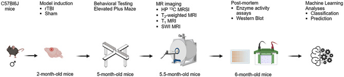
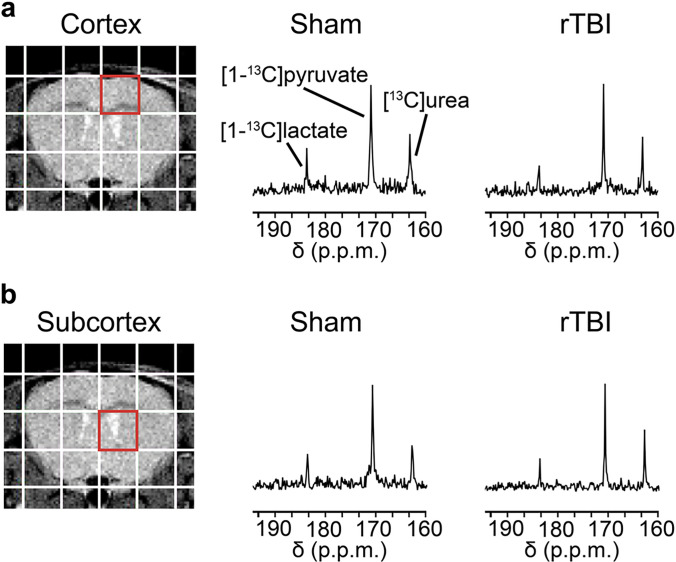
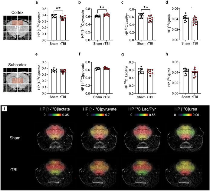
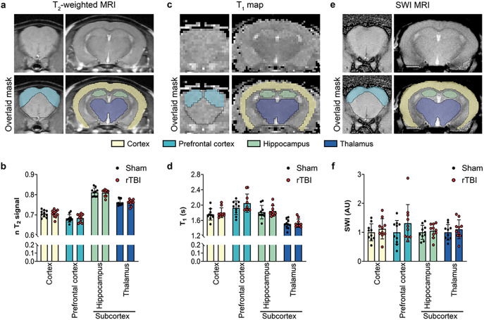
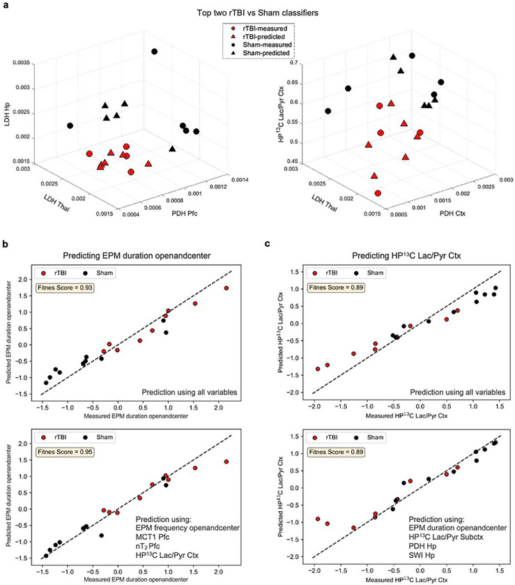
Career athletes, active military, and head trauma victims are at increased risk for mild repetitive traumatic brain injury (rTBI), a condition that contributes to the development of epilepsy and neurodegenerative diseases. Standard clinical imaging fails to identify rTBI-induced lesions, and novel non-invasive methods are needed. Here, we evaluated if hyperpolarized 13C magnetic resonance spectroscopic imaging (HP 13C MRSI) could detect long-lasting changes in brain metabolism 3.5 months post-injury in a rTBI mouse model. Our results show that this metabolic imaging approach can detect changes in cortical metabolism at that timepoint, whereas multimodal MR imaging did not detect any structural or contrast alterations. Using Machine Learning, we further show that HP 13C MRSI parameters can help classify rTBI vs. Sham and predict long-term rTBI-induced behavioral outcomes. Altogether, our study demonstrates the potential of metabolic imaging to improve detection, classification and outcome prediction of previously undetected rTBI.


Conflict of interest statement

Competing interests The authors declare that they have no competing interests.

Figures

Figure 1
Figure 1. Study outline.
Experimental timeline of the study. Two-month old male mice received a rTBI using the CHIMERA device or underwent a Sham procedure (no impact). Risk-taking behavior was evaluated at 3 months post-injury using the Elevated Plus Maze. MR imaging was performed 3.5 months after Sham or rTBI, and included HP 13C MRSI, T2-weighted MRI, T1 mapping MRI, and SWI MRI. Tissue was collected 4 months after Sham or rTBI procedures to evaluate PDH and LDH activities, and expression of MCT1 and MCT4. ML analyses methods were used to identify the best classifiers between rTBI and Sham, and the best predictors of the risk-taking behavior and HP 13C Lac/Pyr in the cortex.
Figure 2
Figure 2. HP 13C spectra following co-injection of HP [1–13C]pyruvate and [13C]urea.
Representative T2-weighted MR image overlaid with the grid used for HP 13C MRSI acquisitions. Representative 13C spectra showing HP [1-13C]pyruvate, HP [13C]urea and HP [1-13C]lactate in the (a) cortex (red voxel) and (b) subcortex (red voxel) for a Sham and a rTBI mouse.
Figure 3
Figure 3. HP 13C MRSI detects long-lasting metabolic alterations following rTBI.
Quantitative analyses of (a) HP [1-13C]lactate levels, (b) HP [1-13C]pyruvate levels, (c) HP 13C Lac/Pyr, and (d) HP [13C]urea for the cortex (highlighted red voxels), revealed lower HP [1-13C]lactate levels (p = 0.0073), higher HP [1-13C]pyruvate levels (p = 0.0073), and lower HP 13C Lac/Pyr (p = 0.0071) in rTBI compared to Sham mice. In contrast, quantitative analyses of (e) HP [1-13C]lactate levels, (f) HP [1–13C]pyruvate levels, (g) HP 13C Lac/Pyr, and (h) HP [13C]urea for the subcortex (highlighted red voxels), did not detect differences between rTBI and Sham mice. (i) Representative HP 13C heatmaps for a Sham and a rTBI mouse, highlighting lower HP [1-13C]lactate, higher HP [1-13C]pyruvate and lower HP 13C Lac/Pyr in cortical areas in rTBI mice. N = 9 rTBI and 10 Sham mice. Unpaired t-test (**p ≤ 0.01); data are expressed as means ± SD.
Figure 4
Figure 4. Multimodal MRI does not detect long-lasting effect of injury in rTBI.
(a) Representative T2-weighted MRI data and corresponding manual brain masking. (b) Quantitative analyses of T2-weighted signal intensity revealed no significant differences for brain subregions between Sham and rTBI. (c) Representative T1 map and corresponding manual brain masking. (d) Quantitative analyses of T1 maps revealed no significant differences for brain subregions between Sham and rTBI. (e) Representative SWI data and corresponding manual brain masking. (f) Quantitative analyses of SWI intensity revealed no significant differences for brain subregions between Sham and rTBI. Brain masking color code: yellow: cortex, green: light blue: prefrontal cortex; hippocampus; dark blue: thalamus. N = 10 rTBI and 10 Sham mice. Unpaired t-test; data are expressed as means ± SD.
Figure 5
Figure 5. ML analyses identify best rTBI and Sham classifiers and best predictors of changes in risk-taking behavior and HP 13C Lac/Pyr Ctx.
(a) Top two triplets that can classify rTBI (red) and Sham (black) mice. Here, circles represent the mice for which all three variables are measured whereas triangles represent mice for which at least one of the three variables were missing and predicted by ML algorithms (see Methods). (b-c) Prediction performance of the best predictors of risk-taking behavior (b, bottom panel), and HP 13C Lac/Pyr Ctx (c, bottom panel) compared to the prediction performance of the case in which all variables are used (b-c top panel). N = 10 rTBI and 10 Sham mice.

References

    1. Brett BL, Gardner RC, Godbout J, Dams-O’Connor K, Keene CD. Traumatic Brain Injury and Risk of Neurodegenerative Disorder. Biol Psychiatry 91, 498–507 (2022). - PMC - PubMed
    1. Ding K, Gupta PK, Diaz-Arrastia R. Epilepsy after Traumatic Brain Injury. In: Translational Research in Traumatic Brain Injury (eds Laskowitz D, Grant G) (2016). - PubMed
    1. Hay J, Johnson VE, Smith DH, Stewart W. Chronic Traumatic Encephalopathy: The Neuropathological Legacy of Traumatic Brain Injury. Annu Rev Pathol 11, 21–45 (2016). - PMC - PubMed
    1. Ng TS, et al. Neuroimaging in repetitive brain trauma. Alzheimers Res Ther 6, 10 (2014). - PMC - PubMed
    1. Asken BM, Rabinovici GD. Identifying degenerative effects of repetitive head trauma with neuroimaging: a clinically-oriented review. Acta Neuropathol Commun 9, 96 (2021). - PMC - PubMed

Publication types