[Preprint]. 2023 Aug 17:2023.08.15.553056.
doi: 10.1101/2023.08.15.553056.

Exploring Temporal and Sex-Linked Dysregulation in Alzheimer's Disease Phospho-Proteome

Affiliations

Exploring Temporal and Sex-Linked Dysregulation in Alzheimer's Disease Phospho-Proteome

Serhan Yılmaz et al. bioRxiv. .

Update in

Abstract

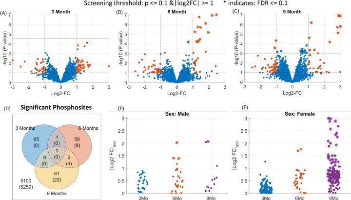
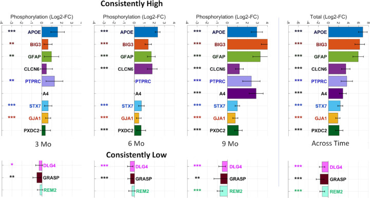
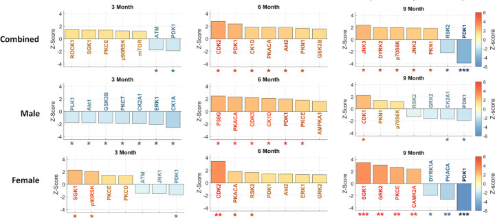
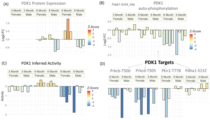
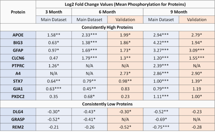
This study aims to characterize dysregulation of phosphorylation for the 5XFAD mouse model of Alzheimer's disease (AD). Employing global phosphoproteome measurements, we analyze temporal (3, 6, 9 months) and sex-dependent effects on mouse hippocampus tissue to unveil molecular signatures associated with AD initiation and progression. Our results indicate 1.9 to 4.4 times higher phosphorylation prevalence compared to protein expression across all time points, with approximately 4.5 times greater prevalence in females compared to males at 3 and 9 months. Moreover, our findings reveal consistent phosphorylation of known AD biomarkers APOE and GFAP in 5XFAD mice, alongside novel candidates BIG3, CLCN6 and STX7, suggesting their potential as biomarkers for AD pathology. In addition, we identify PDK1 as a significantly dysregulated kinase at 9 months in females, and the regulation of gap junction activity as a key pathway associated with Alzheimer's disease across all time points. AD-Xplorer, the interactive browser of our dataset, enables exploration of AD-related changes in phosphorylation, protein expression, kinase activities, and pathways. AD-Xplorer aids in biomarker discovery and therapeutic target identification, emphasizing temporal and sex-specific nature of significant phosphoproteomic signatures. Available at: https://yilmazs.shinyapps.io/ADXplorer.


Figures

Figure 1.
Figure 1.. Phosphosites identified as differentially phosphorylated for Alzheimer’s disease at different time points.
(Panels A-C) Volcano plots indicating the screened phosphosites with high or low phosphorylation at 3/6/9 month time points. The dashed horizontal lines in each plot indicate the statistical significance threshold (p<0.1 and FDR ≤ 0.1) and the vertical line the threshold on fold changes (>2). The statistically significant phosphosites at FDR ≤ 0.1 level are marked as stars (*). (Panel D) Venn diagram showing the number of phosphosites that pass the screening (p ≤ 0.1 and 0.5 ≥ FC ≥ 2) for 3/6/9 months data. Whereas, the numbers in parenthesis indicate the significant ones at FDR ≤ 0.1 level. (Panels E-F) Identified phosphosites that pass the screening in sex-specific analysis. The y-axis indicates a lower-bound of the 95% confidence intervals for each phosphosite.
Figure 2.
Figure 2.. Proteins identified as differentially phosphorylated for Alzheimer’s disease across different time-points.
(Left panel) The bars indicate the mean phosphorylation for each protein identified as consistently phosphorylated and the error bars indicate 95% confidence intervals. Proteins are marked according to their statistical significance levels: * for p ≤ 0.1, ** for FDR ≤ 0.1 and *** for FDR ≤ 0.01.
Figure 3.
Figure 3.. Inferred kinase activities across 3/6/9 months time-points and different sexes.
For each subplot, the top seven most significant kinases are shown. The error bars indicate 95% confidence intervals. Kinases are marked according to their statistical significance levels: * for p ≤ 0.1, ** for FDR ≤ 0.1 and *** for FDR ≤ 0.01.
Figure 4.
Figure 4.. Dysregulation of PDK1 at protein expression and phosphorylation level for different sexes and timepoints of Alzheimer’s disease.
(Panel A) Protein expression (log2-FC) of PDK1 for all timepoints and sexes. (Panel B) The auto-phosphorylation (log2-FC) of PDK1 for all timepoints and sexes. (Panel C) Inferred activity score of PDK1 for all timepoints and sexes. (Panel D) Phosphorylation (log2-FC) of known targets of PDK1 for 9 month data. For all plots, the coloring is done to reflect the degree of statistical significance based on z-scores.
Figure 5.
Figure 5.. Validation data results for candidate proteins.
Proteins are marked according to their statistical significance levels: * for p ≤ 0.1, ** for FDR ≤ 0.1 and *** for FDR ≤ 0.01.

References

    1. [Bilbao, 2014] Bilbao A., Rieker C., Cannella N., Parlato R., Golda S., Piechota M., … & Parkitna J. R. (2014). CREB activity in dopamine D1 receptor expressing neurons regulates cocaine-induced behavioral effects. Frontiers in behavioral neuroscience, 8, 212. - PMC - PubMed
    1. [Bornemann, 2001] Bornemann K. D., Wiederhold K. H., Pauli C., Ermini F., Stald M., Schnell L., et al. (2001). Aβ-induced inflammatory processes in microglia cells of APP23 transgenic mice. The American journal of pathology, 158(1), 63–73. - PMC - PubMed
    1. [Braunstein, 2016] Li T., Braunstein K. E., Zhang J., Lau A., Sibener L., Deeble C., et al. (2016) The neuritic plaque facilitates pathological conversion of tau in an Alzheimer’s disease mouse model. Nat. Commun. 7, 12082. - PMC - PubMed
    1. [Dammer, 2022] Dammer E. B., Ping L., Duong D. M., Modeste E. S., Seyfried N. T., Lah J. J., et al. (2022). Multi-platform proteomic analysis of Alzheimer’s disease cerebrospinal fluid and plasma reveals network biomarkers associated with proteostasis and the matrisome. Alzheimer’s Research & Therapy, 14(1), 1–32. - PMC - PubMed
    1. [Del Greco, 2011] Del Greco M F., Pattaro C., Luchner A., Pichler I., Winkler T., Hicks A. A., et al. (2011). Genome-wide association analysis and fine mapping of NT-proBNP level provide novel insight into the role of the MTHFR-CLCN6-NPPA-NPPB gene cluster. Human molecular genetics, 20(8), 1660–1671. - PMC - PubMed

Publication types