. 2023 Aug 14:14:1206026.
doi: 10.3389/fimmu.2023.1206026. eCollection 2023.

Exacerbated lung inflammation following secondary RSV exposure is CD4+ T cell-dependent and is not mitigated in infant BALB/c mice born to PreF-vaccinated dams

Affiliations

Exacerbated lung inflammation following secondary RSV exposure is CD4+ T cell-dependent and is not mitigated in infant BALB/c mice born to PreF-vaccinated dams

Jessica L Kosanovich et al. Front Immunol. .

Abstract

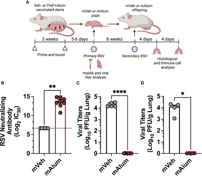
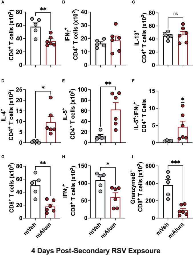
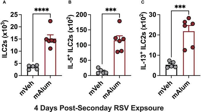
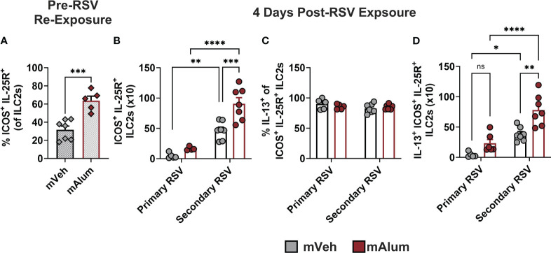
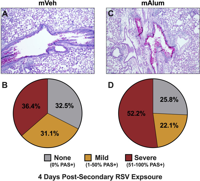
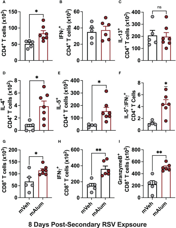
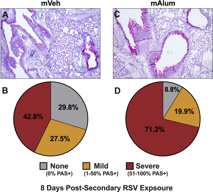
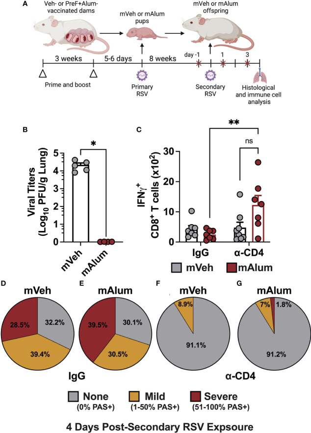
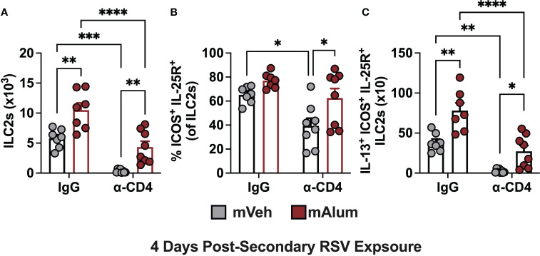
Respiratory syncytial virus (RSV) is the leading cause of childhood hospitalizations due to bronchiolitis in children under 5 years of age. Moreover, severe RSV disease requiring hospitalization is associated with the subsequent development of wheezing and asthma. Due to the young age in which viral protection is needed and risk of vaccine enhanced disease following direct infant vaccination, current approaches aim to protect young children through maternal immunization strategies that boost neutralizing maternal antibody (matAb) levels. However, there is a scarcity of studies investigating the influence of maternal immunization on secondary immune responses to RSV in the offspring or whether the subsequent development of wheezing and asthma is mitigated. Toward this goal, our lab developed a murine model of maternal RSV vaccination and repeat RSV exposure to evaluate the changes in immune response and development of exacerbated lung inflammation on secondary RSV exposure in mice born to immunized dams. Despite complete protection following primary RSV exposure, offspring born to pre-fusion F (PreF)-vaccinated dams had exaggerated secondary ILC2 and Th2 responses, characterized by enhanced production of IL-4, IL-5, and IL-13. These enhanced type 2 cellular responses were associated with exaggerated airway eosinophilia and mucus hyperproduction upon re-exposure to RSV. Importantly, depletion of CD4+ T cells led to complete amelioration of the observed type 2 pathology on secondary RSV exposure. These unanticipated results highlight the need for additional studies that look beyond primary protection to better understand how maternal immunization shapes subsequent immune responses to repeat RSV exposure.

Keywords: Respiratory syncytial virus; T cell-mediated pathology; maternal immunization; secondary RSV exposure; type 2 inflammation.


Conflict of interest statement

SG, DB, and MY are employed by Calder Biosciences. The remaining authors declare that the research was conducted in the absence of any commercial or financial relationships that could be construed as a potential conflict of interest.

Figures

Figure 1
Figure 1
Offspring born to PreF+Alum-vaccinated dams are protected from primary and secondary RSV exposure. Pregnant Balb/c mice completed a 2-dose vaccination series of PBS (mVeh) or preF+Alum 1 week prior to parturition. At PND5-6, a cohort of mVeh and mAlum pups were culled for pre-challenge serum antibody analysis while a second cohort of pups were intranasally exposed to 5x10⁵ PFU/gm RSV L19 and culled a 4-days post-exposure for viral titer analysis. A third cohort of mVeh and mAlum offspring were aged to adulthood, re-exposed to RSV L19 at 9 weeks old (PND63) and culled for sample collection at 4- and 8-days post-exposure (A). Pre-challenge serum from mVeh and mAlum pups was analyzed for RSV neutralizing antibody as described in the methods; since no dilution of Veh control samples achieved a signal less than 80% of the limit of blank-corrected virus-only control, no IC50 could be determined for these samples and the lowest serum dilution used in the assay is reported as the neutralization titer, in this case 1:100 (marked by a dashed line) (B). Left lungs were harvested from pups (C) and aged adults for viral titer analysis (D). Data are represented as mean ± SEM (n=4-9 mice per group). Statistical significance was calculated using an unpaired t-test between mVeh and mAlum offspring. *p ≤ 0.05, **p ≤ 0.01 and ****p ≤ 0.0001.
Figure 2
Figure 2
mAlum offspring have a dominant Th2 phenotype with reduced cytotoxic CD8+ T cell activity. mVeh and mAlum offspring were treated as described in Figure 1A . At 4dpse, total BAL CD4+ T cells (A), as well as intracellular production of IFNγ, (B), IL-13 (C), IL-4 (D), and IL-5 (E) by CD4+ T cells were quantified. From these totals, the ratio of IL-5:IFNγ CD4+ T cells was calculated (F). At the same timepoint, total BAL CD8+ T cells (G), as well as those producing IFNγ (H), and GranzymeB (I) were quantified. Data are represented as mean ± SEM (n=5-6 mice per group). Statistical significance was calculated using an unpaired t-test (A-E, G-I) or Wilcoxon signed-rank test (F). ns – non-significant, *p ≤ 0.05, **p ≤ 0.01 and ***p ≤ 0.001.
Figure 3
Figure 3
Enhanced ILC2 activity in mAlum offspring following secondary RSV exposure. mVeh and mAlum offspring were generated and exposed to RSV as described in Figure 1 . Total lung ILC2s (A), along with those producing IL-5 (B) and IL-13 (C) were quantified at 4dpse. Data are represented as mean ± SEM (n=5-6 mice per group). Statistical significance was calculated using an unpaired t-test. ***p ≤ 0.001 and ****p ≤ 0.0001.
Figure 4
Figure 4
ILC2s with hyperresponsive phenotype are enriched in lungs of mAlum offspring. The frequency of hyperresponsive ILC2s, characterized by surface expression of ICOS and IL-25R, was assessed in the lungs of mVeh and mAlum offspring prior to secondary RSV exposure (A). Total ICOS+ IL-25R+ ILC2s (B), the frequency of IL-13-producing ICOS+ IL-25R+ ILC2s (C) and IL-13+ ICOS+ IL-25R+ ILC2s (D) were quantified at 4 days post-exposure in the lungs of adult mVeh and mAlum offspring following primary (1°) and secondary (2°) RSV exposure. Data are represented as mean ± SEM (n=5-7 mice per group). Statistical significance was calculated using an unpaired t-test (A) or two-way ANOVA with Sidak’s multiple comparison test (B-D) between groups. ns – nonsignificant, *p ≤ 0.05 **p ≤ 0.01, ***p ≤ 0.001 and ****p ≤ 0.0001.
Figure 5
Figure 5
Mucus metaplasia increased in mAlum offspring following secondary RSV exposure. At 4dpse, lungs were formalin-fixed, paraffin-embedded, and processed for PAS staining. Each panel represents an individual mouse from each offspring group (A, C). Lung sections were scored as described in the methods and represented as a proportion of airways scored as having no PAS staining (none), airways with 1-50% PAS staining (mild), and airways with 51-100% PAS staining (severe) (B, D). n=3 mice per group.
Figure 6
Figure 6
Th2 responses are sustained as cytotoxic CD8+ T cells increase in mAlum offspring. At 8dpse, total BAL CD4+ T cells (A), as well as intracellular production of IFNγ, (B), IL-13 (C), IL-4 (D), and IL-5 (E) by CD4+ T cells were quantified in mVeh and mAlum offspring, generated as described in Figure 1 . From these totals, the ratio of IL-5:IFNγ CD4+ T cells was calculated (F). Total BAL CD8+ T cells (G), as well as those producing IFNγ (H), and GranzymeB (I) were quantified. Data are represented as mean ± SEM (n=5-7 mice per group). Statistical significance was calculated using an unpaired t-test (A-E, G-I) or Wilcoxon signed-rank test (F). ns – nonsignificant, *p ≤ 0.05 and **p ≤ 0.01.
Figure 7
Figure 7
Worsening mucus production in mAlum offspring over course of secondary RSV exposure. At 8dpse, lungs were formalin-fixed, paraffin-embedded, and processed for PAS staining. Each panel represents an individual mouse from each offspring group (A, C). Lung sections were scored, and the proportion of airways scored as having no PAS staining (none), airways with 1-50% PAS staining (mild), and airways with 51-100% PAS staining (severe) was calculated (B, D). n=3 mice per group.
Figure 8
Figure 8
CD4+ T cells contribute to mucus production, but not viral control in mAlum offspring. Pregnant dams were vaccinated, and pups were primarily challenged with RSV and aged to adulthood as described in Figure 1 . Adult mVeh and mAlum offspring were administered isotype (IgG) or CD4-depleting (α-CD4) antibody starting 1 day prior to secondary RSV challenge and every 2 days through sacrifice (A). Left lungs were harvested for viral titer analysis (B) while IFNγ+ CD8+ T cells were quantified from the BAL (C). The proportion of airways scored with no, mild, and severe PAS staining was calculated in IgG-treated mVeh (D) and mAlum (E) offspring, as well as CD4-depleted mVeh (F) and mAlum (G) offspring. Data are represented as mean ± SEM (n=5-8 mice per group). Statistical significance was calculated using an unpaired t-test (B) or two-way ANOVA with Sidak’s multiple comparison test (C). *p ≤ 0.05 and **p ≤ 0.01.
Figure 9
Figure 9
Hyperresponsive ILC2s are reduced in the absence of CD4+ T cells. Adult mVeh and mAlum offspring were treated as described in Figure 8 . Total lung ILC2s (A), frequency of lung ILC2s dual positive for ICOS and IL-25 (B), and IL-13+ ICOS+ IL-25R+ ILC2s (C) were quantified from IgG- and α-CD4-treated mVeh and mAlum offspring. Data are represented as mean ± SEM (n=7-8 mice per group). Statistical significance was calculated using two-way ANOVA with Sidak’s multiple comparison test (C). *p ≤ 0.05 **p ≤ 0.01, ***p ≤ 0.001 and ****p ≤ 0.0001.

Similar articles

Cited by

References

    1. Shi T, McAllister DA, O’Brien KL, Simoes EAF, Madhi SA, Gessner BD, et al. . Global, regional, and national disease burden estimates of acute lower respiratory infections due to respiratory syncytial virus in young children in 2015: a systematic review and modelling study. Lancet (2017) 390(10098):946–58. doi: 10.1016/S0140-6736(17)30938-8 - DOI - PMC - PubMed
    1. Stein RT, Sherrill D, Morgan WJ, Holberg CJ, Halonen M, Taussig LM, et al. . Respiratory syncytial virus in early life and risk of wheeze and allergy by age 13 years. Lancet (9178) 1999:541–5:354. doi: 10.1016/S0140-6736(98)10321-5 - DOI - PubMed
    1. Zomer-Kooijker K, van der Ent CK, Ermers MJ, Uiterwaal CS, Rovers MM, Bont LJ, et al. . Increased risk of wheeze and decreased lung function after respiratory syncytial virus infection. PloS One (2014) 9(1):e87162. doi: 10.1371/journal.pone.0087162 - DOI - PMC - PubMed
    1. Glezen WP, Taber LH, Frank AL, Kasel JA. Risk of primary infection and reinfection with respiratory syncytial virus. Am J Dis Child. (1986) 140(6):543–6. doi: 10.1001/archpedi.1986.02140200053026 - DOI - PubMed
    1. Hall CB, Weinberg GA, Iwane MK, Blumkin AK, Edwards KM, Staat MA, et al. . The burden of respiratory syncytial virus infection in young children. N Engl J Med (2009) 360(6):588–98. doi: 10.1056/NEJMoa0804877 - DOI - PMC - PubMed

Publication types