. 2023 Aug 14:14:1225610.
doi: 10.3389/fimmu.2023.1225610. eCollection 2023.

Novel anti-CD30/CD3 bispecific antibodies activate human T cells and mediate potent anti-tumor activity

Affiliations

Novel anti-CD30/CD3 bispecific antibodies activate human T cells and mediate potent anti-tumor activity

Mary L Faber et al. Front Immunol. .

Abstract

CD30 is expressed on Hodgkin lymphomas (HL), many non-Hodgkin lymphomas (NHLs), and non-lymphoid malignancies in children and adults. Tumor expression, combined with restricted expression in healthy tissues, identifies CD30 as a promising immunotherapy target. An anti-CD30 antibody-drug conjugate (ADC) has been approved by the FDA for HL. While anti-CD30 ADCs and chimeric antigen receptors (CARs) have shown promise, their shortcomings and toxicities suggest that alternative treatments are needed. We developed novel anti-CD30 x anti-CD3 bispecific antibodies (biAbs) to coat activated patient T cells (ATCs) ex vivo prior to autologous re-infusions. Our goal is to harness the dual specificity of the biAb, the power of cellular therapy, and the safety of non-genetically modified autologous T cell infusions. We present a comprehensive characterization of the CD30 binding and tumor cell killing properties of these biAbs. Five unique murine monoclonal antibodies (mAbs) were generated against the extracellular domain of human CD30. Resultant anti-CD30 mAbs were purified and screened for binding specificity, affinity, and epitope recognition. Two lead mAb candidates with unique sequences and CD30 binding clusters that differ from the ADC in clinical use were identified. These mAbs were chemically conjugated with OKT3 (an anti-CD3 mAb). ATCs were armed and evaluated in vitro for binding, cytokine production, and cytotoxicity against tumor lines and then in vivo for tumor cell killing. Our lead mAb was subcloned to make a Master Cell Bank (MCB) and screened for binding against a library of human cell surface proteins. Only huCD30 was bound. These studies support a clinical trial in development employing ex vivo-loading of autologous T cells with this novel biAb.

Keywords: antibody heteroconjugation; cytotoxicity; immunotherapy; membrane proteome array; surface plasmon resonance; transplantation; tumor necrosis factor receptor superfamily; xenograft tumor models.


Conflict of interest statement

Authors MF, NS, and JM are co-founders of the company Tundra Targeted Therapeutics, Inc. Author AT is a co-founder of the company Nova Immune Platform LLC, and author LGL is a co-founder of the company Transtarget, Inc., and a member of the SAB of Rapa Therapeutics. The remaining authors declare that the research was conducted in the absence of any commercial or financial relationships that could be construed as a potential conflict of interest.

Figures
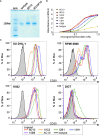
Figure 1
Figure 1
Purification of novel anti-CD30 mAbs and binding to huCD30. (A) Coomassie Blue-stained gel following non-reducing PAGE of purified 8D10 and 8D10F10 (subclone for preparation of a clinical trial MCB). (B) Binding of various dilutions of our novel mAbs to huCD30-GST protein was assessed by ELISA. (C) FCM analyses were carried out for all five novel CD30 mAbs and AC10. Representative FCM histograms are shown.
Figure 2
Figure 2
Categorization of mAbs by sequence and cluster binding. (A) VL and VH protein sequences were compared across all 5 novel mAbs and AC10. Levels of sequence similarity range from white (low) to blue (high). Numbering indicates the sequence identity as a percentage. (B) Cluster mapping via competitive binding. Fluorescently labelled mAbs were incubated with CD30+ SU-DHL-1 cells blocked with excess unlabeled mAbs. FCM analysis was then performed. High inhibition (red) indicates a shared cluster and low inhibition (blue) indicates distinct clusters. Results represent the means of 3 independent experiments (n=3). (C) A depiction of the three known clusters of mAb binding to CD30.
Figure 3
Figure 3
Assessment of biAb binding capabilities. (A) BiAbs capable of binding to CD3 and CD30 were developed by heteroconjugation of mAb 8D10 or 10C2 with OKT3. (B) The ability of biAbs 8D10 and 10C2 to bind to both CD3 and CD30 post-conjugation was determined by FCM. Binding of unconjugated 8D10, 10C2, and OKT3 mAbs and an isotype control is shown for reference. (C) A two-color FCM assay was developed to determine T cell-Raji LV30 conjugate formation in the presence of biAbs. (D) Assessment of percent conjugation of Raji and Raji LV30 cells with unarmed or biAb-armed T cells was measured by detection of two-color events and plotted as a percentage of all events. Results represent means ± SD of 3 independent experiments (n = 3). Representative FCM plots from this assay are shown in (E). n.s, not significant, ** = p < 0.01 between the indicated groups, calculated using a one-way ANOVA with Tukey’s multiple comparisons test.
Figure 4
Figure 4
Cytokine production by biAb-armed T cells after co-culture with tumor cells. (A) Co-culture conditions included no target (0), a CD30low cell line (Raji), CD30+ cell lines (Raji LV30 and RPMI 6666), and a CD30 cell line (OCIAML2). Production of IFN-γ, TNF-α, IL-2, GM-CSF, and IL-4 was assessed. (B) Co-culture conditions included no target (0), a CD30low cell line (Raji), and a CD30+ cell line (HH). Results represent means ± SD of 3 independent experiments (n = 3). * = p < 0.05, ** = p < 0.01, *** = p < 0.001 between the indicated groups, calculated using a two-way ANOVA with Dunnett’s multiple comparisons test.
Figure 5
Figure 5
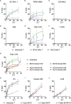
Cytotoxicity of CD30/CD3 biAb-armed T cells. (A–F) Cytotoxicity of 8D10 & 10C2 biAb-armed T cells against CD30 and CD30+ tumor cells was assessed using a standard 4-hour 51Cr release assay. Lysis of two tumor cell lines with endogenous CD30 expression, (A) SU-DHL-1 and (B) RPMI 6666, was measured, in addition to (C) OCIAML2, a CD30 cell line, (D) NT Raji, a CD30low cell line, and (E) Raji LV30 cells, which are CD30high. (F) Activated T cells, which express low levels of CD30, were also assessed for lysis. Cytotoxicity of unarmed, 8D10, and AC10 biAb-armed T cells was compared at 24 hours against Raji LV30 cells (G) after T cells were armed with varying levels (50-500ng/million cells) of biAb. Cytotoxicity of 8D10 and 8D10F10 biAb-armed human T cells used fresh or after cryopreservation against CD30+ cell lines: (H) HH, (I) Karpas, and (J) L428 was ascertained. Results represent means ± SD of 3-4 independent experiments per cell line (n = 3-4). * = p < 0.05, ** = p < 0.01, *** = p < 0.001, **** = p < 0.0001 between biAb-armed T cells and unarmed T cells. # = p < 0.05, ## = p < 0.01, #### = p < 0.0001 between 8D10 and 10C2 biAb-armed T cells (A–F) and between 8D10 and AC10 biAb-armed T cells (G). Statistical significance was calculated using a two-way ANOVA with Tukey’s multiple comparisons test.
Figure 6
Figure 6
Membrane Proteome Array (MPA) analysis and in vivo tumor inhibition in NSG mice. (A) Purified 8D10F10 was tested for binding to the MPA from Integral Molecular at a previously determined optimal concentration. (B) Validation results. Serial ligand dilutions were incubated with newly identified targets and binding was measured by FCM. (C) In vivo survival studies. NSG mice (8 per group from 2 independent experiments) were injected with HH tumor cells (day 0) and then with PBS, unarmed, or 8D10 biAb-armed human T cells twice per week for 3 weeks from day 10. *p < 0.05 calculated using log-rank (Mantel-Cox) test.

References

    1. Gruss HJ, DaSilva N, Hu ZB, Uphoff CC, Goodwin RG, Drexler HG. Expression and regulation of CD30 ligand and CD30 in human leukemia-lymphoma cell lines. Leukemia (1994) 8:2083–94. - PubMed
    1. Durkop H, Foss HD, Eitelbach F, Anagnostopoulos I, Latza U, Pileri S, et al. Expression of the CD30 antigen in non-lymphoid tissues and cells. J Pathol (2000) 190:613–8. doi: 10.1002/(SICI)1096-9896(200004)190:5<613::AID-PATH559>3.0.CO;2-0 - DOI - PubMed
    1. Kim WY, Nam SJ, Kim S, Kim TM, Heo DS, Kim CW, et al. Prognostic implications of CD30 expression in extranodal natural killer/T-cell lymphoma according to treatment modalities. Leuk Lymphoma (2015) 56:1778–86. doi: 10.3109/10428194.2014.974048 - DOI - PubMed
    1. Cheng J, Zhu H, Choi JK. CD30 expression in pediatric neoplasms, study of 585 cases. Pediatr Dev Pathol (2017) 20:191–6. doi: 10.1177/1093526616689185 - DOI - PubMed
    1. van der Weyden CA, Pileri SA, Feldman AL, Whisstock J, Prince HM. Understanding CD30 biology and therapeutic targeting: a historical perspective providing insight into future directions. Blood Cancer J (2017) 7:e603. doi: 10.1038/bcj.2017.85 - DOI - PMC - PubMed

Publication types

Supplementary concepts