. 2023 Oct;146(4):631-645.
doi: 10.1007/s00401-023-02625-6. Epub 2023 Aug 30.

Analysis of inflammatory markers and tau deposits in an autopsy series of nine patients with anti-IgLON5 disease

Affiliations

Analysis of inflammatory markers and tau deposits in an autopsy series of nine patients with anti-IgLON5 disease

Evelyn Berger-Sieczkowski et al. Acta Neuropathol. 2023 Oct.

Abstract

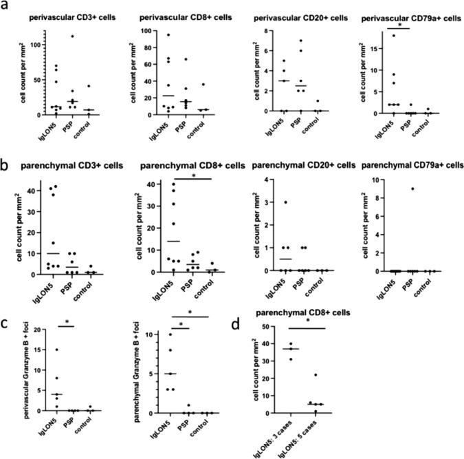
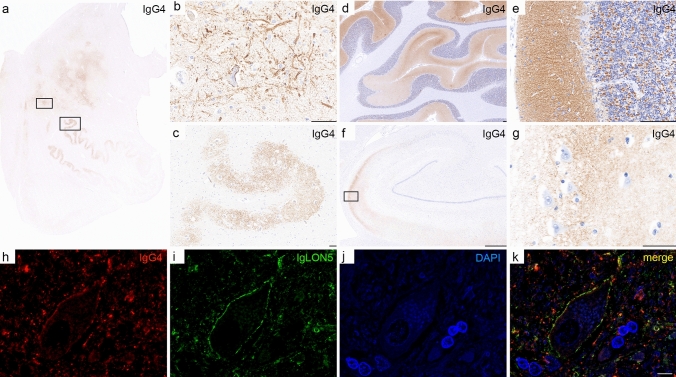
Anti-IgLON5 disease is a rare neurological, probably autoimmune, disorder associated in many cases with a specific tauopathy. Only a few post-mortem neuropathological studies have been reported so far. Little is known about the pathogenic mechanisms that result in neurodegeneration. We investigated the neuropathology of anti-IgLON5 disease and characterized cellular and humoral inflammation. We included nine cases (six of them previously published). Median age of patients was 71 years (53-82 years), the median disease duration was 6 years (0.5-13 years), and the female to male ratio was 5:4. Six cases with a median disease duration of 9 years presented a prominent tauopathy. Five of them had a classical anti-IgLON5-related brainstem tauopathy and another presented a prominent neuronal and glial 4-repeat tauopathy, consistent with progressive supranuclear palsy (PSP). Three cases with short disease duration (median 1.25 years) only showed a primary age-related neurofibrillary pathology. Inflammatory infiltrates of T and B cells were mild to moderate and did not significantly differ between anti-IgLON5 disease cases with or without tauopathy. In contrast, we found an extensive neuropil deposition of IgG4 in the tegmentum of the brainstem, olivary nucleus, and cerebellar cortex that was most prominent in two patients with short disease duration without the typical IgLON5-related tauopathy. The IgG4 deposits were particularly prominent in the cerebellar cortex and in these regions accompanied by mild IgG1 deposits. Activated complement deposition (C9neo) was absent. Our study indicates that IgLON5-related tau pathology occurs in later disease stages and may also present a PSP-phenotype with exclusively 4-repeat neuronal and glial tau pathology. The prominent deposition of anti-IgLON5 IgG4 at predilection sites for tau pathology suggests that anti-IgLON5 antibodies precede the tau pathology. Early start of immunotherapy might prevent irreversible neuronal damage and progression of the disease, at least in a subgroup of patients.


Conflict of interest statement

E. Berger-Sieczkowski, V. Endmayr, C. Haider, G. Ricken, P. Jauk, S. Macher, Schnider, E. Bradley-Zechmeister, R. Reinecke, Reinecke Raphael, G. Kasprian, C. Weber, M. Bergmann, C. Gaig, L. Sabater, and E. Gelpi report no relevant disclosures. I. Koneczny has participated in meetings sponsored by Alexion. I. Milenkovic has participated in meetings sponsored by and received honoraria (lectures, advisory boards, consultations) from: UCB, Sanofi-Genzyme, BioMarin and Takeda. A. Heidbreder reports speaker honoraria for UCB, Bioprojet, Idorsia, Medice, Jazz Pharmaceuticals. B. Högl reports speaker honoraria Jazz and Abbvie and advisor feed from Lundbeck. S. Mariotto reports speaker honoraria from Biogen, Sanofy, and Novartis. T. Berger has participated in meetings sponsored by and received honoraria (lectures, advisory boards, consultations) from pharmaceutical companies marketing treatments for MS: Allergan, Biogen, Biologix, Bionorica, BMS/Celgene, Eisei, Janssen-Cilag, MedDay, Merck, Novartis, Roche, Sandoz, Sanofi-Genzyme, Teva, UCB. His institution has received financial support in the past 12 months by unrestricted research grants (Bayer, Biogen, BMS/Celgene, Merck, Novartis, Roche, Sanofi-Genzyme, Teva) and for participation in clinical trials in multiple sclerosis sponsored by Alexion, Bayer, Biogen, BMS/Celgene, Merck, Novartis, Roche, Sanofi-Aventis, Teva. R. Höftberger reports speaker honoraria from Novartis and Biogen. The Medical University of Vienna (Austria; employer of Dr. Höftberger) receives payment for antibody assays and for antibody validation experiments organized by Euroimmun (Lübeck, Germany). Dr. F. Graus holds a patent licensed to Euroimmun for the use of IgLON5 in an autoantibody test, for which he receives royalties, and receives honoraria from MedLink Neurology for his role as associate editor. W. Pirker attended advisory boards by Stada, AbbVie Pharma, Merz and Bial. He received travel grants and educational support from Boehringer Ingelheim, AOP Pharma as well as lecturing honoraria from GE, UCB, Bial, Stada, AOP Orphan, AbbVie, Medtronic and Merz Pharma.

Figures

Fig. 1
Fig. 1
Brain MRI scan of patient 1 with PSP neuropathology. Brain MRI scan (axial, fluid-attenuated recovery sequences (FLAIR)) performed 8 years after symptom onset shows cortical and subcortical atrophy (a). Sagittal FLAIR image reveals reduced size of the mesencephalic tegmentum leading to the hummingbird appearance of brainstem (b; red arrow). Axial FLAIR image of the brainstem shows an increased signal hyperintensity of the periaqueductal grey matter (c, white arrow). Furthermore, atrophy of the mesencephalic and pontine tegmentum is evident (c, d; red arrows)
Fig. 2
Fig. 2
PSP neuropathology in anti-IgLON5 disease (patient 1). Neuropathology shows neuronal loss and the presence of neurofibrillary tangles in the tegmentum of the midbrain, pons, and medulla oblongata (ac; AT8), subthalamic nucleus (d; H&E), substantia nigra pars compacta (e; H&E), locus coeruleus (f; AT8; upper rectangle in b enlarged in f), and magnocellular nuclei of the medulla oblongata (g; AT8, upper rectangle in c enlarged in g) that were strongly immunoreactive for hTau, particularly for 4-repeat tau isoforms (h, substantia nigra; RD4) and negative for 3-repeat tau isoforms (i, substantia nigra; RD3). In addition, neurofibrillary tangles and neuropil threads were visible in the pontine base (j; AT8; lower rectangle in b enlarged in j), synaptic glomeruli of the cerebellar cortex (k; AT8; arrows), and olivary nucleus (l; AT8; lower rectangle in c enlarged in l). Glial pathology in basal ganglia consists of tufted astrocytes (m; AT8), some granular fuzzy astrocytes (n; GFAP), and oligodendroglial coiled bodies (o; AT8). Scale bars: d, e, k, m, n, o: 50 μm; fj, l: 100 μm
Fig. 3
Fig. 3
Cellular inflammation in anti-IgLON5 disease. Cellular inflammation was mild to moderate and mainly composed of perivascular and parenchymal CD3 (a) and CD8 positive T cells (b) and few perivascular CD79a positive B cells/plasma cells (c). Parenchymal CD8 T cells were granzyme B positive and granules showed a polarization towards neurons (d; arrows). In addition, neurons showed an upregulation of MHC class I in the reticular formation and olivary nuclei (e, rectangle enlarged in f). Marked microglia activation was found in the HLA-DR staining in the tegmentum of medulla oblongata and nucleus olivaris (g), including the formation of microglial nodules (h; rectangle in g enlarged in h). Images are depicted from patient 1. Scale bars: 50 μm
Fig. 4
Fig. 4
Inflammation in anti-IgLON5 disease. Scatter dot plots with an overview of CD3 + , CD8 + T cells and CD20 + , CD79a + B-/plasma cells either perivascular (a) or in the parenchyma (b) of the brainstem of definite anti-IgLON5 patients. CD8/Granzyme B + T cells were significantly more frequent in the parenchyma of IgLON5 patients (IgLON5 vs. PSP p = 0.03; IgLON5 vs. control p = 0,01) (c). Three cases showed significantly (p = 0.04) more parenchymal CD8 + T cells among them two cases with short disease duration and no tauopathy and one case with PSP-like pathology but no immunotherapy compared to the other IgLON5 patients (d). A non-parametric Kruskal–Wallis or Mann–Whitney test has been performed, using a post-hoc Dunn`s multiple comparison correction, *can be assumed as p < 0.05 if not otherwise stated, not significant differences are not illustrated
Fig. 5
Fig. 5
IgG4 deposits in anti-IgLON5 disease. IgG4 deposits were prominent in the tegmentum of the brainstem and olivary nucleus (ac; rectangles in an enlarged in b and c), molecular layer and synaptic glomeruli in the granule cell layer of the cerebellar cortex (d, e), and one case also showed prominent IgG4 deposition in the CA1-CA2 sector of the hippocampus (f, g; rectangle in f enlarged in g). Immunofluorescence double labelling revealed a co-localisation of IgG4 deposits (h, red) with IgLON5 (i, green) on neuronal membranes and the neuropil (j, DAPI; k, merge). Images a-c and hk are depicted from patient 3, and images d-g are depicted from patient 7; Scale bars: b, c, g: 50 μm; d, e: 150 μm; f: 1 mm; k: 10 μm

References

    1. Altendorfer B, Unger MS, Poupardin R, Hoog A, Asslaber D, Gratz IK, et al. Transcriptomic profiling identifies CD8(+) t cells in the brain of aged and Alzheimer’s Disease transgenic mice as tissue-resident memory T cells. J Immunol. 2022;209:1272–1285. doi: 10.4049/jimmunol.2100737. - DOI - PMC - PubMed
    1. Attems J, Toledo JB, Walker L, Gelpi E, Gentleman S, Halliday G, et al. Neuropathological consensus criteria for the evaluation of Lewy pathology in post-mortem brains: a multi-centre study. Acta Neuropathol. 2021;141:159–172. doi: 10.1007/s00401-020-02255-2. - DOI - PMC - PubMed
    1. Bruggemann N, Wandinger KP, Gaig C, Sprenger A, Junghanns K, Helmchen C, et al. Dystonia, lower limb stiffness, and upward gaze palsy in a patient with IgLON5 antibodies. Mov Disord. 2016;31:762–764. doi: 10.1002/mds.26608. - DOI - PubMed
    1. Cagnin A, Mariotto S, Fiorini M, Gaule M, Bonetto N, Tagliapietra M, et al. Microglial and neuronal TDP-43 pathology in Anti-IgLON5-related tauopathy. J Alzheimers Dis. 2017;59:13–20. doi: 10.3233/jad-170189. - DOI - PubMed
    1. Erro ME, Sabater L, Martinez L, Herrera M, Ostolaza A, Garcia de Gurtubay I, et al. Anti-IGLON5 disease: A new case without neuropathologic evidence of brainstem tauopathy. Neurol Neuroimmunol Neuroinflamm. 2020 doi: 10.1212/nxi.0000000000000651. - DOI - PMC - PubMed

Publication types

Supplementary concepts