. 2023 Sep;483(3):333-348.
doi: 10.1007/s00428-023-03616-4. Epub 2023 Aug 30.

Cytotoxic peripheral T-cell lymphomas and EBV-positive T/NK-cell lymphoproliferative diseases: emerging concepts, recent advances, and the putative role of clonal hematopoiesis. A report of the 2022 EA4HP/SH lymphoma workshop

Affiliations

Cytotoxic peripheral T-cell lymphomas and EBV-positive T/NK-cell lymphoproliferative diseases: emerging concepts, recent advances, and the putative role of clonal hematopoiesis. A report of the 2022 EA4HP/SH lymphoma workshop

Fina Climent et al. Virchows Arch. 2023 Sep.

Abstract

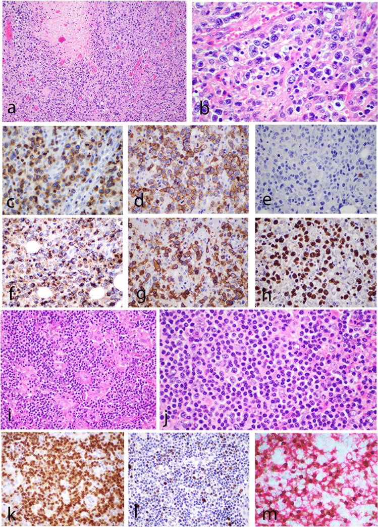
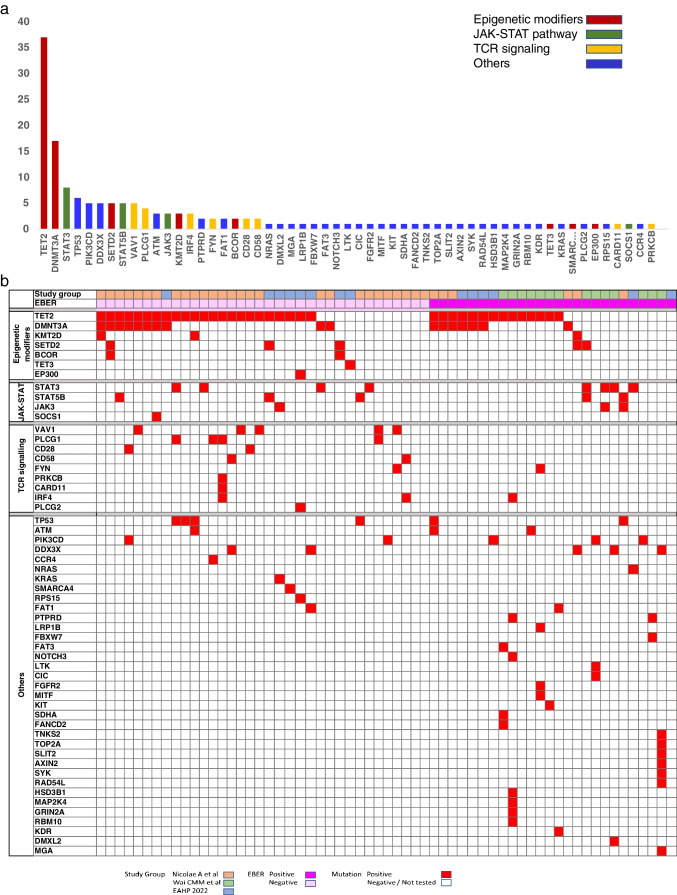
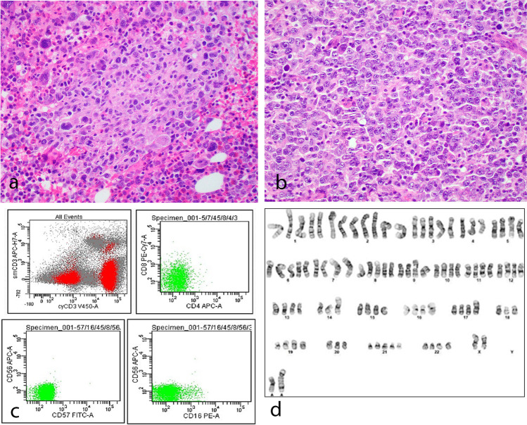
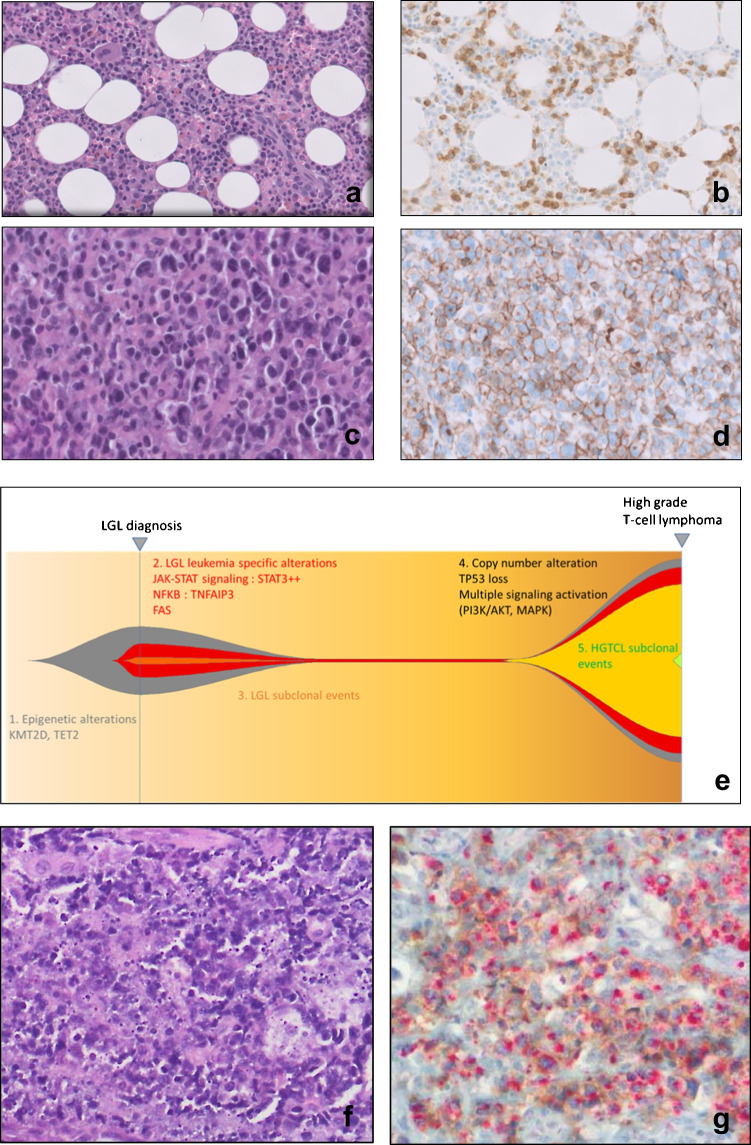
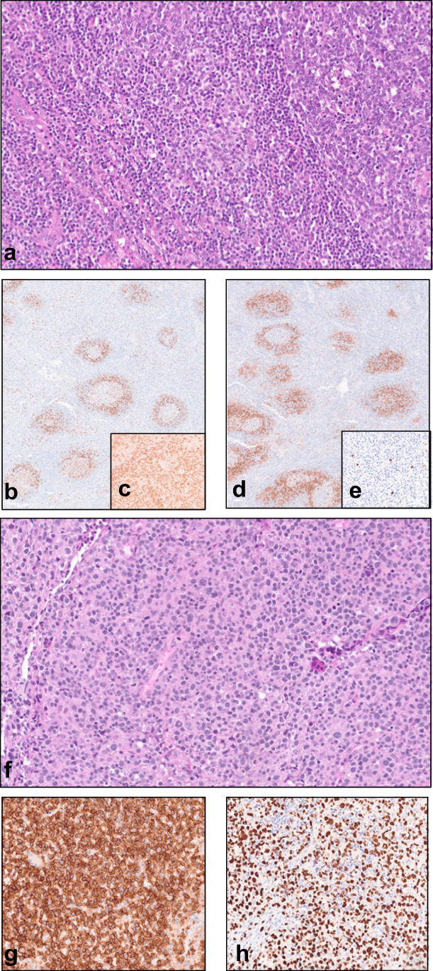
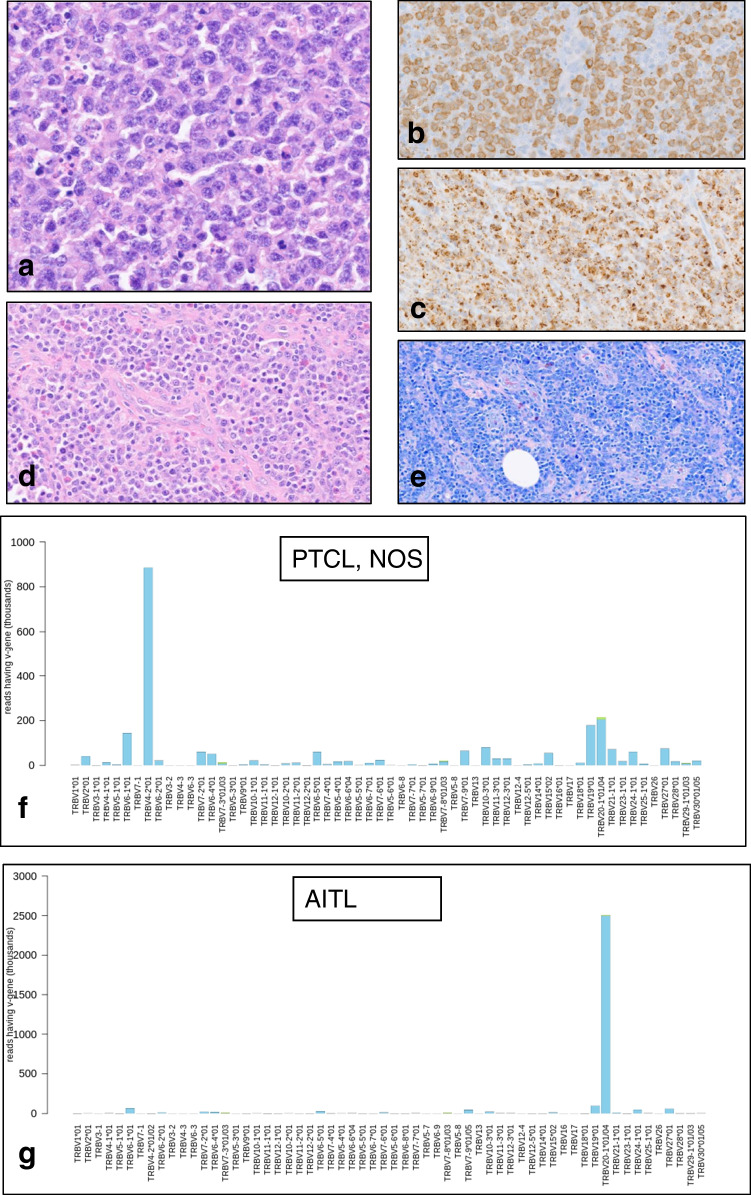
Cytotoxic peripheral T-cell lymphomas and EBV-positive T/NK-cell lymphoproliferative diseases were discussed at the 2022 European Association for Haematopathology/Society for Hematopathology lymphoma workshop held in Florence, Italy. This session focused on (i) primary nodal EBV-positive T and NK-cell lymphomas (primary nodal-EBV-TNKL), (ii) extranodal EBV-positive T/NK lymphoproliferative diseases (LPD) in children and adults, (iii) cytotoxic peripheral T-cell lymphomas, NOS (cPTCL-NOS), EBV-negative, and (iv) miscellaneous cases. Primary nodal-EBV-TNKL is a newly recognized entity which is rare, aggressive, and associated with underlying immune deficiency/immune dysregulation. All cases presented with lymphadenopathy but some demonstrated involvement of tonsil/Waldeyer's ring and extranodal sites. The majority of tumors are of T-cell lineage, and the most frequent mutations involve the epigenetic modifier genes, such as TET2 and DNMT3A, and JAK-STAT genes. A spectrum of EBV-positive T/NK LPD involving extranodal sites were discussed and highlight the diagnostic challenge with primary nodal-EBV-TNKL when these extranodal EBV-positive T/NK LPD cases demonstrate predominant nodal disease either at presentation or during disease progression from chronic active EBV disease. The majority of cPTCL-NOS demonstrated the TBX21 phenotype. Some cases had a background of immunosuppression or immune dysregulation. Interestingly, an unexpected association of cPTCL-NOS, EBV-positive and negative, with TFH lymphomas/LPDs was observed in the workshop cases. Similar to a published literature, the genetic landscape of cPTCL-NOS from the workshop showed frequent mutations in epigenetic modifiers, including TET2 and DNMT3A, suggesting a role of clonal hematopoiesis in the disease pathogenesis.

Keywords: Clonal hematopoiesis; Cytotoxic T-cell lymphoma; EA4HP workshop; Epstein–Barr virus; NK-cell lymphoma; T-cell lymphoma.


Conflict of interest statement

The authors declare no competing interests.

Figures

Fig. 1
Fig. 1
Histologic features of nodal EBV-positive T and NK-cell lymphoma. af Case LYWS-1138, courtesy of L. Goh. a The tumor shows areas of necrosis and diffuse sheets of neoplastic cells. b The tumor cells are large with irregular vesicular nuclei, coarse chromatin, and prominent nucleoli. Immunohistochemistry reveals positive expression for c CD3, d CD8, f TIA1, g TCRgamma, and h EBER and negativity for e CD56. im Case LYWS-1190, courtesy of R.K.H. Au-Yeung. i The tumor reveals predominantly small cells with abundant histiocytes in the background, resembling lymphoepithelioid (Lennert) lymphoma. j Tumor cells display small monotonous nuclei with mild nuclear atypia and indistinct nucleoli. They are positive for k TCRβF1. l Ki67 proliferation index is low. m EBER/CD8 double stain demonstrates that the neoplastic cells are positive for CD8 and EBER
Fig. 2
Fig. 2
Mutational landscape of nodal cytotoxic peripheral T-cell lymphomas (PTCL) based on workshop cases and cases from recent literature with next generation sequencing data (Wai CMM. Haematologica (2022) 1;107(8):1864, Nicolae A. Modern Pathology (2022) 35:1126–1136). Histogram plot (a) and heatmap (b) illustrate the frequency of mutations in all cases of cytotoxic PTCL, both EBV-positive and EBV-negative. A total of 61 cases analyzed revealed that the most common mutations involve epigenetic modifiers, such as TET2 and DNMT3A, JAK/STAT pathway and TCR signaling genes
Fig. 3
Fig. 3
Case LYWS-1181 submitted by I Obiorah was an example of aggressive NK-cell leukemia (ANKL). The patient presented with multiple lymphadenopathy, fever, HLH, and involvement of the BM and spleen. The tumor cells in the BM (a) and LN (b) were large and pleomorphic. Flow cytometry (c) revealed an infiltrate which was cCD3+, sCD3−, CD4−, CD8−, CD56−, CD57−, and CD16−. Cytogenetics demonstrated hyperploidy with multiple tetraploid clones and complex cytogenetic anomalies (d)
Fig. 4
Fig. 4
LGL transformation case (LYWS-1416 submitted by F. Gutierrez-Llamas). a The BM biopsy in this 78-year-old male shows interstitial infiltrate of small lymphocytes b expressing CD3. The lymphocytes are also positive for CD8 and Granzyme B. c The lymph node shows an atypical infiltrate of large cells (H&E), that are positive for d CD8, CD56, CD30, Granzyme B, and p53. Clonal analysis revealed the same TR gamma rearrangement in both samples. e NGS confirms the presence of the same TET2 mutations in both sites, without the STAT3 mutation and the presence of new mutations (JAK3 and KRAS) in the transformation sample (f and g). Case LYWS-1200 submitted by G Frigola displays the co-expression of cytotoxic and TFH markers in the same cells [double stain PD1 (brown) TIA-1 (red)]
Fig. 5
Fig. 5
Case LYWS-1396, presented by L Wang, shows the association of a cytotoxic nodal EBV+ T and NK-cell lymphoma (primary nodal-EBV-NKTL) and an angioimmunoblastic T-cell lymphoma (AITL) in a 48-year-old female. The initial LN biopsy shows an AITL pattern 1 with reactive follicles (a). The tumor displays a T-follicular helper (TFH) phenotype with positive expression of PD1 (b, perifollicular pattern), expression of IDH2R172K (c), and CD10 (d). Scattered large B blasts are highlighted by EBER (e). The second lymphoma displays a diffuse proliferation of atypical medium-large cells (f). Immunohistochemistry shows positivity for CD8 (g) and EBER (h)
Fig. 6.
Fig. 6.
Case LYWS-1094, presented by A. Volgelsberg, describes the divergent evolution of two clonally unrelated T-cell lymphomas from clonal hematopoiesis. A 71-year-old woman presented with enlarged cervical LNs. The biopsy shows diffuse infiltration of large cells (a). Immunohistochemistry illustrates positivity for CD3 (b), TIA-1 (c), CD8 (not shown), and βF1 (not shown). The patient received CHOP therapy and achieved complete remission. Five months later, the patient relapsed and the lymph node reveals a mixed infiltration of lymphoid cells (d) and proliferation of high endothelial venules (e). TR beta sequencing of both lymphomas confirms the presence of distinctly different rearrangements (f and g)

References

    1. Paul S, Lal G. The molecular mechanism of natural killer cells function and its importance in cancer immunotherapy. Front Immunol. 2017;8:1124. - PMC - PubMed
    1. Oh DY, Fong L. Cytotoxic CD4 T cells in cancer: expanding the immune effector toolbox. Immunity. 2021;54:2701–2711. - PMC - PubMed
    1. Cachot A, Bilous M, Liu Y-C, et al. Tumor-specific cytolytic CD4 T cells mediate immunity against human cancer. Sci Adv. 2021;7:eabe3348. - PMC - PubMed
    1. Alaggio R, Amador C, Anagnostopoulos I, et al. The 5th edition of the World Health Organization Classification of Haematolymphoid Tumours: lymphoid neoplasms. Leukemia. 2022;36:1720–1748. - PMC - PubMed
    1. Campo E, Jaffe ES, Cook JR, et al. The International Consensus Classification of Mature Lymphoid Neoplasms: a report from the Clinical Advisory Committee. Blood. 2022;140:1229–1253. - PMC - PubMed