. 2023 Oct;38(7):2427-2442.
doi: 10.1007/s11011-023-01277-5. Epub 2023 Aug 30.

Hordeum vulgare ethanolic extract mitigates high salt-induced cerebellum damage via attenuation of oxidative stress, neuroinflammation, and neurochemical alterations in hypertensive rats

Affiliations

Hordeum vulgare ethanolic extract mitigates high salt-induced cerebellum damage via attenuation of oxidative stress, neuroinflammation, and neurochemical alterations in hypertensive rats

O A Ahmed-Farid et al. Metab Brain Dis. 2023 Oct.

Abstract

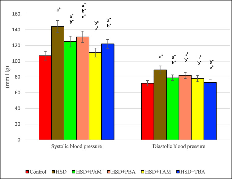
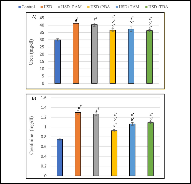
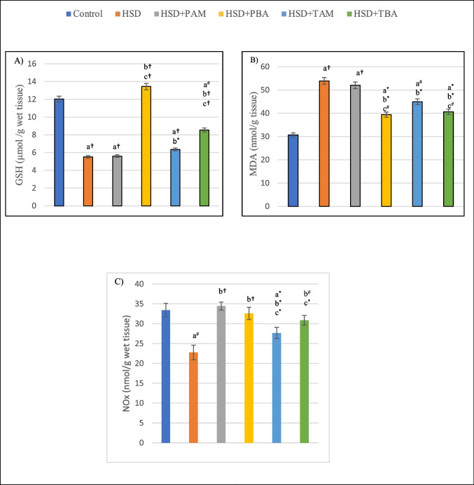
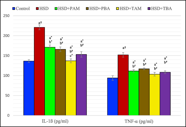
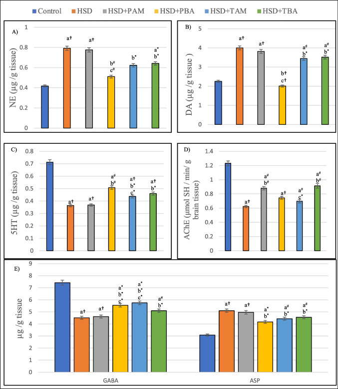
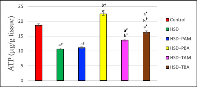
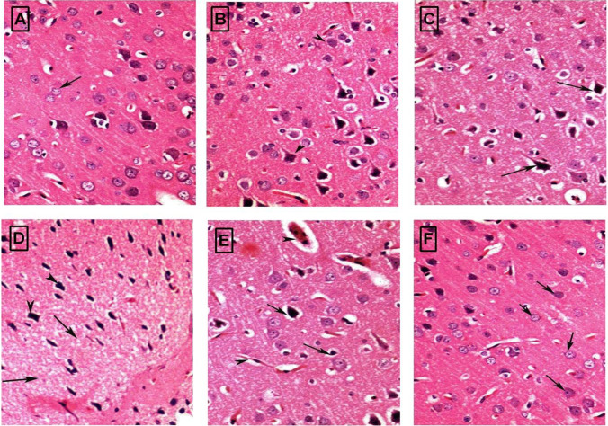
High salt intake increases inflammatory and oxidative stress responses and causes an imbalance of neurotransmitters involved in the pathogenesis of hypertension that is related to the onset of cerebral injury. Using natural compounds that target oxidative stress and neuroinflammation pathways remains a promising approach for treating neurological diseases. Barley (Hordeum vulgare L.) seeds are rich in protein, fiber, minerals, and phenolic compounds, that exhibit potent neuroprotective effects in various neurodegenerative diseases. Therefore, this work aimed to investigate the efficacy of barley ethanolic extract against a high salt diet (HSD)-induced cerebellum injury in hypertensive rats. Forty-eight Wistar rats were divided into six groups. Group (I) was the control. The second group, the HSD group, was fed a diet containing 8% NaCl. Groups II and III were fed an HSD and simultaneously treated with either amlodipine (1 mg /kg b.wt p.o) or barley extract (1000 mg /kg b.wt p.o) for five weeks. Groups IV and V were fed HSD for five weeks, then administered with either amlodipine or barley extract for another five weeks. The results revealed that barley treatment significantly reduced blood pressure and effectively reduced oxidative stress and inflammation in rat's cerebellum as indicated by higher GSH and nitric oxide levels and lower malondialdehyde, TNF-α, and IL-1ß levels. Additionally, barley restored the balance of neurotransmitters and improved cellular energy performance in the cerebellum of HSD-fed rats. These findings suggest that barley supplementation exerted protective effects against high salt-induced hypertension by an antioxidant, anti-inflammatory, and vasodilating effects and restoring neurochemical alterations.

Keywords: Barley; Cerebellum; Inflammation; Neurotransmitters; Oxidative Stress; Rats.


Conflict of interest statement

The authors have no relevant financial or non-financial interests to disclose.

Figures

Fig. 1
Fig. 1
Systolic and diastolic blood pressure of rats on the normal salt diet (negative control), or 8% salt diet (HSD group), HSD + amlodipine for five weeks (HSD + PAM), HSD and barley extract for five weeks (HSD + PBA), HSD for five weeks then amlodipine for another five weeks (HSD + TAM) or HSD for five weeks then barley extract for another five weeks (HSD + TBA). Data are presented as mean values ± SEM; (n = 8). a means a significant difference from the control negative group, b means a significant difference from HSD group, and c means a significant difference from HSD + PAM group. *, # and † represent statistical significance at p < 0.05, p < 0.01 and p < 0.001, respectively (one-way ANOVA followed by Post hoc test)
Fig. 2
Fig. 2
Serum levels of urea (A) and creatinine (B) of rats on the normal salt diet (negative control), or 8% salt diet (HSD group), HSD + amlodipine for five weeks (HSD + PAM), HSD and barley extract for five weeks (HSD + PBA), HSD for five weeks then amlodipine for another five weeks (HSD + TAM) or HSD for five weeks then barley extract for another five weeks (HSD + TBA). Data are presented as mean values ± SEM; (n = 8). a means a significant difference from the negative control group, b means a significant difference from the HSD group, and c means a significant difference from the HSD + PAM group. *, # and † represent statistical significance at p < 0.05, p < 0.01 and p < 0.001, respectively (one-way ANOVA followed by Post hoc test)
Fig. 3
Fig. 3
Cerebral levels of (A) reduced glutathione (GSH), (B) malondialdehyde (MDA), and (C) nitric oxide (NOx) of rats on the normal salt diet (negative control), or 8% salt diet (HSD group), HSD + amlodipine for five weeks (HSD + PAM), HSD and barley extract for five weeks (HSD + PBA), HSD for five weeks then amlodipine for another five weeks (HSD + TAM) or HSD for five weeks then barley extract for another five weeks (HSD + TBA). Data are presented as mean values ± SEM; (n = 8). a means a significant difference from the negative control group, b means a significant difference from the HSD group, and c means a significant difference from the HSD + PAM group. *, # and † represent statistical significance at p < 0.05, p < 0.01 and p < 0.001, respectively (one-way ANOVA followed by Post hoc test)
Fig. 4
Fig. 4
Cerebral levels of interleukin-1 ß (IL- ß) and tumor necrosis factor- α (TNF- α) in rats on the normal salt diet (negative control), or 8% salt diet (HSD group), HSD + amlodipine for five weeks (HSD + PAM), HSD and barley extract for five weeks (HSD + PBA), HSD for five weeks then amlodipine for another five weeks (HSD + TAM) or HSD for five weeks then barley extract for another five weeks (HSD + TBA). Data are presented as mean values ± SEM; (n = 8). a means a significant difference from the negative control group, b means a significant difference from the HSD group, and c means a significant difference from HSD + PAM group. *, # and † represent statistical significance at p < 0.05, p < 0.01 and p < 0.001, respectively (one-way ANOVA followed by Post hoc test)
Fig. 5
Fig. 5
Cerebral levels of (A) norepinephrine (NE), (B) dopamine (DA), (C) 5-hydroxytryptamine (5-HT), (D) acetylcholinesterase activity, and (E) gamma-aminobutyric acid (GABA) and aspartate (ASP) of rats on normal salt diet (negative control), or 8% salt diet (HSD group), HSD + amlodipine for five weeks (HSD + PAM), HSD and barley extract for five weeks (HSD + PBA), HSD for five weeks then amlodipine for another five weeks (HSD + TAM) or HSD for five weeks then barley extract for another five weeks (HSD + TBA). Data are presented as mean values ± SEM; (n = 8). a means a significant difference from negative control group, b means asignificant difference from HSD group, and c means significant difference from HSD + PAM group. *, # and † represent statistical significance at p < 0.05, p < 0.01 and p < 0.001, respectively (one-way ANOVA followed by Post hoc test)
Fig. 6
Fig. 6
ATP level in the brain of rats on the normal salt diet (negative control), or 8% salt diet (HSD group), HSD + amlodipine for five weeks (HSD + PAM), HSD and barley extract for five weeks (HSD + PBA), HSD for five weeks then amlodipine for another five weeks (HSD + TAM) or HSD for five weeks then barley extract for another five weeks (HSD + TBA). Data are presented as mean values ± SEM; (n = 8). a means a significant difference from the negative control group, b means a significant difference from HSD group, and c means a significant difference from HSD + PAM group. *, # and † represent statistical significance at p < 0.05, p < 0.01 and p < 0.001, respectively (one-way ANOVA followed by Post hoc test)
Fig. 7
Fig. 7
Histological changes of (H & E) stained sections from the rat brain (Cerebral cortex) were obtained from (A) control group showing no histopathological changes; (B) high salt (HS) group showing necrosis of neurons and neuronophagia; (C) HSD + PAM showing mild improvement but apoptotic (head) nuclei seeing; (D) HSD + PBA group showing improvement with mild cytoplasmic vacuolation (E) HSD + TAM group showing congestion of cerebral blood vessel, neuronal edema, necrosis of neurons and neuronophagia and (F) HSD + TBA group showing pronounced improvement of neurocytes with normal neurons (H & E × 400)

Similar articles

Cited by

References

    1. Badrasawi MM, Shahar S, AbdManaf Z, Haron H. Effect of Talbinah food consumption on depressive symptoms among elderly individuals in long term care facilities, randomized clinical trial. Clin Interv Aging. 2013;8:279–285. doi: 10.2147/CIA.S37586. - DOI - PMC - PubMed
    1. Bhusal A, Rahman MH, Suk K. Hypothalamic inflammation in metabolic disorders and aging. Cell Mol Life Sci. 2021;79(1):32. doi: 10.1007/s00018-021-04019-x. - DOI - PMC - PubMed
    1. Biod T, Sirota B, et al. In: Practical clinical biochemistry. 4. Watson A, et al., editors. Prentice-Hall of India Private Ltd: New Delhi; 1948. pp. 142–145.
    1. Borrelli S, Provenzano M, Gagliardi I, Michael A, Liberti ME, De Nicola L, Conte G, Garofalo C, Andreucci M. Sodium intake and chronic kidney disease. Int J Mol Sci. 2020;21(13):4744. doi: 10.3390/ijms21134744. - DOI - PMC - PubMed
    1. Darwish IE, Maklad HM, Diab IH. Behavioral and neuronal biochemical possible effects in experimental induced chronic mild stress in male albino rats under the effect of oral barley administration in comparison to venlafaxine. Int J Physiol Pathophysiol Pharmacol. 2013;5(2):128–36. - PMC - PubMed

Publication types