. 2023 Oct;73(9-10):724-737.
doi: 10.1007/s12031-023-02151-w. Epub 2023 Aug 30.

Early Alterations of PACAP and VIP Expression in the Female Rat Brain Following Spinal Cord Injury

Affiliations

Early Alterations of PACAP and VIP Expression in the Female Rat Brain Following Spinal Cord Injury

Sarah Thomas Broome et al. J Mol Neurosci. 2023 Oct.

Abstract

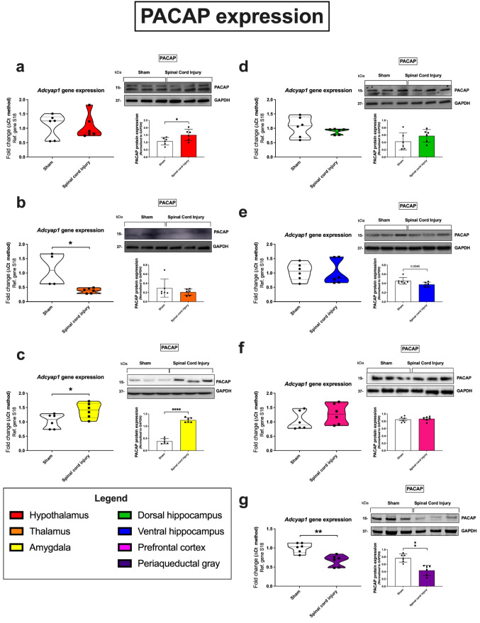
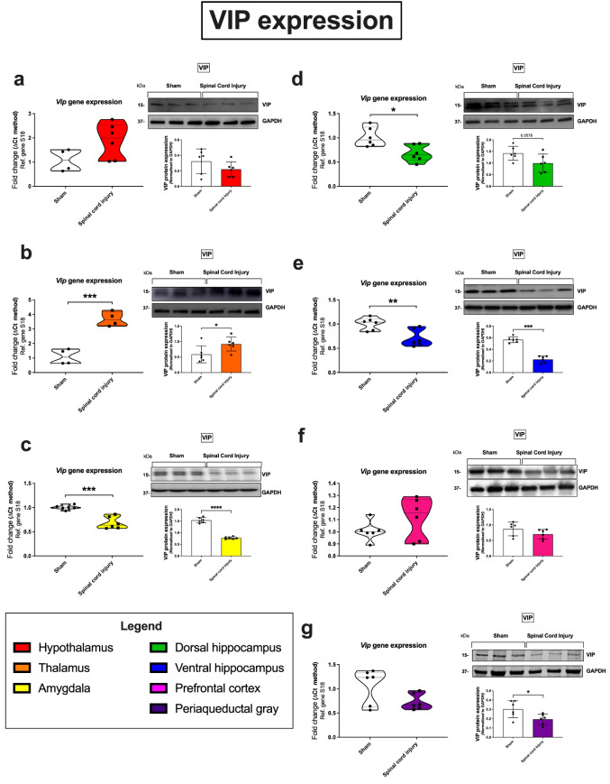
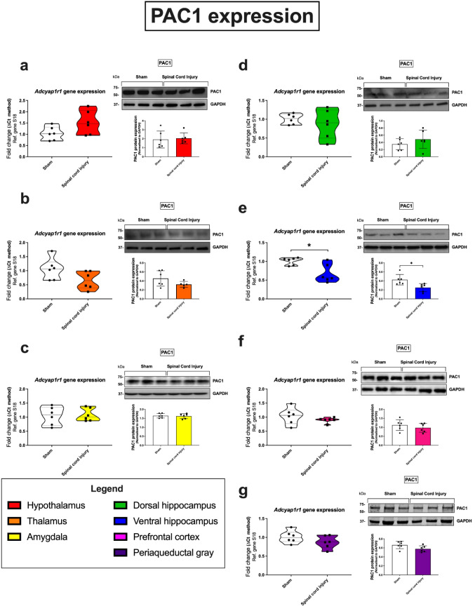
Previous evidence shows that rapid changes occur in the brain following spinal cord injury (SCI). Here, we interrogated the expression of the neuropeptides pituitary adenylyl cyclase-activating peptide (PACAP), vasoactive intestinal peptides (VIP), and their binding receptors in the rat brain 24 h following SCI. Female Sprague-Dawley rats underwent thoracic laminectomy; half of the rats received a mild contusion injury at the level of the T10 vertebrate (SCI group); the other half underwent sham surgery (sham group). Twenty-four hours post-surgery, the hypothalamus, thalamus, amygdala, hippocampus (dorsal and ventral), prefrontal cortex, and periaqueductal gray were collected. PACAP, VIP, PAC1, VPAC1, and VPAC2 mRNA and protein levels were measured by real-time quantitative polymerase chain reaction and Western blot. In SCI rats, PACAP expression was increased in the hypothalamus (104-141% vs sham) and amygdala (138-350%), but downregulated in the thalamus (35-95%) and periaqueductal gray (58-68%). VIP expression was increased only in the thalamus (175-385%), with a reduction in the amygdala (51-68%), hippocampus (40-75%), and periaqueductal gray (74-76%). The expression of the PAC1 receptor was the least disturbed by SCI, with decrease expression in the ventral hippocampus (63-68%) only. The expression levels of VPAC1 and VPAC2 receptors were globally reduced, with more prominent reductions of VPAC1 vs VPAC2 in the amygdala (21-70%) and ventral hippocampus (72-75%). In addition, VPAC1 downregulation also extended to the dorsal hippocampus (69-70%). These findings demonstrate that as early as 24 h post-SCI, there are region-specific disruptions of PACAP, VIP, and related receptor transcript and protein levels in supraspinal regions controlling higher cognitive functions.

Keywords: Central nervous system (CNS); Pituitary adenylate cyclase-activating polypeptide (PACAP); Spinal cord injury (SCI); Vasoactive intestinal peptide (VIP).


Conflict of interest statement

The authors declare no competing interests.

Figures

Fig. 1
Fig. 1
Gene and protein expression of the PACAP neuropeptide in distinct brain regions 24 h following SCI. Real-time qPCR analyses with representative Western blots and densitometry of PACAP expression in the hypothalamus (a), thalamus (b), amygdala (c), dorsal hippocampus (d), ventral hippocampus (e), prefrontal cortex (f), and periaqueductal gray (g) isolated from female rat brains subjected to SCI or sham surgery. Fold changes in gene expression were calculated using the ΔΔCt method after normalization to s18 (ribosomal protein s18), used as the housekeeping gene. Protein expression was normalized to GAPDH, the loading control. Densitometric results are expressed as the mean ± SEM from n = 4-6 rats per group. *p < 0.05 or **p < 0.01, compared to sham controls, as determined by unpaired Student’s t test. PACAP pituitary adenylate cyclase-activating peptide, s18 ribosomal protein s18, GAPDH glyceraldehyde-3-phosphate dehydrogenase
Fig. 2
Fig. 2
Gene and protein expression of the VIP neuropeptide in distinct brain regions 24 h following SCI. Real-time qPCR analyses with representative Western blots and densitometry of VIP expression in the hypothalamus (a), thalamus (b), amygdala (c), dorsal hippocampus (d), ventral hippocampus (e), prefrontal cortex (f), and periaqueductal gray (g) isolated from female rat brains subjected to SCI or sham surgery. Fold changes in gene expression were calculated using the ΔΔCt method after normalization to s18 (ribosomal protein s18). Protein expression was normalized to GAPDH, the loading control. Densitometric results are expressed as the mean ± SEM from n = 4-6 rats per group. *p < 0.05 or **p < 0.01, compared to sham controls, as determined by unpaired Student’s t test. VIP vasoactive intestinal peptide, s18 ribosomal protein s18, GAPDH glyceraldehyde-3-phosphate dehydrogenase
Fig. 3
Fig. 3
Gene and protein expression of the PAC1 receptor in distinct brain regions 24 h following SCI. Real-time qPCR analyses with representative Western blots and densitometry of PAC1 expression in the hypothalamus (a), thalamus (b), amygdala (c), dorsal hippocampus (d), ventral hippocampus (e), prefrontal cortex (f), and periaqueductal gray (g) isolated from female rat brains subjected to SCI or sham surgery. Fold changes in gene expression were calculated using the ΔΔCt method after normalization to s18 (ribosomal protein s18). Protein expression was normalized to GAPDH, the loading control. Densitometric results are expressed as the mean ± SEM from n = 6 rats per group. *p < 0.05 or **p < 0.01, compared to sham controls, as determined by unpaired Student’s t test. PAC1 PAC1 receptor, s18 ribosomal protein s18, GAPDH glyceraldehyde-3-phosphate dehydrogenase
Fig. 4
Fig. 4
Gene and protein expression of the VPAC1 receptor in distinct brain regions 24 h following SCI. Real-time qPCR analyses with representative Western blots and densitometry of VPAC1 expression in the hypothalamus (a), thalamus (b), amygdala (c), dorsal hippocampus (d), ventral hippocampus (e), prefrontal cortex (f), and periaqueductal gray (g) isolated from female rat brains subjected to SCI or sham surgery. Fold changes in gene expression were calculated using the ΔΔCt method after normalization to s18 (ribosomal protein s18). Protein expression was normalized to GAPDH, the loading control. Densitometric results are expressed as the mean ± SEM from n = 6 rats per group. *p < 0.05 or **p < 0.01, compared to sham controls, as determined by unpaired Student’s t test. VPAC1 VPAC1 receptor, s18 ribosomal protein s18, GAPDH glyceraldehyde-3-phosphate dehydrogenase
Fig. 5
Fig. 5
Gene and protein expression of the VPAC2 receptor in distinct brain regions 24 h following SCI. Real-time qPCR analyses with representative Western blots and densitometry of VPAC2 expression in the hypothalamus (a), thalamus (b), amygdala (c), dorsal hippocampus (d), ventral hippocampus (e), prefrontal cortex (f), and periaqueductal gray (g) isolated from female rat brains subjected to SCI or sham surgery. Fold changes in gene expression were calculated using the ΔΔCt method after normalization to s18 (ribosomal protein s18). Protein expression was normalized to GAPDH, the loading control. Densitometric results are expressed as the mean ± SEM from n = 6 rats per group. *p < 0.05 or **p < 0.01, compared to sham controls, as determined by unpaired Student’s t test. VPAC2 VPAC2 receptor, s18 ribosomal protein s18, GAPDH glyceraldehyde-3-phosphate dehydrogenase

Similar articles

Cited by

References

    1. Abad C, Tan YV. Immunomodulatory roles of PACAP and VIP: lessons from knockout mice. J Mol Neurosci. 2018;66(1):102–113. doi: 10.1007/s12031-018-1150-y. - DOI - PubMed
    1. Alizadeh A, Dyck SM, Karimi-Abdolrezaee S. Traumatic spinal cord injury: an overview of pathophysiology, models and acute injury mechanisms. Front Neurol. 2019;10:282. doi: 10.3389/fneur.2019.00282. - DOI - PMC - PubMed
    1. Atlasz T, Szabadfi K, Kiss P, Racz B, Gallyas F, Tamas A, Gaal V, Marton Z, Gabriel R, Reglodi D (2010) Pituitary adenylate cyclase activating polypeptide in the retina: focus on the retinoprotective effects. Ann NY Acad Sci 1200:128–139. 10.1111/j.1749-6632.2010.05512.x - PubMed
    1. Banks WA, Kastin AJ, Arimura A. Effect of spinal cord injury on the permeability of the blood-brain and blood-spinal cord barriers to the neurotropin PACAP. Exp Neurol. 1998;151(1):116–123. doi: 10.1006/exnr.1998.6786. - DOI - PubMed
    1. Baxter MG, Croxson PL. Facing the role of the amygdala in emotional information processing. Proc Natl Acad Sci U S A. 2012;109(52):21180–21181. doi: 10.1073/pnas.1219167110. - DOI - PMC - PubMed

MeSH terms

Substances