. 2023 Aug;169(8):001389.
doi: 10.1099/mic.0.001389.

Timing of dense granule biogenesis in asexual malaria parasites

Affiliations

Timing of dense granule biogenesis in asexual malaria parasites

Tansy Vallintine et al. Microbiology (Reading). 2023 Aug.

Abstract

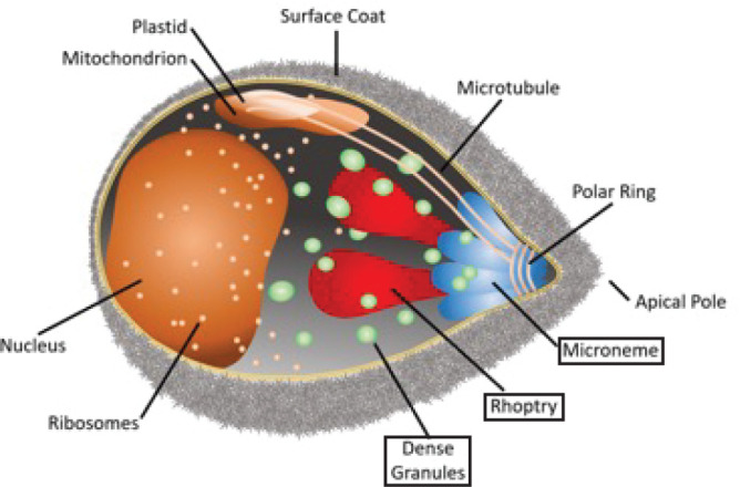
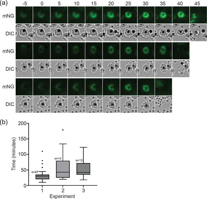
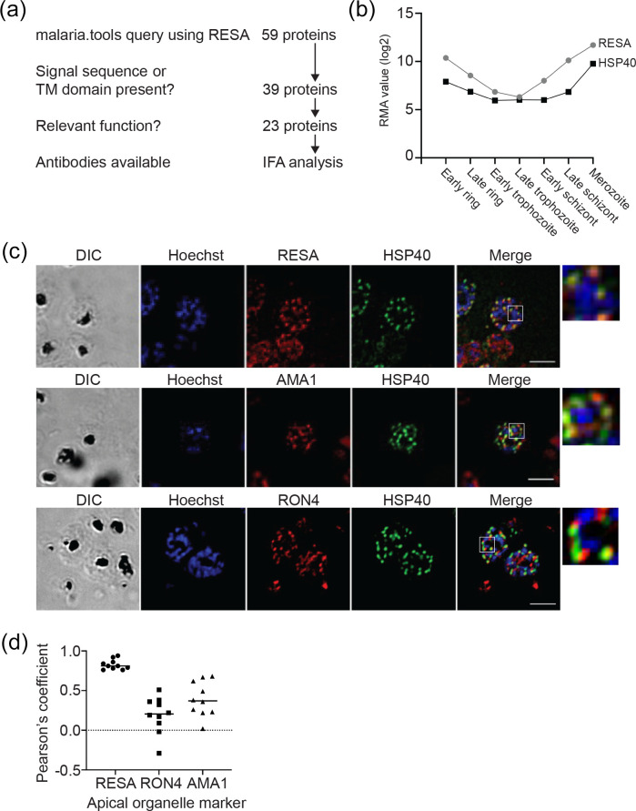
Malaria is an important infectious disease that continues to claim hundreds of thousands of lives annually. The disease is caused by infection of host erythrocytes by apicomplexan parasites of the genus Plasmodium. The parasite contains three different apical organelles - micronemes, rhoptries and dense granules (DGs) - whose contents are secreted to mediate binding to and invasion of the host cell and the extensive remodelling of the host cell that occurs following invasion. Whereas the roles of micronemes and rhoptries in binding and invasion of the host erythrocyte have been studied in detail, the roles of DGs in Plasmodium parasites are poorly understood. They have been proposed to control host cell remodelling through regulated protein secretion after invasion, but many basic aspects of the biology of DGs remain unknown. Here we describe DG biogenesis timing for the first time, using RESA localization as a proxy for the timing of DG formation. We show that DG formation commences approximately 37 min prior to schizont egress, as measured by the recruitment of the DG marker RESA. Furthermore, using a bioinformatics approach, we aimed to predict additional cargo of the DGs and identified the J-dot protein HSP40 as a DG protein, further supporting the very early role of these organelles in the interaction of the parasite with the host cell.

Keywords: Plasmodium; apical organelles; apicomplexa; dense granules; malaria.


Conflict of interest statement

The authors declare that there is no conflict of interest.

Figures

Fig. 1.
Fig. 1.
A Plasmodium falciparum merozoite. Indicated are the different organelles inside the parasite, with the names of the apical organelles in boxes.
Fig. 2.
Fig. 2.
Generation of an mNeonGreen-RESA-expressing parasite. (a) Integration strategy using selection-linked integration (SLI). mNG, mNeonGreen; T, T2A peptide; neo, amino 3′-glycosylphosphotransferase (neomycin resistance gene); 5′ and 3′, 5′ and 3′ regulatory regions, respectively. The plasmid pTV002, containing a fusion of the 3′ region of the RESA gene and the gene encoding mNG, was introduced into 3D7 parasites. Transfectants were selected with WR and integrants were subsequently selected with G418. (b) Integration PCR of genomic DNA from wild-type (3D7, left) and integrant (INT, right) parasites using the indicated primer pairs. See panel (a) for the binding sites of the primers. The expected sizes of the PCR products are indicated at the bottom. (c) Anti-mNeonGreen immunoblot of 3D7 (right) and RESA-mNG integrant parasite extracts (left). The band of the expected size is indicated (*). The same extracts were probed with anti-aldolase antibodies as loading control (right). (d) Live-cell fluorescence imaging of parasites expressing RESA-mNG. Top, schizont; bottom, ring. Bars, 5 µm.
Fig. 3.
Fig. 3.
Video microscopy of dense granule formation. (a) Highly synchronized RESA-mNG-expressing parasites were observed using live video microscopy at intervals of 5 min. Parasites were imaged on three separate occasions; each row is taken from a different experiment. The formation of punctate spots of fluorescence was designated as the start of dense granule formation (time point 0). mNG, mNeonGreen fluorescence; DIC, differential interference contrast. (b) Quantification of dense granule formation for the three different experiments. The boxes represent the interquartile range, where 50% of the data points are found. The horizontal line crossing the box represents the median. The y-axis represents the time (in minutes) from the first detection of clustering of RESA-mNG fluorescence in spots to egress of the parasites.
Fig. 4.
Fig. 4.
Identification of additional dense granule proteins. (a) Outline of malaria.tools query using RESA (PF3D7_0102200). (b) Comparison of expression pattern of RESA and HSP40. Robust multi-array averaging (RMA) values of RESA (grey) and HSP40 (black) expression levels at different stages of the intra-erythrocytic life cycle are shown. Data were obtained from Le Roch et al. [54]. (c) Co-staining of P. falciparum parasites using anti-HSP40 and the dense granule marker RESA (top), the microneme marker AMA1 (middle) and the rhoptry neck marker RON4 (bottom). Samples were also stained with Hoechst 33 342 to visualize DNA. Panels on the far right show an expanded view of the region indicated in the white boxes. Bar, 5 µm. (d) Pearson’s correlation coefficient (PCC) of the colocalization of the anti-HSP40 staining and the staining using the indicated antibodies that recognize apical organelle markers. For each combination, the PCC was determined using ten clearly labelled merozoites in different schizonts.

Similar articles

References

    1. Carruthers VB, Sibley LD. Sequential protein secretion from three distinct organelles of Toxoplasma gondii accompanies invasion of human fibroblasts. Eur J Cell Biol. 1997;73:114–123. - PubMed
    1. Singh S, Alam MM, Pal-Bhowmick I, Brzostowski JA, Chitnis CE. Distinct external signals trigger sequential release of apical organelles during erythrocyte invasion by malaria parasites. PLoS Pathog. 2010;6:e1000746. doi: 10.1371/journal.ppat.1000746. - DOI - PMC - PubMed
    1. Kats LM, Cooke BM, Coppel RL, Black CG. Protein trafficking to apical organelles of malaria parasites - building an invasion machine. Traffic. 2008;9:176–186. doi: 10.1111/j.1600-0854.2007.00681.x. - DOI - PubMed
    1. Gubbels MJ, Duraisingh MT. Evolution of apicomplexan secretory organelles. Int J Parasitol. 2012;42:1071–1081. doi: 10.1016/j.ijpara.2012.09.009. - DOI - PMC - PubMed
    1. Cao J, Kaneko O, Thongkukiatkul A, Tachibana M, Otsuki H, et al. Rhoptry neck protein RON2 forms a complex with microneme protein AMA1 in Plasmodium falciparum merozoites. Parasitol Int. 2009;58:29–35. doi: 10.1016/j.parint.2008.09.005. - DOI - PubMed

Publication types

LinkOut - more resources