. 2023 Jun;11(6):e006863.
doi: 10.1136/jitc-2023-006863. Epub 2023 Jun 23.

BiTE secretion by adoptively transferred stem-like T cells improves FRα+ ovarian cancer control

Affiliations

BiTE secretion by adoptively transferred stem-like T cells improves FRα+ ovarian cancer control

A J Robert McGray et al. J Immunother Cancer. 2023 Jun.

Abstract

Background: Cancer immunotherapies can produce complete therapeutic responses, however, outcomes in ovarian cancer (OC) are modest. While adoptive T-cell transfer (ACT) has been evaluated in OC, durable effects are rare. Poor therapeutic efficacy is likely multifactorial, stemming from limited antigen recognition, insufficient tumor targeting due to a suppressive tumor microenvironment (TME), and limited intratumoral accumulation/persistence of infused T cells. Importantly, host T cells infiltrate tumors, and ACT approaches that leverage endogenous tumor-infiltrating T cells for antitumor immunity could effectively magnify therapeutic responses.

Methods: Using retroviral transduction, we have generated T cells that secrete a folate receptor alpha (FRα)-directed bispecific T-cell engager (FR-B T cells), a tumor antigen commonly overexpressed in OC and other tumor types. The antitumor activity and therapeutic efficacy of FR-B T cells was assessed using FRα+ cancer cell lines, OC patient samples, and preclinical tumor models with accompanying mechanistic studies. Different cytokine stimulation of T cells (interleukin (IL)-2+IL-7 vs IL-2+IL-15) during FR-B T cell production and the resulting impact on therapeutic outcome following ACT was also assessed.

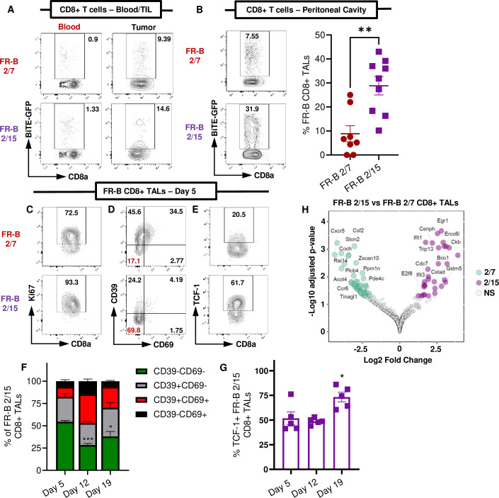
Results: FR-B T cells efficiently lysed FRα+ cell lines, targeted FRα+ OC patient tumor cells, and were found to engage and activate patient T cells present in the TME through secretion of T cell engagers. Additionally, FR-B T cell therapy was effective in an immunocompetent in vivo OC model, with response duration dependent on both endogenous T cells and FR-B T cell persistence. IL-2/IL-15 preconditioning prior to ACT produced less differentiated FR-B T cells and enhanced therapeutic efficacy, with mechanistic studies revealing preferential accumulation of TCF-1+CD39-CD69- stem-like CD8+ FR B T cells in the peritoneal cavity over solid tumors.

Conclusions: These findings highlight the therapeutic potential of FR-B T cells in OC and suggest FR-B T cells can persist in extratumoral spaces while actively directing antitumor immunity. As the therapeutic activity of infused T cell therapies in solid tumor indications is often limited by poor intratumoral accumulation of transferred T cells, engager-secreting T cells that can effectively leverage endogenous immunity may have distinct mechanistic advantages for enhancing therapeutic responses rates.

Keywords: Cell Engineering; Immunity, Cellular; Immunotherapy; Immunotherapy, Adoptive; Tumor Microenvironment.


Conflict of interest statement

Competing interests: AJRM, TT, and KO are inventors on provisional patents pertaining to the development and use of BiTE-secreting T cells in cancer. KO is a co-founder of Tactiva Therapeutics. All other authors have no financial conflicts of interest to disclose.

Figures

Figure 1
Figure 1
FR-Bh T cells target FRα+ tumor cells and initiate antitumor immune responses against patient with OC specimens. (A) Representative fluorescence activated cell sorting plots demonstrating efficient production of FR-Bh T cells via retroviral transduction. (B) % SKOV-6 target cell lysis (left) and IFN-γ production (right) following 24 hours co-culture with FR-Bh or CONT-ENG T cells at specified E:T ratios (n=3/condition). (C) % FRα+ cancer cells across tested patients with OC (n=10). (D–F) FRα+ (■) and FRα− (■) tumor cell number and corresponding IFN-γ production (●) from 48 hour OC patient co-cultures following the addition of CONT-ENG or FR-Bh T cells. Baseline tumor cell number and IFN-γ (co-cultures containing endogenous TALs only) shown for comparison. Individual patients and FRα status as shown. (G) Heatmap showing relative changes and average log10 fold change for inflammatory factors following addition of CONT-ENG or FR-Bh T cells to patient co-cultures (n=4 patients). Data presented as mean±SEM. Data in (B) is from one representative experiment (three independent studies for target cell lysis, two independent studies for IFN-γ production), (D–F) was conducted once for each individual patient with OC. Data in (B) two-way analysis of variance and (G) paired t-test (two-tailed), p<0.05, ***p<0.001. BiTEs, bispecific T-cell engagers; CONT-ENG, control engager; E:T, effector to target; FC, Fold Change; FR-Bh, folate receptor alpha bispecific T-cell engager-secreting T cell; FRα, folate receptor alpha; GFP, green fluorescent protein; GM-CSF, granulocyte-macrophage colony-stimulating factor; hFRa, human FRα; IFN, interferon; IL, interleukin; IP, intraperitoneal; MIP-1, macrophage inflammatory protein 1; OC, ovarian cancer; TALs, tumor associated lymphocytes; TNF, tumor necrosis factor.
Figure 2
Figure 2
FR-Bh T cells and endogenous OC patient T cells are activated by bispecific T-cell engagers when directed against FRα+ OC patient samples. (A) Representative fluorescence activated cell sorting (FACs) plots showing surface expression of activation markers (CD25, CD69, CD137, PD-1) for CD8+ CONT ENG or FR-Bh T cells following 48 hours co-culture with a FRα+ OC patient sample. (B) Graphical representation of data in (A) across all FRα+ patients (upper, n=7) and FRαlo patients (lower, n=3). (C) Data as in (A) for endogenous CD8+ tumor associated lymphocytes (TALs). (D) Graphical representation of data in (C) across all FRα+ patients (n=7). Baseline activation (endogenous T cells only) was used for comparison. For data in (B) and (D), connected data points and unique colors correspond to individual patients with OC. Data in (B) unpaired and (D) paired t-test (two-tailed), *p<0.05, **p<0.01, ***p<0.001.CONT-ENG, control engager; FR-Bh, folate receptor alpha bispecific T-cell engager-secreting T cell; FRα, folate receptor alpha; OC, ovarian cancer; PD-1, Programmed cell death protein 1.
Figure 3
Figure 3
Therapeutic delivery of murine FR-B T cells improves tumor control and survival in ovarian cancer tumor-bearing mice. (A) Representative FACs plots demonstrating efficient production of FR-B CD8+T cells via retroviral transduction. Untransduced (UTD) CD8+T cells shown as a control. (B) IE9-mp1-hFRa target cell lysis (left, n=4 ROI/condition) and IFN-γ production (right, n=3 replicates/condition) following 24 hours co-culture with FR-B or unarmed control T cells at specified E:T ratios. Parental IE9-mp1 cells (hFRa−) were used as a target antigen negative control. (C) Activation of GFP+ transduced T cells (FR-B or Luc/GFP) and GFP− UTD bystander T cells based on CD69 surface staining 24 hours following co-culture with IE9-mp1-hFRa cells. (D) Experimental design (left) and survival (right) of IE9-mp1-hFRa tumor bearing mice treated locoregionally with 3×10⁶ FR-B T cells or unarmed control T cells (two doses) by IP injection (n=10–11/group). Data presented as mean±SEM. Data in (A–C) is from one representative experiment (minimum of two independent studies for each experiment). Data in (D) compiled from two independent experiments. Data in (B) two-way analysis of variance and (D) log-rank test, *p<0.05, **p<0.01, ***p<0.001. E:T, effector to target; FR-B T, folate receptor alpha bispecific T-cell engager-secreting T cell; FRα, folate receptor alpha; GFP, green fluorescent protein; hFRa, human FRα; IFN, interferon; IP, intraperitoneal; Luc, luciferase; ROI, Region of Interest
Figure 4
Figure 4
Preconditioning FR-B T cells with IL-2 and IL-15 produces stem-like FR-B T cells with reduced effector function but enhanced persistence and antitumor activity when adoptively transferred: Representative fluorescence activated cell sorting (FACs) plots showing (A) similar transduction efficiencies between FR-B 2/7 and FR-B 2/15 CD8+T cells using retroviral transduction with untransduced (UTD) CD8+T cells shown as a control and (B) increased frequency of TCF-1hi FR-B CD8+and CD4+ cells in 2/15 conditioned T cells. (C) Mitochondrial respiration of FR-B 2/7 and FR-B 2/15 T cells measured by Mitochondrial Stress Test using the Seahorse XFe96 Analyzer (n=8/group). (D) IFN-γ production (left, n=3 replicates/condition) and IE9-mp1-hFRa target cell lysis (right, n=4 ROI/condition) following 48 hours co-culture with FR-B 2/7, FR-B 2/15, or unarmed control T cells. (E) Enumeration of FR-B T cells following serial co-culture with IE9-mp1-hFRa target cells at a fixed 6:1 E:T ratio (n=6 wells/time point). T cells were harvested, counted and replated on fresh tumor cells as indicated (●). (F) IE9-mp1-hFRa target cell counts (n=8–9 ROI/condition) following final 72 hours co-culture with FR-B 2/7, FR-B 2/15, or unarmed control T cells (serial co-culture stress test). (G) Survival of IE9-mp1-hFRa tumor bearing mice treated locoregionally with 8.33×10⁵ FR-B T cells or unarmed control T cells (one dose, day 5) by IP injection (n=10/group). Data presented as mean±SEM. Data in (A–D) and (F) is from one representative experiment (at least two independent studies for each experiment). Data in (E) and (G) compiled from two independent experiments. Data in (C and E) two-way analysis of variance (ANOVA), (D and F) one-way ANOVA, (G) log-rank test, *p<0.05, **p<0.01, ***p<0.001, ****p<0.0001. BiTEs, bispecific T-cell engagers; E:T, effector to target; FR-B T, folate receptor alpha bispecific T-cell engager-secreting T cell; GFP, green fluorescent protein; hFRa, human folate receptor alpha; IFN, interferon; IL, interleukin; IP, intraperitoneal; OCR, Oxygen Consumption Rate; ROI, Region of Interest
Figure 5
Figure 5
IL-2/IL-15 preconditioning increases the persistence of stem-like FR-B CD8+ TALs in the ovarian cancer peritoneal TME: Representative FACs plots quantifying the frequency of FR-B CD8+T cells (GFP+) in the blood and tumor (A) or peritoneal TME (B left) 5 days post ACT with 8.4×10⁵ FR-B 2/7 or 2/15 T cells. (B right) Graphical representation of FR-B CD8+TAL frequency (n=8–9/group). (C–E) Representative FACs plots showing the frequency of Ki67+ (C), CD39+/CD69+ (D) and TCF-1+ (E) FR-B CD8+TALs following 2/7 or 2/15 preconditioning. (F–G) Analysis of CD39 and CD69 (F) or TCF-1+ (G) positivity in FR-B 2/15 CD8+TALs 5, 12, and 19 days post ACT (n=5/group). (H) Volcano plot of differentially expressed genes between FR-B 2/7 and FR-B 2/15 CD8+TALs (n=2–3/group). Data presented as mean±SEM. Data in (B) compiled from two independent experiments. Data in (F & G) are from one experiment. Data in (B) unpaired t-test (two-tailed), data in (F & G) one-way analysis of variance, *p<0.05, **p<0.01, ***p<0.001. ACT, adoptive T cell transfer; FACs, fluorescence activated cell sorting; FRα, folate receptor alpha; GFP, green fluorescent protein; IL, interleukin; TAL, tumor associated lymphocyte; TIL, tumor-infiltrating lymphocyte; TME, tumor microenvironment.
Figure 6
Figure 6
Proposed mechanism of action for durable antitumor immunity following locoregional infusion of FR-B T cells in ovarian cancer: (Left) FR-B 2/7 T cells have robust effector function, but limited persistence in either the peritoneal TME or solid tumor lesions, leading to short-term BiTE-mediated antitumor immunity and therapeutic failure. (Right) FR-B 2/15 T cells develop a stem-like phenotype and effectively persist with high frequency within the extratumoral peritoneal TME to functionally direct antitumor immune responses, resulting in prolonged BiTE activity and durable antitumor immunity. BiTE, bispecific T-cell engager; FR-B T, folate receptor alpha bispecific T-cell engager-secreting T cell; TME, tumor microenvironment.

Similar articles

Cited by

References

    1. Rafiq S, Hackett CS, Brentjens RJ. Engineering strategies to overcome the current roadblocks in CAR T cell therapy. Nat Rev Clin Oncol 2020;17:147–67. 10.1038/s41571-019-0297-y - DOI - PMC - PubMed
    1. Rosenberg SA, Yang JC, Sherry RM, et al. . Durable complete responses in heavily pretreated patients with metastatic melanoma using T-cell transfer immunotherapy. Clin Cancer Res 2011;17:4550–7. 10.1158/1078-0432.CCR-11-0116 - DOI - PMC - PubMed
    1. Creelan BC, Wang C, Teer JK, et al. . Tumor-infiltrating lymphocyte treatment for anti-PD-1-resistant metastatic lung cancer: a phase 1 trial. Nat Med 2021;27:1410–8. 10.1038/s41591-021-01462-y - DOI - PMC - PubMed
    1. Kverneland AH, Chamberlain CA, Borch TH, et al. . Adoptive cell therapy with tumor-infiltrating lymphocytes supported by checkpoint inhibition across multiple solid cancer types. J Immunother Cancer 2021;9:e003499. 10.1136/jitc-2021-003499 - DOI - PMC - PubMed
    1. Kandalaft LE, Dangaj Laniti D, Coukos G. Immunobiology of high-grade serous ovarian cancer: lessons for clinical translation. Nat Rev Cancer 2022;22:640–56. 10.1038/s41568-022-00503-z - DOI - PubMed

Publication types