. 2023 Aug 30;19(8):e1011598.
doi: 10.1371/journal.ppat.1011598. eCollection 2023 Aug.

Characterization of molecular mechanisms driving Merkel cell polyomavirus oncogene transcription and tumorigenic potential

Affiliations

Characterization of molecular mechanisms driving Merkel cell polyomavirus oncogene transcription and tumorigenic potential

June F Yang et al. PLoS Pathog. .

Abstract

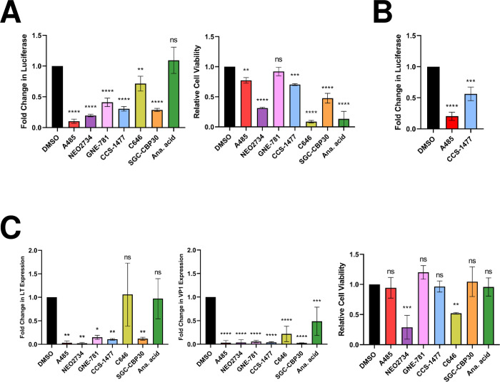
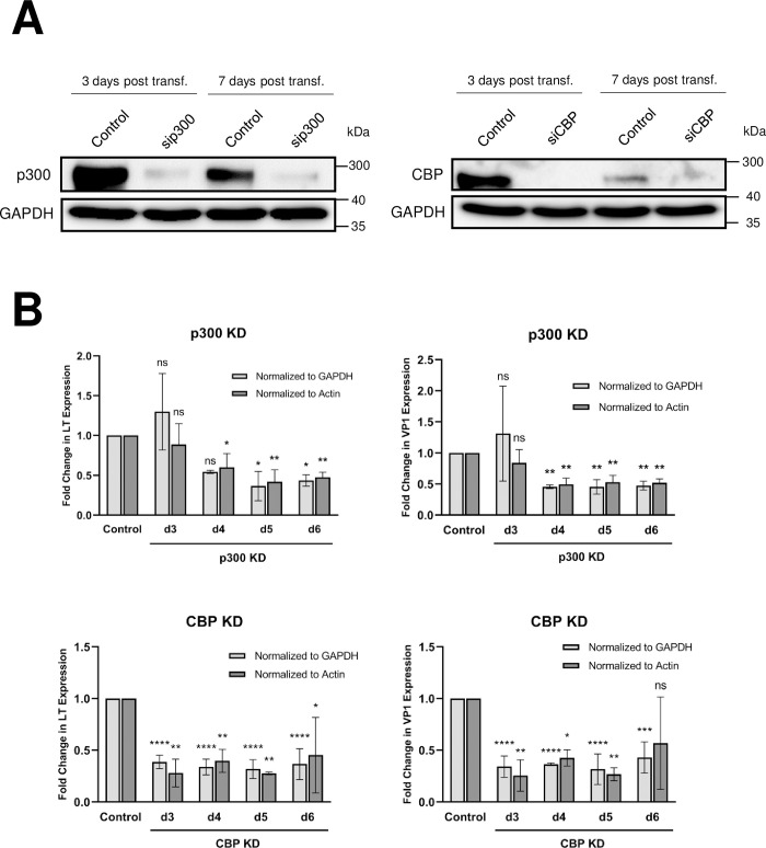
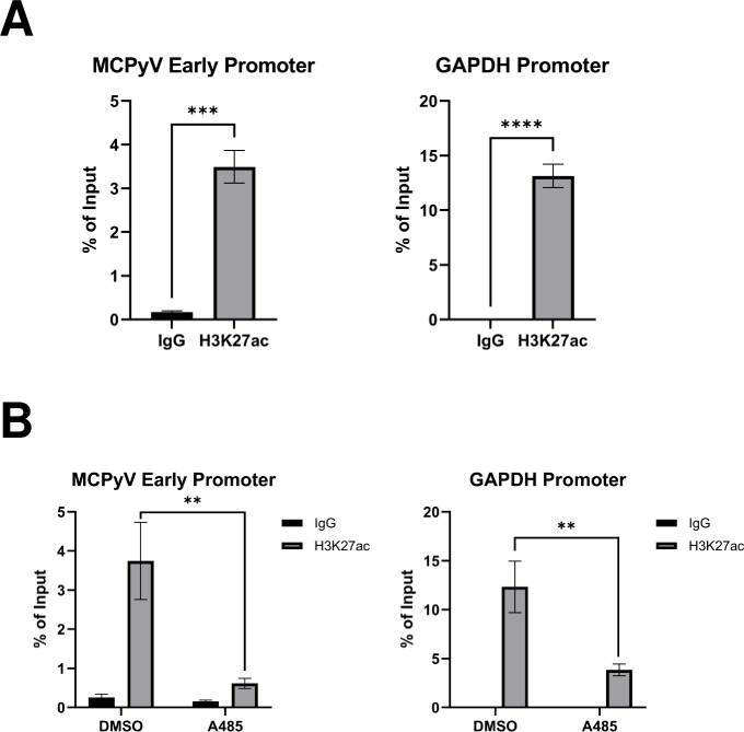
Merkel cell polyomavirus (MCPyV) is associated with approximately 80% of cases of Merkel cell carcinoma (MCC), an aggressive type of skin cancer. The incidence of MCC has tripled over the past twenty years, but there are currently very few effective targeted treatments. A better understanding of the MCPyV life cycle and its oncogenic mechanisms is needed to unveil novel strategies for the prevention and treatment of MCC. MCPyV infection and oncogenesis are reliant on the expression of the early viral oncoproteins, which drive the viral life cycle and MCPyV+ MCC tumor cell growth. To date, the molecular mechanisms regulating the transcription of the MCPyV oncogenes remain largely uncharacterized. In this study, we investigated how MCPyV early transcription is regulated to support viral infection and MCC tumorigenesis. Our studies established the roles of multiple cellular factors in the control of MCPyV gene expression. Inhibitor screening experiments revealed that the histone acetyltransferases p300 and CBP positively regulate MCPyV transcription. Their regulation of viral gene expression occurs through coactivation of the transcription factor NF-κB, which binds to the viral genome to drive MCPyV oncogene expression in a manner that is tightly controlled through a negative feedback loop. Furthermore, we discovered that small molecule inhibitors specifically targeting p300/CBP histone acetyltransferase activity are effective at blocking MCPyV tumor antigen expression and MCPyV+ MCC cell proliferation. Together, our work establishes key cellular factors regulating MCPyV transcription, providing the basis for understanding the largely unknown mechanisms governing MCPyV transcription that defines its infectious host cell tropism, viral life cycle, and oncogenic potential. Our studies also identify a novel therapeutic strategy against MCPyV+ MCC through specific blockage of MCPyV oncogene expression and MCC tumor growth.


Conflict of interest statement

The authors have declared that no competing interests exist.

Figures

Fig 1
Fig 1. MCPyV EP is specifically activated in the MCPyV+ MCC cell line MKL-1 and normal HDFs, but not in keratinocytes.
Lentiviruses carrying MCPyV EP-RFP or HPV11 LCR-RFP were used to infect MKL-1, HDFs and the keratinocyte cell line HaCaT. The stable cells were imaged using an inverted fluorescence microscope (IX81; Olympus). HPV11 LCR-RFP preferentially expressed in keratinocytes serves as a control for keratinocyte viability. Bar: 20μm.
Fig 2
Fig 2. HATi treatment represses MCPyV EP-driven transcription.
(A) HEK293 cells stably expressing an MCPyV EP-luciferase reporter were treated with DMSO, 2 μM A485, 1 μM NEO2734, 1 μM GNE-781, 1 μM CCS-1477, 10 μM C646, 10 μM SGC-CBP30, or 20 μM anacardic acid for 72h, then collected for luciferase or CellTiterGlo 3D assays. (B) HDFs stably expressing an MCPyV EP-luciferase reporter were treated with DMSO, 250 nM A485, or 250 nM CCS-1477 for 72h, then collected for luciferase assays. For both (A) and (B), fold changes in Luciferase were calculated after luciferase readings were normalized to the total protein concentration of each sample. (C) MCPyV-infected HDFs were treated with DMSO, 2 μM A485, 1 μM NEO2734, 1 μM GNE-781, 1 μM CCS-1477, 10 μM C646, 10 μM SGC-CBP30, or 20 μM anacardic acid on day 2 post-infection. Cells were collected on day 5 post-infection for CellTiterGlo 3D assays or RT-qPCR analysis of viral mRNA. RT-qPCR quantifications of viral mRNA expression were normalized to levels of cellular GAPDH mRNA. Error bars represent the standard deviation of three independent experiments. ****p<0.0001; ***p<0.001; **p<0.01; *p<0.05; ns = not significant.
Fig 3
Fig 3. p300 and CBP are important for supporting MCPyV transcription during infection.
(A) Whole cell lysates of HDFs transfected with siRNA targeting p300 (sip300), CBP (siCBP), or a scrambled control were collected at d3 and d7 post-transfection for Western blot analysis. (B) HDFs were transfected with siRNA against p300 (sip300), CBP (siCBP), or a scrambled control 24h prior to infection with 108 viral genome equivalents of MCPyV. RT-qPCR analysis of viral mRNA expression in MCPyV-infected HDFs was performed on days 3 through 6 post-infection. Changes in LT and VP1 expression in the KD cells relative to the levels of viral transcription in control siRNA-transfected HDFs were calculated and normalized to cellular levels of GAPDH or actin mRNA as indicated. Error bars represent the standard deviation of three independent experiments. ****p<0.0001; ***p<0.001; **p<0.01; *p<0.05; ns = not significant.
Fig 4
Fig 4. Detection of p300/CBP-specific histone acetylation marks on the MCPyV EP.
ChIP was performed with MKL-1 cells (A) or MKL-1 cells that have been treated for 1h with DMSO or 2 uM A485 (B) using 0.5 μg normal rabbit IgG or antibody recognizing the p300/CBP-specific histone acetylation mark H3K27ac. qPCR was performed on the ChIP samples using primers recognizing the MCPyV EP or the GAPDH promoter. Error bars represent the standard deviation of three independent experiments. ****p<0.0001; ***p<0.001; **p<0.01.
Fig 5
Fig 5. Inhibition of NF-κB activity represses MCPyV EP-driven viral oncogene expression, which is lethal in MCPyV+ MCC.
(A) HEK293 cells stably expressing an MCPyV EP-luciferase reporter were treated with DMSO or 25 μM JSH-23 for 72h before EP-driven luciferase expression was measured by luciferase assay. (B) PETA and (C) MKL-1 cells were treated with DMSO or 25 μM JSH-23 for up to 9 days. At 24h and 72h post-treatment, RT-qPCR analysis was performed to measure relative changes in MCPyV LTT expression during treatment; LTT mRNA levels were normalized to the levels of cellular GAPDH mRNA. The viability of the cells was measured during treatment using the CellTiterGlo 3D assay. The % viability of the cells in each condition is expressed as the fold change in the sample’s CellTiterGlo reading relative to its d0 measurement. Error bars represent the standard deviation of three independent experiments. ****p<0.0001; ***p<0.001.
Fig 6
Fig 6. NF-κB p65 binds directly to the MCPyV NCRR.
(A) NCRR-specific DNA binding activity is detected in HEK293 nuclear extracts containing overexpressed p65. EMSA was performed using a set of positive control probes and nuclear extract provided in the LightShift Chemiluminescent EMSA kit (“Control EMSA”) or using full NCRR probes and nuclear extracts from HEK293 cells transfected with a p65-expressing plasmid (“NCRR EMSA”). (B) Schematic of the biotinylated DNA pulldown assay. Biotinylated (blue stars) NCRR probes were bound to streptavidin-coated magnetic beads (brown), then incubated with nuclear extracts containing the protein of interest (in yellow; other nuclear proteins are indicated in gray). Alternatively, the nuclear extracts are pre-incubated with an excess amount of unlabeled NCRR probe before being incubated with the bead-bound probes. Protein-probe complexes (protein of interest [yellow] bound to biotinylated [blue stars] probes) are eluted off the beads for analysis by SDS-PAGE/Western blot or agarose gel electrophoresis. (C) NF-κB p65 binds the MCPyV NCRR. Biotinylated NCRR pulldown assays were performed with nuclear extracts from untreated HEK293 cells (“Untreated”), or cells overexpressing p65 (“p65”), and biotinylated-NCRR probes in the presence or absence of an excess of unlabeled NCRR competitor (“Competitor”). The left panel depicts the detection of p65 by Western blotting in the input (1%) and pulldown samples, while the right panel demonstrates that comparable amounts of biotinylated probe were bound to the beads in each pulldown experiment.
Fig 7
Fig 7. NF-κB p65 regulates MCPyV EP-driven transcription in a tightly controlled manner.
HEK293 cells were transfected with an MCPyV EP-luciferase reporter, a control reporter expressing Renilla luciferase, and the indicated amounts of a p65 expression plasmid. Cells were collected 24h after transfection for Western blot analysis (A), luciferase assay (B), or RT-qPCR analysis for IκB mRNA (C). Luciferase readings were normalized to the Renilla luciferase values for each sample. Changes in IκB mRNA level were normalized to cellular GAPDH mRNA. Error bars represent the standard deviation of three independent experiments. ****p<0.0001; ***p<0.001; **p<0.01; ns = not significant.
Fig 8
Fig 8. NF-κB p65 functions downstream of p300/CBP to modulate MCPyV gene expression.
(A) Biotinylated NCRR pulldown assays were performed with nuclear extracts from cells pre-treated with DMSO or 2 μM A485 for 20h before transfection with a p65-expressing plasmid. Nuclear extracts were collected 24h after transfection and incubated with biotinylated-NCRR probes attached to streptavidin magnetic beads, in the presence or absence of an excess of unlabeled NCRR competitor (“Competitor”). Protein and DNA were eluted from the beads for Western blot or agarose gel analysis. The upper panel depicts the detection of p65 by Western blotting in the input (1%) and pulldown samples, with band intensities for the Pulldown and Competitor lanes relative to the DMSO Pulldown condition. The lower left panel presents the Western blotting analysis of the nuclear extracts, while the lower right panel demonstrates that comparable amounts of biotinylated probe were bound to the beads in each pulldown experiment. (B) HEK293 cells were pre-treated with DMSO or 25 μM JSH for 16h before being transfected with an MCPyV EP-luciferase reporter plasmid. 8h after transfection, cells were treated with DMSO or 1 μM SAHA, and collected for luciferase assay 20h later. Luciferase values were normalized to the total protein concentration of each sample. Error bars represent the standard deviation of three independent experiments. ***p<0.001; ns = not significant.
Fig 9
Fig 9. Inhibition of p300/CBP activity represses MCPyV LTT expression to specifically kill MCPyV+ MCC.
(A) PETA and MKL-1 cells were treated with DMSO, 2 μM A485, 1 μM NEO2734, 1 μM GNE-781, 1 μM CCS-1477, 10 μM C646, 10 μM SGC-CBP30, or 20 μM anacardic acid. After 3 and 7 days, cell lysates were subjected to Western blotting analysis to detect MCPyV LTT and GAPDH expression. (B) PETA, MKL-1, MCC-13, and HDFs were treated with the indicated inhibitors for up to 9 days. Cell viability on days 0, 3, 6, and 9 was measured using the CellTiterGlo 3D assay. The % viability of the cells in each condition is expressed as the fold change in the sample’s CellTiterGlo reading relative to its d0 measurement. In each plot, the % viability of DMSO-treated cells is represented by a dotted black line, while the % viability of HATi-treated cells is represented by a solid colored line. Error bars represent the standard deviation of three independent experiments.
Fig 10
Fig 10. Molecular mechanisms regulating the NCRR-driven transcriptional program of MCPyV.
p300/CBP upregulate MCPyV gene expression primarily through acetylation of the p65 subunit of NF-κB, which binds directly to kB site(s) on the NCRR. p300/CBP may also acetylate the histones associated with the viral NCRR to stimulate MCPyV transcription. In addition to p300/CBP-mediated acetylation, NF-κB activity is also stimulated by MCPyV infection in HDFs. Overstimulation of NF-κB induces the expression of IκBα, which in turn inhibits NF-κB activity through a negative feedback mechanism. The accumulation of NF-κB acetylation within the cell by HDAC inhibition upregulates the p300/CBP-mediated stimulation of viral transcription, while HAT inhibition robustly represses viral transcription downstream of p300/CBP.

Similar articles

Cited by

References

    1. Toker C. Trabecular Carcinoma of the Skin. Archives of Dermatology. 1972;105(1):107–10. doi: 10.1001/archderm.1972.01620040075020 - DOI - PubMed
    1. Tang CK, Toker C. Trabecular carcinoma of the skin: an ultrastructural study. Cancer. 1978;42(5):2311–21. Epub 1978/11/01. doi: 10.1002/1097-0142(197811)42:5&lt;2311::aid-cncr2820420531&gt;3.0.co;2-l - DOI - PubMed
    1. Paulson KG, Park SY, Vandeven NA, Lachance K, Thomas H, Chapuis AG, et al.. Merkel cell carcinoma: Current US incidence and projected increases based on changing demographics. Journal of the American Academy of Dermatology. 2018;78(3):457–63.e2. Epub 20171102. doi: 10.1016/j.jaad.2017.10.028 ; PubMed Central PMCID: PMC5815902. - DOI - PMC - PubMed
    1. Agelli M, Clegg LX, Becker JC, Rollison DE. The Etiology and Epidemiology of Merkel Cell Carcinoma. Current Problems in Cancer. 2010;34(1):14–37. 10.1016/j.currproblcancer.2010.01.001. doi: 10.1016/j.currproblcancer.2010.01.001 - DOI - DOI - PubMed
    1. Fitzgerald TL, Dennis S, Kachare SD, Vohra NA, Wong JH, Zervos EE. Dramatic Increase in the Incidence and Mortality from Merkel Cell Carcinoma in the United States. Am Surg. 2015;81(8):802–6. doi: 10.1177/000313481508100819 . - DOI - PubMed

Publication types