. 2023 Aug 30;15(711):eabo1557.
doi: 10.1126/scitranslmed.abo1557. Epub 2023 Aug 30.

A blood-based marker of mitochondrial DNA damage in Parkinson's disease

Affiliations

A blood-based marker of mitochondrial DNA damage in Parkinson's disease

Rui Qi et al. Sci Transl Med. .

Abstract

Parkinson's disease (PD) is the most common neurodegenerative movement disorder, and neuroprotective or disease-modifying interventions remain elusive. High-throughput markers aimed at stratifying patients on the basis of shared etiology are required to ensure the success of disease-modifying therapies in clinical trials. Mitochondrial dysfunction plays a prominent role in the pathogenesis of PD. Previously, we found brain region-specific accumulation of mitochondrial DNA (mtDNA) damage in PD neuronal culture and animal models, as well as in human PD postmortem brain tissue. To investigate mtDNA damage as a potential blood-based marker for PD, we describe herein a PCR-based assay (Mito DNADX) that allows for the accurate real-time quantification of mtDNA damage in a scalable platform. We found that mtDNA damage was increased in peripheral blood mononuclear cells derived from patients with idiopathic PD and those harboring the PD-associated leucine-rich repeat kinase 2 (LRRK2) G2019S mutation in comparison with age-matched controls. In addition, mtDNA damage was elevated in non-disease-manifesting LRRK2 mutation carriers, demonstrating that mtDNA damage can occur irrespective of a PD diagnosis. We further established that Lrrk2 G2019S knock-in mice displayed increased mtDNA damage, whereas Lrrk2 knockout mice showed fewer mtDNA lesions in the ventral midbrain, compared with wild-type control mice. Furthermore, a small-molecule kinase inhibitor of LRRK2 mitigated mtDNA damage in a rotenone PD rat midbrain neuron model and in idiopathic PD patient-derived lymphoblastoid cell lines. Quantifying mtDNA damage using the Mito DNADX assay may have utility as a candidate marker of PD and for measuring the pharmacodynamic response to LRRK2 kinase inhibitors.


Conflict of interest statement

Competing interests: L.H.S. and S.S. are coinventors on US patent application number 17/227,186 entitled “Mitochondrial health parameters as clinical predictors of Parkinson’s disease.” L.H.S. is on the scientific advisory board of Lucy Therapeutics. R.N.A. received consultation fees from Avrobio, Biohaven, Capsida, Caraway, Gain Therapeutics, Genzyme/Sanofi, and Takeda. C.M. acted as advisor to the MJFF for the creation and management of FBN. C.M. received funding from the MJFF as the principal investigator of FBN. T.S. has served as a consultant for AcureX, Adamas, AskBio, Amneal, Blue Rock Therapeutics, Critical Path for Parkinson’s Consortium (CPP), Denali, MJFF, Neuroderm, Sanofi, Sinopia, Roche, Takeda, and Vanqua Bio. T.S. served on the ad board for AcureX, Adamas, AskBio, Denali, and Roche. T.S. has served as a member of the scientific advisory board of Neuroderm, Sanofi, and UCB. T.S. has received research funding from Amneal, Biogen, Neuroderm, Prevail, Roche, and UCB and is an investigator for National Institute of Neurological Disorders and Stroke (NINDS), MJFF, and the Parkinson’s Foundation. The other authors declare that they have no competing interests.

Figures

Fig. 1.
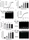
Fig. 1.. The Mito DNADX assay measures the frequency of mtDNA lesions in real time.
HEK293 cells were exposed to increasing concentrations of H2O2 for 1 hour at 37°C, and total cellular DNA was isolated. (A) The amount of ResoLight dye fluorescence associated with each 8.9-kb amplification PCR product relative to the vehicle treated control is plotted. (B) The decrease in relative amplification from (A) was then converted to lesion frequency using the newly developed equation as described in Materials and Methods and then the Poisson equation applied. (C) Representative 0.6% agarose gel depicting the specificity of the mitochondrial PCR end product. (D) The amount of SYBR Green dye fluorescence associated with each 248-bp amplification PCR product relative to the vehicle-treated control is plotted. (E) The mtDNA copy number relative to control for increasing concentrations of H2O2. (F) Representative 2.0% agarose gel depicting the specificity of the 248-bp mitochondrial amplicon. (G) The mtDNA lesion frequency from (B) was then normalized both to mtDNA copy in (E) and 10 kb to generate the final mtDNA lesion/10-kb frequency. (H) Representative 2.0% agarose gel of extracted DNA using the Autogen 610L protocol. The Mito DNADX assay was performed in technical triplicate for each biological replicate. [*P < 0.05 and ***P < 0.0001, determined by one-way analysis of variance (ANOVA) with Dunnett’s multiple comparison test]. n = 3 to 6 biological replicates. Data are presented as means ± SEM.
Fig. 2.
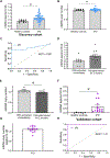
Fig. 2.. LRRK2 kinase inhibition reverts mtDNA damage in idiopathic PD patient–derived lymphoblastoid cells.
(A) The frequency of mtDNA lesions was analyzed in idiopathic PD patient–derived LCLs (n = 21), PD patient–derived LCLs from G2019S LRRK2 carriers (n = 2), and age-matched healthy controls (n = 12). *P < 0.01, determined by one-way ANOVA with Bonferroni’s multiple comparison test. (B) mtDNA copy number for LCLs in (A). (C) The frequency of mtDNA lesions of idiopathic PD patient–derived LCLs (n = 2 lines, two independent determinations each) treated with DMSO or the LRRK2 kinase inhibitor, MLi-2 (10 or 30 nM for 24 hours). *P < 0.001, determined by one-way ANOVAwith Dunnett’s multiple comparison test. (D) mtDNA copy number for treatment groups in (C). iPD, idiopathic PD. Data are presented as means ± SEM.
Fig. 3.
Fig. 3.. LRRK2 function alters mtDNA lesion frequency in mice.
(A) The frequency of mtDNA lesions was analyzed in the ventral midbrain derived from both heterozygous and homozygous Lrrk2 G2019S knock-in (GKI) (n = 14) mice relative to wild-type (WT) mice (n = 9). (B) mtDNA copy number for animals used in (A). (C) The frequency of mtDNA lesions was analyzed in the ventral midbrain derived from Lrrk2 KO mice (n = 6) relative to WT mice (n = 5). (D) mtDNA copy number for animals used in (C). *P < 0.05 determined by Student’s t test. Data are presented as means ± SEM.
Fig. 4.
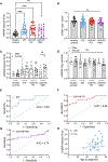
Fig. 4.. The frequency of mtDNA lesions in PBMCs can predict PD status in a discovery and validation cohort.
(A) The frequency of mtDNA lesions in PBMCs derived from patients with idiopathic PD (n = 53) and age-matched healthy controls (n = 10) (*P < 0.001, determined by Mann-Whitney test). (B) mtDNA copy number for PBMCs analyzed in (A). (C) ROC curve showing prediction success for PD diagnosis (*P < 0.001). AUC, area under the curve. (D) The impact of storage on mtDNA lesion frequency. (E) The impact of storage on mtDNA copy number (*P < 0.05, determined by Student’s t test). (F) The frequency of mtDNA lesions in PBMCs from a validation cohort derived from patients with idiopathic PD (n = 14) and age-matched healthy controls (n = 6) (*P < 0.0001, determined by Mann-Whitney test). (G) mtDNA copy number for (F). (H) ROC curve showing prediction success for PD diagnosis (P < 0.001). The Mito DNADX assay was performed in technical triplicate for each biological replicate. ns, P > 0.05. Data are presented as means ± SEM. All mtDNA damage quantifications were performed blinded before unblinding for clinical and demographic data.
Fig. 5.
Fig. 5.. mtDNA lesion frequency is increased in idiopathic PD with acute washout of PD-related medications.
(A) The frequency of mtDNA lesions in PBMCs derived from patients with idiopathic PD (n = 16) collected during a washout period and age-matched healthy controls (n = 21) (*P < 0.0001, determined by Mann-Whitney test). (B) Steady-state mtDNA copy number for (A). (C) ROC curve showing prediction success for PD (P < 0.0001). The Mito DNADX assay was performed in technical triplicate for each biological replicate. Data are presented as means ± SEM. All mtDNA damage quantifications were performed blinded before unblinding for clinical and demographic data.
Fig. 6.
Fig. 6.. mtDNA damage is increased in both idiopathic and LRRK2 PD.
(A) Frequency of mtDNA lesions in PBMCs derived from participants with idiopathic PD, LRRK2-G2019S carriers with PD, or LRRK2-G2019S nonmanifesting (NMC) (***P < 0.001 and **P < 0.01, determined by one-way ANOVA with a Tukey’s post hoc comparison). (B) mtDNA copy number for groups in (A). (C) Frequency of mtDNA lesions for groups stratified by sex (*P < 0.01 determined by one-way ANOVA with a Šidák’s post hoc comparison). (D) mtDNA copy number for groups in (C). (E and F) ROC curve for idiopathic and LRRK2-G2019S PD compared with unaffected controls (P < 0.0001). (G) ROC curve for LRRK2 G2019S NMC from healthy controls (P < 0.01). (H) Correlation between age at diagnosis and mtDNA lesion frequency (P < 0.05, Pearson correlation coefficient). The Mito DNADX assay was performed in technical triplicate for each biological replicate. Data are presented as means ± SEM. All mtDNA damage quantification was performed blinded before unblinding for clinical, demographic data and LRRK2 G2019S mutation status for the grouped analysis.

References

    1. Fearnley JM, Lees AJ, Ageing and Parkinson’s disease: Substantia nigra regional selectivity. Brain 114 (Pt 5), 2283–2301 (1991). - PubMed
    1. Cheng HC, Ulane CM, Burke RE, Clinical progression in Parkinson disease and the neurobiology of axons. Ann. Neurol. 67, 715–725 (2010). - PMC - PubMed
    1. Marras C, Lang A, Parkinson’s disease subtypes: Lost in translation? J. Neurol. Neurosurg. Psychiatry 84, 409–415 (2013). - PubMed
    1. Mestre TA, Fereshtehnejad SM, Berg D, Bohnen NI, Dujardin K, Erro R, Espay AJ, Halliday G, van Hilten JJ, Hu MT, Jeon B, Klein C, Leentjens AFG, Marinus J, Mollenhauer B, Postuma R, Rajalingam R, Rodriguez-Violante M, Simuni T, Surmeier DJ, Weintraub D, McDermott MP, Lawton M, Marras C, Parkinson’s disease subtypes: Critical appraisal and recommendations. J. Parkinsons Dis. 11, 395–404 (2021). - PMC - PubMed
    1. Espay AJ, Schwarzschild MA, Tanner CM, Fernandez HH, Simon DK, Leverenz JB, Merola A, Chen-Plotkin A, Brundin P, Kauffman MA, Erro R, Kieburtz K, Woo D, Macklin EA, Standaert DG, Lang AE, Biomarker-driven phenotyping in Parkinson’s disease: A translational missing link in disease-modifying clinical trials. Mov. Disord. 32, 319–324 (2017). - PMC - PubMed

Publication types

Substances