. 2023 Sep;9(35):eadh4890.
doi: 10.1126/sciadv.adh4890. Epub 2023 Aug 30.

Structures of wild-type and selected CMT1X mutant connexin 32 gap junction channels and hemichannels

Affiliations

Structures of wild-type and selected CMT1X mutant connexin 32 gap junction channels and hemichannels

Chao Qi et al. Sci Adv. 2023 Sep.

Abstract

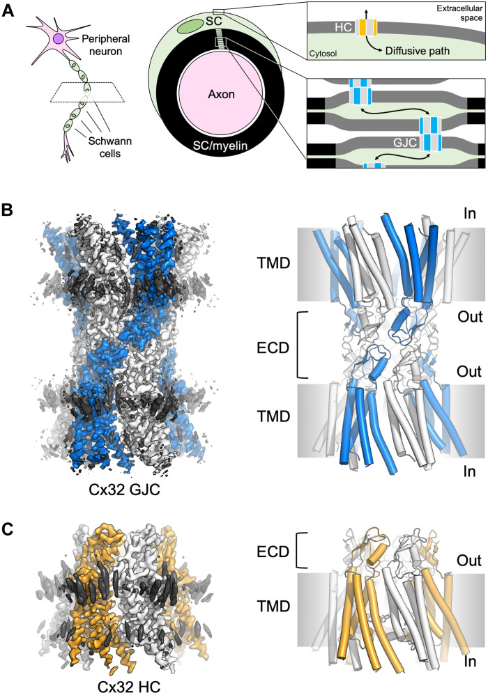
In myelinating Schwann cells, connection between myelin layers is mediated by gap junction channels (GJCs) formed by docked connexin 32 (Cx32) hemichannels (HCs). Mutations in Cx32 cause the X-linked Charcot-Marie-Tooth disease (CMT1X), a degenerative neuropathy without a cure. A molecular link between Cx32 dysfunction and CMT1X pathogenesis is still missing. Here, we describe the high-resolution cryo-electron cryo-myography (cryo-EM) structures of the Cx32 GJC and HC, along with two CMT1X-linked mutants, W3S and R22G. While the structures of wild-type and mutant GJCs are virtually identical, the HCs show a major difference: In the W3S and R22G mutant HCs, the amino-terminal gating helix partially occludes the pore, consistent with a diminished HC activity. Our results suggest that HC dysfunction may be involved in the pathogenesis of CMT1X.


Figures

Fig. 1.
Fig. 1.. Cryo-EM structures of Cx32 GJC and HC.
(A) Illustration of Cx32 GJCs and HCs in distinct membrane compartments of the myelinating Schwann cells (SC). (B) Cryo-EM map and model of Cx32 GJC. Cx32 is surrounded by lipid-like molecules at the outer leaflet and inner leaflet of the membrane (black density). (C) Cryo-EM map and model of Cx32 HC.
Fig. 2.
Fig. 2.. Comparisons of Cx32 GJC and HC.
(A and B) Cryo-EM map (bottom view) and model (side view) of Cx32 GJC. The NTH (red and black) inserts to the channel pore, representing the open state of GJC. The lipid-1 density is labeled using pink color. (C and D) Cx32 HC map (bottom view) and model (side view). The NTH (red and black) is parallel to the membrane layer, shrinking the pore of Cx32 HC to about 11 Å. Lipid-2 (cyan) is stabilized by NTH, TM1, and TM2. (E) Electrostatic map of Cx32 HC. Cholesterol (CLR) molecules (cyan) are fitted to the lipid-2 position. (F) Detailed view of the interaction between CLR and NTH, TM1, and TM2. All the residues within 4 Å to CLR are shown as sticks. Two important residues, W3 and R22, are shown as green color.
Fig. 3.
Fig. 3.. Comparison of the WT Cx32 HC structure to the cryo-EM structures of W3S and R22G Cx32 mutants.
(A) Substrate conduction pathways for Cx32, W3S, and R22G HCs, calculated using Hole software. The arrow indicates the narrowest part of the path. Hollow arrow for Cx32 and black arrow for mutants. (B) The pore radius along the substrate conduction pathway was calculated using hole in (A). (C) Structural alignment of NTH between Cx32 HC (orange), W3S HC (pink), and R22G HC (green). Key residues, W3 and R22, are labeled as spheres. (D and E) Detailed view of NTH in Cx32, W3S, and R22G. The key residues within 4 Å close to CLR are shown as sticks. The pink and green arrows indicate the movement of NTH in W3S and R22G compared to Cx32 HC.
Fig. 4.
Fig. 4.. Functional properties of GJCs and HCs formed by WT, W3S, or R22G Cx32 in HEK293F cells.
(A) A sketch illustrating the GJC activity measurements using dual patch-clamp; black circles indicate Cx32 connexons (left). Boxplot of the GJ conductance (gj) values measured by dual patch-clamp in untransfected (control) HEK293F cells or transfected with WT, R22G, or W3S constructs. Cell pairs were selected on the basis of similar expression of the cytosolic YFP. At the end of each experiment, gj was lowered to zero by a CO2-saturated extracellular solution to confirm that the cells were connected by GJs and not by cytoplasmic bridges. gj values of untransfected cells (n = 14) resulted significantly lower (P < 0.05) than WT (n = 14) but not than R22G (n = 13) and W3S (n = 13). Statistical analysis was performed using the Kruskal-Wallis test. (B) Left: FRAP experiments (sketch indicating dye transfer between two coupled cells). Intercellular diffusion of the fluorescent tracer SR101 (molecular weight, 606.7 Da) was assessed in control (n = 10) and transfected cells expressing Cx32 WT (n = 20), W3S (n = 13), or R22G (n = 13). (C) Left: Sketch of a whole-cell patch-clamp experiment with Cx32 HCs at the cell surface. Whole-cell patch-clamp experiments performed in control (n = 5) or transfected cells with Cx32 WT (orange, n = 13), W3S (pink, n = 6), or R22G (green, n = 10). The currents mediated by mutant HCs are lower with respect to the WT. (D) SR101 dye uptake experiments performed in control cells (n = 935) or cells expressing Cx32 WT (n = 1136), W3S (n = 702), or R22G (n = 905); mutant HCs showed reduced dye uptake; asterisks indicate statistical significance (one-way ANOVA: ****P < 0.0001; ***P < 0.001; *P < 0.1; ns, no significant difference). a.f.u., arbitrary fluorescence units.
Fig. 5.
Fig. 5.. A schematic illustration of the key features observed in the Cx32 structures.
(A) Cx32 GJC is determined in an apparently open state, with a central pore of ~15-Å diameter; this estimate does not take into account the flexible NTH domains, as well as the lipids that may occlude the pore under physiological conditions, in the cellular plasma membrane. (B) The open state of the Cx32 HC features a central pore about 11 Å. The NTH domains are parallel to the membrane plane in an iris-like arrangement. (C and D) The W3S and R22G HC structures feature a closed state, where the NTH rearrangement reduces the pore diameter to ~6 Å.

Similar articles

Cited by

References

    1. M. Goldberg, M. De Pitta, V. Volman, H. Berry, E. Ben-Jacob, Nonlinear gap junctions enable long-distance propagation of pulsating calcium waves in astrocyte networks. PLOS Comput. Biol. 6, e1000909 (2010). - PMC - PubMed
    1. S. Rohr, Role of gap junctions in the propagation of the cardiac action potential. Cardiovasc. Res. 62, 309–322 (2004). - PubMed
    1. I. Epifantseva, R. M. Shaw, Intracellular trafficking pathways of Cx43 gap junction channels. Biochim. Biophys. Acta Biomembr. 1860, 40–47 (2018). - PMC - PubMed
    1. D. C. Spray, R. Iglesias, N. Shraer, S. O. Suadicani, V. Belzer, R. Hanstein, M. Hanani, Gap junction mediated signaling between satellite glia and neurons in trigeminal ganglia. Glia 67, 791–801 (2019). - PMC - PubMed
    1. P. Meda, Gap junction proteins are key drivers of endocrine function. Biochim. Biophys. Acta Biomembr. 1860, 124–140 (2018). - PubMed