. 2023 Nov 30;35(12):4217-4237.
doi: 10.1093/plcell/koad227.

The plant-unique protein DRIF1 coordinates with sorting nexin 1 to regulate membrane protein homeostasis

Affiliations

The plant-unique protein DRIF1 coordinates with sorting nexin 1 to regulate membrane protein homeostasis

Ying Zhu et al. Plant Cell. .

Abstract

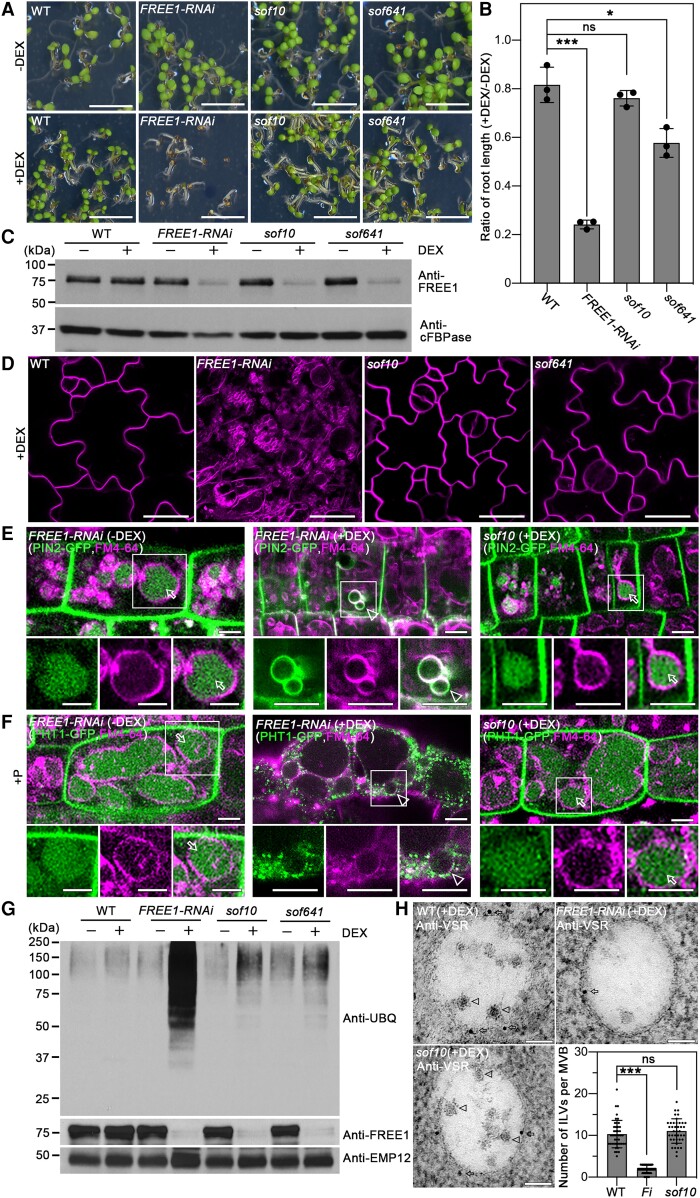
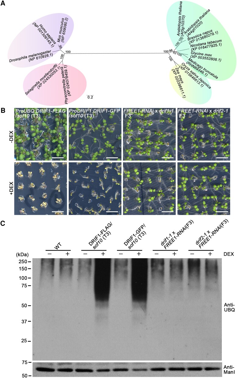
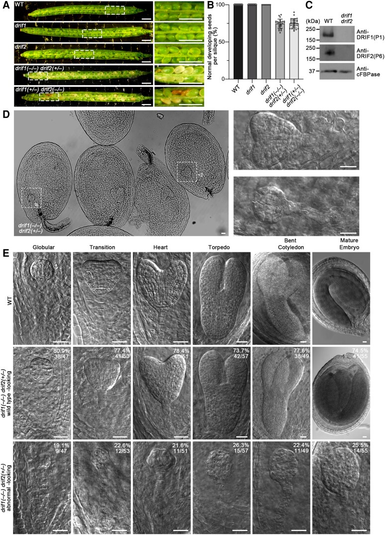
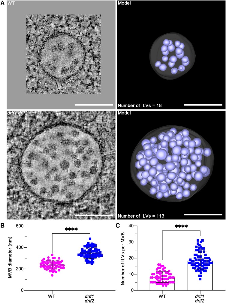
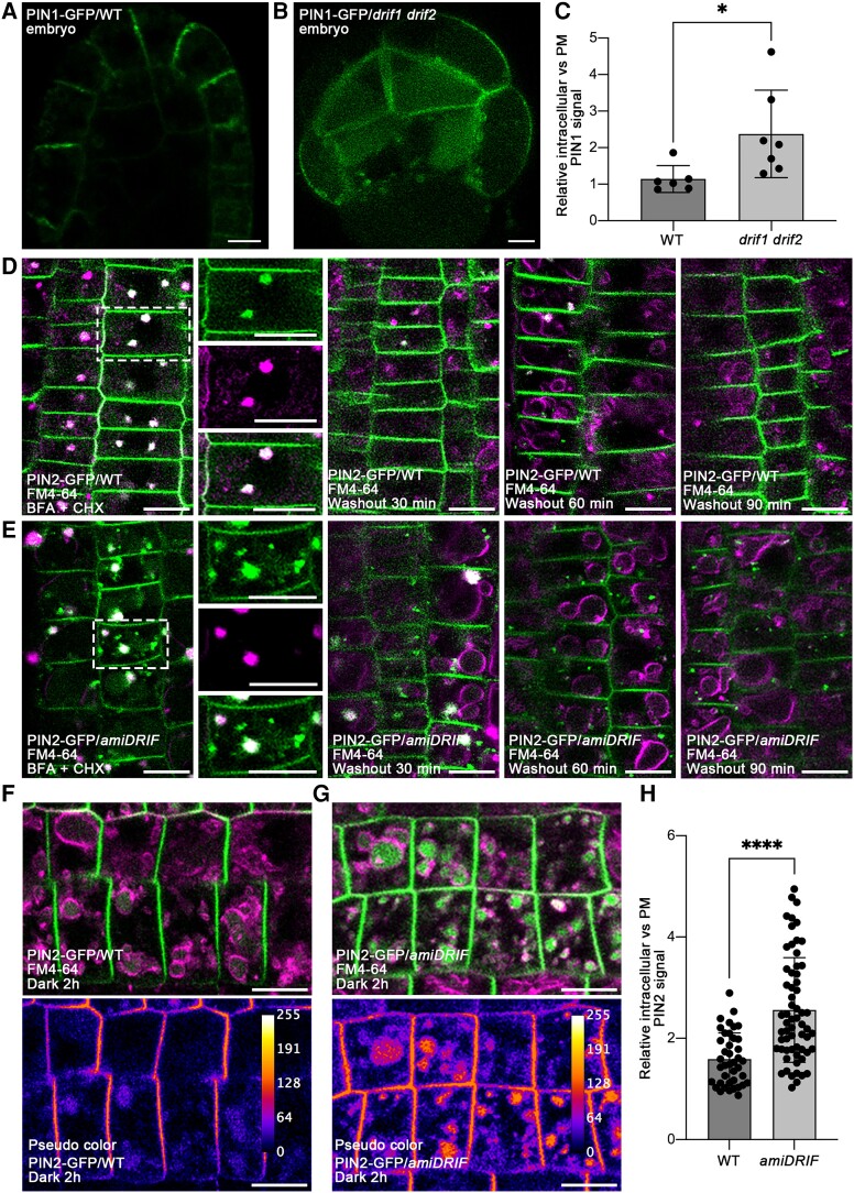
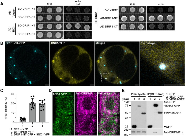
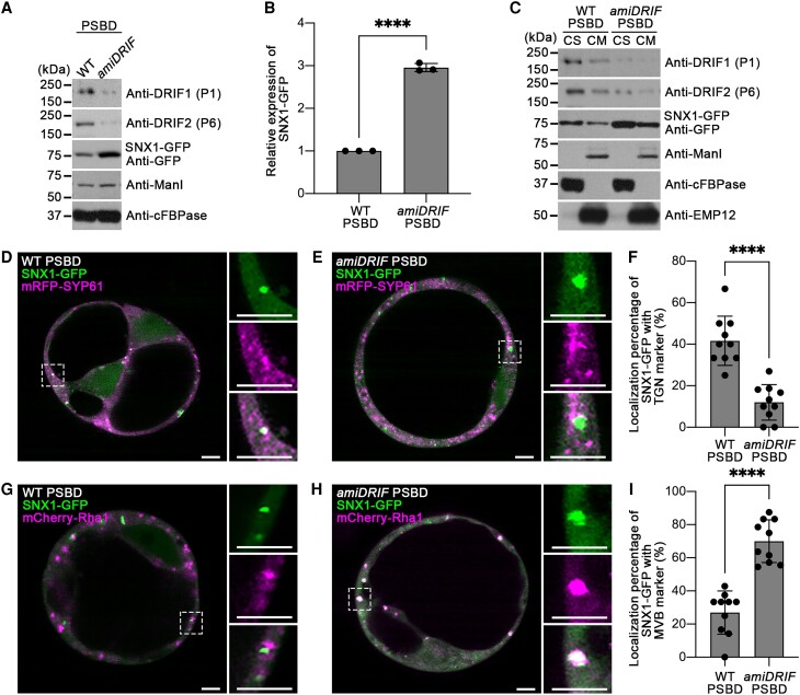
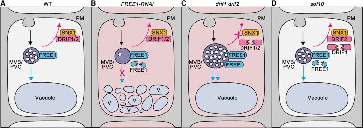
Membrane protein homeostasis is fine-tuned by the cellular pathways for vacuolar degradation and recycling, which ultimately facilitate plant growth and cell-environment interactions. The endosomal sorting complex required for transport (ESCRT) machinery plays important roles in regulating intraluminal vesicle (ILV) formation and membrane protein sorting to vacuoles. We previously showed that the plant-specific ESCRT component FYVE DOMAIN PROTEIN REQUIRED FOR ENDOSOMAL SORTING1 (FREE1) performs multiple functions in plants, although the underlying mechanisms remain elusive. In this study, we performed a suppressor screen of the FREE1-RNAi mutant and identified and characterized 2 suppressor of free1 (sof) mutants in Arabidopsis (Arabidopsis thaliana). These mutants, sof10 and sof641, result in a premature stop codon or a missense mutation in AT5G10370, respectively. This gene was named DEAH and RING domain-containing protein as FREE1 suppressor 1 (DRIF1). DRIF1 has a homologous gene, DRIF2, in the Arabidopsis genome with 95% identity to DRIF1. The embryos of drif1 drif2 mutants arrested at the globular stage and formed enlarged multivesicular bodies (MVBs) with an increased number of ILVs. DRIF1 is a membrane-associated protein that coordinates with retromer component sorting nexin 1 to regulate PIN-FORMED2 recycling to the plasma membrane. Altogether, our data demonstrate that DRIF1 is a unique retromer interactor that orchestrates FREE1-mediated ILV formation of MVBs and vacuolar sorting of membrane proteins for degradation in plants.


Conflict of interest statement

Conflict of interest statement. The authors declare no conflict of interest.

Figures

Figure 1.
Figure 1.
The sof10 mutant converts the seedling lethality of FREE1-RNAi. A) The lethal phenotype of DEX-inducible FREE1-RNAi is rescued in the sof10 and sof641 mutants. M3 seeds were sown on MS plates without DEX (DEX) or with 10 μm DEX (+DEX) and grown for 7 d before phenotypic analysis. Scale bar, 5 mm. B) Quantification of the root length ratio for 7-d-old seedlings of WT, FREE1-RNAi, sof10, and sof641 mutants grown on MS with (+) or without (−) DEX as shown in A). Data are shown as mean ± Sd, and results from 3 individual experiments are plotted. For each genotype from each independent experiment, at least 20 seedlings were measured. ***P < 0.001, *P < 0.05, ns, no significance in 1-way ANOVA followed by Turkey's multiple test. For each experiment, n = 16 roots. C)sof10 and sof641 mutants show reduced FREE1 protein levels as FREE1-RNAi. Total proteins were extracted from 7-d-old seedlings of WT, FREE1-RNAi, sof10, and sof641 grown on MS with (+) or without (−) DEX, followed by western blot analysis using FREE1 antibodies. The cytosolic protein cFBPase was used as a loading control. D) The sof10 and sof641 mutants recover vacuolar defects compared with FREE1-RNAi when grown on MS with DEX (+DEX). The 7-d-old seedlings from indicated genotypes were stained with 4 μm FM4-64 dye for 3 h, and the labeled tonoplast was visualized by confocal imaging analysis in magenta. Scale bars, 25 μm. E) The mislocalization of auxin efflux carrier PIN2-GFP is converted in sof10 mutant. Seven-day-old seedlings of PIN2-GFP/FREE1-RNAi or PIN2-GFP/sof10 with or without DEX induction were stained with FM4-64 for the tonoplast and visualized after 6 h dark treatment. Arrows and arrowheads indicate vacuolar and tonoplast localized GFP signals, respectively. Images in white squared boxes are shown by separated channels (from left to right: GFP, RFP, and merged). Scale bars, 5 μm. F) The mislocalization of phosphate transporter PHT1-GFP is reversed in sof10 mutant. Seven-d-old seedlings of PHT1-GFP/FREE1-RNAi or PHT1-GFP/sof10 with or without DEX were stained with FM4-64 and incubated in +P liquid medium for 3 h dark treatment before visualization. Arrows and arrowheads indicate vacuolar and tonoplast localized GFP signals, respectively. Images in white squared boxes are shown by separated channels (from left to right: GFP, RFP, and merged). Scale bars, 5 μm. G) The sof10 and sof641 mutants convert the ubiquitin conjugates accumulation phenotype in FREE1-RNAi. Membrane proteins extracted from indicated 7-d-old seedlings grown on MS with (+) or without (−) DEX were subjected to immunoblot analysis with UBQ and FREE1 antibodies. EMP12 antibody was used as loading control. H) The formation of ILVs in MVB is restored in sof10 mutants. Ultrathin sections were prepared from HPF/FS roots of WT, FREE1-RNAi, and sof10 grown on MS with (+) DEX, followed by immunogold labeling using VSR antibodies. Arrows and arrowheads indicate the gold particles and ILVs, respectively. The number of ILVs per MVB were statistically analyzed, and individual data points were plotted with mean ± Sd ***P < 0.001, ns, no significance in 1-way ANOVA followed by Turkey's multiple test. For each sample, n = 40. Scale bars, 100 nm. FREE1-RNAi, ProDEX:FREE1-RNAi.
Figure 2.
Figure 2.
The sof10 and sof641 mutants affect DRIF1 protein. A) Phylogenetic analysis of DRIF1 homologs by neighbor-joining method. The percentage of replicate trees in which the associated taxa clustered together in the bootstrap test (1,000 replicates) are shown next to the branches. The tree is drawn to scale, with the computed evolutionary distance shown as branch length. Major groupings are indicated by different colors. Note that the DRIF1 protein has a homolog DRIF2 in Arabidopsis. Scale bar, 0.2 substitutions per site. B) Complementation of sof10 with indicated fusion proteins or phenotype of indicated genotype mutants on MS plates with (+) or without (−) DEX. The seedling phenotype of sof10 could be complemented by ProUBQ:DRIF1-FLAG and ProDRIF1:DRIF1-GFP. Note that both drif1-1 and drif2-1 T-DNA insertions could revert the lethal phenotype of FREE1-RNAi seedlings in DEX treatment. Scale bars, 5 mm. C) Analysis of ubiquitin conjugates in indicated genotypes. Membrane proteins were extracted from 7-d-old seedlings of WT and various complementary lines as indicated grown on MS with (+) or without (−) DEX, followed by immunoblot analysis with UBQ antibody. Note that the lines exhibited a lethal phenotype in the DEX treatment also accumulated ubiquitinated membrane proteins at higher levels than the lines that exhibited a survival phenotype as shown in B). Anti-ManI antibody was used as loading control.
Figure 3.
Figure 3.
Embryos of drif1 drif2 mutants are arrested at the globular stage. A) Seed development phenotype in siliques of indicated genotypes viewed under a stereomicroscope. Images in the white dashed boxes are enlarged on the right panels. Note the whitish and shrunken seeds. Scale bars, 1 mm. B) Quantification of percentage of normal developing seeds in progenies of the indicated genotypes. For each genotype, siliques from 3 individual plants were analyzed with all data points plotted with mean ± Sd For WT, n = 9; for drif1, n = 8; for drif2, n = 9; for drif1(−/−) drif2(+/−), n = 24; and for drif1(+/−) drif2(−/−), n = 29 siliques. C) Immunoblot analysis of DRIF1 and DRIF2 proteins in WT and drif1 drif2 mutant seeds. D) Heart stage embryos in progenies of drif1(−/−) drif2(+/−) plants. Details of embryos in the white dashed boxes are enlarged and shown on the right panels. Arrow and arrowhead indicate examples of the normal developing heart stage embryo and the abnormal embryo, respectively, with corresponding enlargements shown on right panels. Scale bars, 25 μm. E) Embryos at different stages in progenies of WT and drif1(−/−) drif2(+/−) plants. Up panel shows WT embryos. Middle and low panels show WT-looking embryos and abnormal embryos in progenies of drif1(−/−) drif2(+/−) plants, respectively. Values and percentages indicate the number and percentages of representative embryos from different stages segregated from the drif1(−/−) drif2(+/−) plants. Scale bars, 25 μm.
Figure 4.
Figure 4.
Electron tomography (ET) analysis of MVBs in embryos of WT and drif1 drif2 mutant. A) Representative tomographic slices (left panel) from 3 serial sections (300-nm thick) with corresponding 3D ET models (right panel) revealed the morphology of MVBs in the embryos of WT and drif1 drif2 mutant, respectively.Scale bars, 200 nm. B and C) Quantification of MVB structural features (MVB diameter and number of ILVs per MVB) from 2D TEM images of WT and drif1 drif2 mutant embryos, respectively, shown in A). Mean ± Sd with all data points plotted. For WT, n = 62 MVBs from at least 20 cells. For drif1 drif2, n = 72 MVBs from at least 20 cells. ****P < 0.0001, ns, no significance in Student's t-test.
Figure 5.
Figure 5.
Dysfunction of the DRIF1 and DRIF2 proteins affects PIN proteins recycling and degradation. A and B) The polar localization of PIN1-GFP in WT A) or drif1 drif2B) embryos. Bars, 5 μm. C) Quantification of relative intracellular versus PM-localized PIN1-GFP signals in WT and drif1 drif2 embryos shown in A) and B). For each genotype, data from 6 embryos were used for quantification and statistical analysis. Mean ± Sd with all data points plotted. *P < 0.05 in Student's t-test. D to E) The DRIF1 and DRIF2 proteins affect PIN2-GFP recycling. Five-day-old WT or amiDRIF seedlings were pretreated with 50 μm CHX for 60 min and then stained with 4 μm FM4-64 in the presence of 50 μm BFA and 50 μm CHX for 5 min, followed by transferred into liquid medium containing 50 μm BFA and 50 μm CHX for 60 min. BFA-induced aggregations of PIN2-GFP in WT D) and amiDRIF mutant E) seedlings were examined after BFA treatment (D/E, left panels). BFA washout was performed by transferring seedlings to fresh liquid medium with 50 μm CHX. The subcellular localization of PIN2-GFP was examined at the indicated time points after washout. PIN2-GFP recycling to the PM is arrested upon BFA removal in amiDRIF mutant E) compared with WT D) (as indicated by 30, 60, and 90 min washout) (D/E, right panels). Images in the white dashed boxes are shown by separated channels (from top to bottom: GFP, RFP, and merged). Note the defects of polar recycling in amiDRIF background. Bars, 10 μm. F to G) The PM-localized PIN2-GFP accumulated in the vacuole of amiDRIFG) compared with WT F) after 2 h dark treatment. Vacuoles are outlined with FM4-64 dye (4 μm) after 2 h uptake in dark. Pseudo color image was used for PIN2-GFP intensity visualization. The calibration bar on up-right corner indicates the level of intensity from min to max. Bars, 10 μm. H) Quantification of relative intracellular versus PM-localized PIN2-GFP signals in WT and amiDRIF background. Mean ± Sd with all data points plotted. Forty and 62 cells from at least 3 individual plants were used for quantification and statistical analysis in WT and amiDRIF, respectively. ****P < 0.0001 in Student's t-test.
Figure 6.
Figure 6.
The NT of DRIF1 interacts with the retromer complex component SNX1. A) Y2H analysis of the binary interactions of full-length SNX1 with DRIF1-NT (1 to 943 amino acids) and DRIF1-CT (944 to 1775 amino acids). Transformed yeast cells were grown on synthetic complete medium + His (SD/Leu/Trp) as a transformation control, -His (SD/His/Leu/Trp) for protein interaction. Two millimolar 3-AT were added to suppress BD autonomous-activation. NT (1 to 943 amino acids of DRIF1); CT, C-terminus (944 to 1775 amino acids of DRIF1); SD, synthetic defined bases; BD, binding domain. B and C) Fluorescence resonance energy transfer (FRET) analysis shows interaction between DRIF1-NT-CFP and SNX1-YFP in vivo. Arabidopsis protoplasts transiently expressing DRIF1-NT (1 to 943 amino acids)-CFP and full-length SNX1-YFP fusions were subjected to photobleaching and FRET analysis. Images of DRIF1-NT-CFP and SNX1-YFP as well as 5× enlarged images in dashed box before FRET are shown B). FRET efficiency was quantified by using the acceptor photobleaching approach C). FRET efficiency was calculated as FRETeff = (Dpost−Dpre)/Dpost, where Dpre and Dpost stand for the donor intensities before and after acceptor bleaching, respectively. For each group, 10 individual protoplasts were used for FRET efficiency quantification and statistical analysis. Mean ± Sd with all data points plotted. Bar, 25 μm. NT (1 to 943 amino acids of DRIF1). D) Partial colocalization of SNX1-GFP with DRIF1 (arrows) in plant. Five-day-old SNX1-GFP seedlings were subjected to PFA fixation for subsequent immunolabeling with DRIF1 (P1) antibody and confocal imaging analysis. Bar, 10 μm. E) IP assay shows association of SNX1 with DRIF1 in plant. GFP, SNX1-GFP, or VPS29-GFP transgenic plants were subjected to total protein extraction and IP with GFP-trap, followed by immunoblotting analysis with indicated antibodies.
Figure 7.
Figure 7.
DRIF proteins regulate the protein level and subcellular localization of SNX1. A) Immunoblot analysis of SNX1-GFP protein. Arabidopsis protoplasts transiently expressing SNX1-GFP or SNX1-GFP and amiDRIF were subjected to total protein extraction, followed by immunoblot analysis with indicated antibodies. P1 and P6 antibodies were applied to detect DRIF1 and DRIF2 proteins, respectively. Anti-cFBPase and anti-ManI antibodies were used as loading controls to exclude the possibility of nonspecific effects of the microRNA knock down, respectively. B) Quantification analysis of protein level of SNX1-GFP. The relative percentages of band intensity of the WT and amiDRIF were calculated and normalized to the anti-cFBPase loading control. The normalized band intensity of SNX1-GFP in WT Arabidopsis PSBD was defined as 1. Three independent immunoblots (n =3) were used for the quantification. Mean ± Sd with all data points plotted, ****P < 0.0001 in Student's t-test. C) Immunoblot analysis of SNX1-GFP distribution in CS and CM fractions. The CS and CM fractions were prepared from Arabidopsis protoplasts transiently expressing SNX1-GFP or SNX1-GFP and amiDRIF. P1 and P6 antibodies were applied to detect DRIF1 and DRIF2 proteins, respectively. GFP antibody was used to detect transiently expressed SNX1-GFP protein. Anti-ManI was used to exclude the possibility of nonspecific effects of the microRNA knock down. Anti-cFBPase and anti-EMP12 were used as loading controls for CS and CM fractions, respectively. D and E) Localization of SNX1-GFP and TGN marker mRFP-SYP61 in Arabidopsis protoplasts D) and Arabidopsis protoplasts expressing amiDRIFE). Images in white squared boxes are shown by separated channels (from top to bottom: GFP, RFP, and merged). Scale bars, 5 μm. F) Quantification of localization percentage of SNX1-GFP with TGN marker mRFP-SYP61. Similar results were observed in 2 independent experiments, and 10 individual protoplasts were used for quantification and statistical analysis. Mean ± Sd with all data points plotted, ****P < 0.0001 in Student's t-test. G and H) Localization of SNX1-GFP and MVB marker mCherry-Rha1 in Arabidopsis protoplasts G) and Arabidopsis protoplasts expressing amiDRIFH). Images in white squared boxes are shown by separated channels (from top to bottom: GFP, RFP, and merged). Scale bars, 5 μm. I) Quantification of localization percentage of SNX1-GFP with MVB marker mCherry-Rha1. Similar results were observed in 2 independent experiments, and 10 individual protoplasts were used for quantification and statistical analysis. Mean ± Sd with all data points plotted, ****P < 0.0001 in Student's t-test.
Figure 8.
Figure 8.
Working model of DRIF proteins function in regulating membrane protein recycling and degradation. The endocytosed PM proteins are subjected to vacuolar degradation (blue arrow) and/or saved from degradation by recycling from MVB/PVC (magenta arrow). A) In WT plants, the membrane proteins are ubiquitinated and sorted into ILVs of MVBs/PVCs for degradation (blue arrow) with the normal function of FREE1 and recycling back to PM (magenta arrow) with the proper function of DRIF1 or DRIF2 and SNX1 interactor. B) In the FREE1-RNAi lethal cells (indicated by red background), the decreased FREE1 proteins level leads to the defects of ILVs formation in MVBs/PVCs and vacuolar degradation (indicated by a magenta cross on blue arrow), while the DRIF1- or DRIF2-SNX1 interaction maintains the normal recycling of membrane proteins (magenta arrow). C) In the drif1 drif2 double mutants, the recycling of membrane proteins was blocked (indicated by a magenta cross on magenta arrow) and induced enlarged MVBs/PVCs with increased number of ILVs, which finally enhanced the degradation of membrane proteins into vacuole (blue arrows). The drif1 drif2 double mutant is embryo lethal (indicated by red background). D) In the sof10 mutant, dysfunction of DRIF1 partially blocks the recycling of membrane proteins (magenta arrow) and recovers the normal morphology and ILVs of MVBs/PVCs as well as the vacuolar degradation (blue arrow) of membrane proteins, resulting in the recovery of the lethal phenotype of FREE1-RNAiB). Therefore, it is proposed that the DRIF1 is an important regulator of the FREE1-mediated vacuolar sorting pathway for membrane proteins in Arabidopsis.

Similar articles

Cited by

References

    1. Ambrose C, Ruan Y, Gardiner J, Tamblyn LM, Catching A, Kirik V, Marc J, Overall R, Wasteneys GO. CLASP Interacts with sorting nexin 1 to link microtubules and auxin transport via PIN2 recycling in Arabidopsis thaliana. Dev Cell. 2013:24(6):649–659. 10.1016/j.devcel.2013.02.007 - DOI - PubMed
    1. Barberon M, Dubeaux G, Kolb C, Isono E, Zelazny E, Vert G. Polarization of IRON-REGULATED TRANSPORTER 1 (IRT1) to the plant-soil interface plays crucial role in metal homeostasis. Proc Natl Acad Sci U S A. 2014:111(22):8293–8298. 10.1073/pnas.1402262111 - DOI - PMC - PubMed
    1. Bayle V, Arrighi J-F, Creff A, Nespoulous C, Vialaret J, Rossignol M, Gonzalez E, Paz-Ares J, Nussaume L. Arabidopsis thaliana high-affinity phosphate transporters exhibit multiple levels of posttranslational regulation. Plant Cell. 2011:23(4):1523–1535. 10.1105/tpc.110.081067 - DOI - PMC - PubMed
    1. Bouché N, Bouchez D. Arabidopsis gene knockout: phenotypes wanted. Curr Opin Plant Biol. 2001:4(2):111–117. 10.1016/S1369-5266(00)00145-X - DOI - PubMed
    1. Brumbarova T, Bauer P, Ivanov R. Molecular mechanisms governing Arabidopsis iron uptake. Trends Plant Sci. 2015:20(2):124–133. 10.1016/j.tplants.2014.11.004 - DOI - PubMed

MeSH terms

Substances

LinkOut - more resources