

Modifications in ocular microperfusion after transcatheter aortic valve implantation
- PMID: 37648792
- PMCID: PMC10468531
- DOI: 10.1038/s41598-023-41054-z
Modifications in ocular microperfusion after transcatheter aortic valve implantation
Abstract
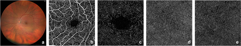
Cerebral embolization is a known complication of transcatheter aortic valve implantation (TAVI) but the effect of the procedure on the ocular perfusion is currently unclear. Thus, we investigated post-procedural morphologic and perfusion changes of the retina and choroid, using optical coherence tomography angiography (OCTA) and color fundus photography (CFP) in a prospective cohort study. Ophthalmic examinations were conducted pre- and post-TAVI. OCTA images were analyzed quantitatively based on vessel density and skeleton density of the superficial and deep retinal plexus as well as the signal intensity and flow deficits in the choriocapillaris. CFP images were assessed for presence of acute retinal ischemia, optic nerve swelling, vessel emboli, hemorrhages and cotton wool spots. Data was analyzed using linear mixed models. Twenty patients (9 women; 11 men) at a mean age of 81 ± 6 years were included. Pre- and post-interventional ocular imaging data were available for 32 eyes. The analysis revealed a significant impairment of the choriocapillaris perfusion after TAVI with an increased proportion of flow deficits (p = 0.044). When controlling for blood pressure, the average size of choriocapillaris flow voids was significantly increased (systolic and diastolic, p = 0.039 and 0.029). Qualitatively, focal areas of retinal ischemia were detected on OCTA in 33% of participants. Silent emboli or cotton wool spots were identified on CFP in 21%. Our findings indicate a reduced choroidal perfusion as well as areas of retinal ischemia and embolization in a considerable proportion of patients following TAVI. Pending confirmation in a larger sample, these complications merit monitoring as well as inclusion in consent procedures for TAVI.
© 2023. Springer Nature Limited.
Conflict of interest statement
JHT: Heidelberg Engineering (Funding), Optos (Funding), Zeiss (Funding), CenterVue (Funding), Novartis (Recipient), Okko (Recipient). MWMW: Novartis Pharma GmbH: honoraria, grant; DigiSight Technologies: travel grant, D-EYE Srl: imaging devices, Heine Optotechnik GmbH: research funding, imaging devices, travel reimbursements, consultant; Eyenuk, Inc: free trial analysis; ASKIN & CO GmbH: travel reimbursement, honoraria; Berlin-Chemie AG: grant, travel reimbursements; CenterVue SpA: grant and imaging devices; Imaging devices were provided by Heidelberg Engineering, Optos, and Carl Zeiss Meditec. RPF: Bayer (Consultant), Ellex (Consultant), Novartis (Consultant), Novartis (Funding), Ophtea (Consultant), Alimera (Consultant), Santhera (Consultant), Roche/Genentech (Consultant), CentreVue (Funding), Zeiss (Funding). ACW, AS, TW and BAK declare no competing interests.
Figures

References
Publication types
MeSH terms
LinkOut - more resources
Full Text Sources
Miscellaneous
