

The microsporidian polar tube: origin, structure, composition, function, and application
- PMID: 37649053
- PMCID: PMC10468886
- DOI: 10.1186/s13071-023-05908-9
The microsporidian polar tube: origin, structure, composition, function, and application
Abstract
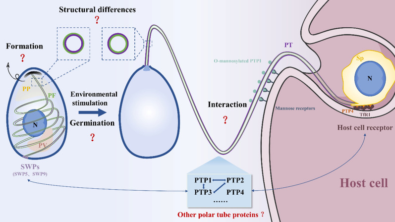
Microsporidia are a class of obligate intracellular parasitic unicellular eukaryotes that infect a variety of hosts, even including humans. Although different species of microsporidia differ in host range and specificity, they all share a similar infection organelle, the polar tube, which is also defined as the polar filament in mature spores. In response to the appropriate environmental stimulation, the spore germinates with the polar filament everted, forming a hollow polar tube, and then the infectious cargo is transported into host cells via the polar tube. Hence, the polar tube plays a key role in microsporidian infection. Here, we review the origin, structure, composition, function, and application of the microsporidian polar tube, focusing on the origin of the polar filament, the structural differences between the polar filament and polar tube, and the characteristics of polar tube proteins. Comparing the three-dimensional structure of PTP6 homologous proteins provides new insight for the screening of additional novel polar tube proteins with low sequence similarity in microsporidia. In addition, the interaction of the polar tube with the spore wall and the host are summarized to better understand the infection mechanism of microsporidia. Due to the specificity of polar tube proteins, they are also used as the target in the diagnosis and prevention of microsporidiosis. With the present findings, we propose a future study on the polar tube of microsporidia.
Keywords: Diagnosis; Interaction; Microsporidia; Polar filament; Polar tube; Polar tube proteins; Structure.
© 2023. BioMed Central Ltd., part of Springer Nature.
Conflict of interest statement
The authors declare that they have no competing interests.
Figures
Similar articles
-
Microsporidia dressing up: the spore polaroplast transport through the polar tube and transformation into the sporoplasm membrane.mBio. 2024 Feb 14;15(2):e0274923. doi: 10.1128/mbio.02749-23. Epub 2024 Jan 9. mBio. 2024. PMID: 38193684 Free PMC article.
-
Proteomic profile of polar filament and polar tube from fungal pathogen microsporidium Nosema bombycis provides new insights into its unique invasion organelle.J Proteomics. 2022 Jul 15;263:104617. doi: 10.1016/j.jprot.2022.104617. Epub 2022 May 18. J Proteomics. 2022. PMID: 35595055
-
The Function and Structure of the Microsporidia Polar Tube.Exp Suppl. 2022;114:179-213. doi: 10.1007/978-3-030-93306-7_8. Exp Suppl. 2022. PMID: 35544004 Free PMC article.
-
The roles of microsporidia spore wall proteins in the spore wall formation and polar tube anchorage to spore wall during development and infection processes.Exp Parasitol. 2018 Apr;187:93-100. doi: 10.1016/j.exppara.2018.03.007. Epub 2018 Mar 6. Exp Parasitol. 2018. PMID: 29522765 Review.
-
Microsporidia: emerging pathogenic protists.Acta Trop. 2001 Feb 23;78(2):89-102. doi: 10.1016/s0001-706x(00)00178-9. Acta Trop. 2001. PMID: 11230819 Review.
Cited by
-
Molecular characterization and phylogenetic analyses of MetAP2 gene and protein of Nosema bombycis isolated from Guangdong, China.Front Vet Sci. 2024 Jun 28;11:1429169. doi: 10.3389/fvets.2024.1429169. eCollection 2024. Front Vet Sci. 2024. PMID: 39005720 Free PMC article.
-
Establishment and application of a loop-mediated isothermal amplification method based on MetAP2 gene for the detection of Nosema bombycis in silkworms (Bombyx mori).Front Vet Sci. 2025 Mar 10;12:1549224. doi: 10.3389/fvets.2025.1549224. eCollection 2025. Front Vet Sci. 2025. PMID: 40129576 Free PMC article.
-
Microsporidia dressing up: the spore polaroplast transport through the polar tube and transformation into the sporoplasm membrane.mBio. 2024 Feb 14;15(2):e0274923. doi: 10.1128/mbio.02749-23. Epub 2024 Jan 9. mBio. 2024. PMID: 38193684 Free PMC article.
References
-
- Vossbrinck CR, Maddox JV, Friedman S, Debrunner-Vossbrinck BA, Woese CR. Ribosomal RNA sequence suggests microsporidia are extremely ancient eukaryotes. Nature. 1987;326:411–414. - PubMed
-
- Keeling PJ, Doolittle WF. Alpha-tubulin from early-diverging eukaryotic lineages and the evolution of the tubulin family. Mol Biol Evol. 1996;13:1297–1305. - PubMed
-
- Keeling PJ, Luker MA, Palmer JD. Evidence from beta-tubulin phylogeny that microsporidia evolved from within the fungi. Mol Biol Evol. 2000;17:23–31. - PubMed
Publication types
MeSH terms
Grants and funding
LinkOut - more resources
Full Text Sources
