. 2023 Aug 30;13(1):159.
doi: 10.1186/s13578-023-01104-5.

Hepatocyte-specific Sox9 knockout ameliorates acute liver injury by suppressing SHP signaling and improving mitochondrial function

Affiliations

Hepatocyte-specific Sox9 knockout ameliorates acute liver injury by suppressing SHP signaling and improving mitochondrial function

Dan Qin et al. Cell Biosci. .

Abstract

Background and aims: Sex determining region Y related high-mobility group box protein 9 (Sox9) is expressed in a subset of hepatocytes, and it is important for chronic liver injury. However, the roles of Sox9+ hepatocytes in response to the acute liver injury and repair are poorly understood.

Methods: In this study, we developed the mature hepatocyte-specific Sox9 knockout mouse line and applied three acute liver injury models including PHx, CCl4 and hepatic ischemia reperfusion (IR). Huh-7 cells were subjected to treatment with hydrogen peroxide (H2O2) in order to induce cellular damage in an in vitro setting.

Results: We found the positive effect of Sox9 deletion on acute liver injury repair. Small heterodimer partner (SHP) expression was highly suppressed in hepatocyte-specific Sox9 deletion mouse liver, accompanied by less cell death and more cell proliferation. However, in mice with hepatocyte-specific Sox9 deletion and SHP overexpression, we observed an opposite phenotype. In addition, the overexpression of SOX9 in H2O2-treated Huh-7 cells resulted in an increase in cytoplasmic SHP accumulation, accompanied by a reduction of SHP in the nucleus. This led to impaired mitochondrial function and subsequent cell death. Notably, both the mitochondrial dysfunction and cell damage were reversed when SHP siRNA was employed, indicating the crucial role of SHP in mediating these effects. Furthermore, we found that Sox9, as a vital transcription factor, directly bound to SHP promoter to regulate SHP transcription.

Conclusions: Overall, our findings unravel the mechanism by which hepatocyte-specific Sox9 knockout ameliorates acute liver injury via suppressing SHP signaling and improving mitochondrial function. This study may provide a new treatment strategy for acute liver injury in future.

Keywords: Acute liver injury; Hepatocyte; Mitochondria; SHP; Sox9.


Conflict of interest statement

The authors declare that they have no competing interests.

Figures
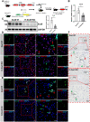
Fig. 1
Fig. 1
Generation and identification of the tamoxifen-inducible Alb-Cre transgenic mouse line Alb-CreERT2/+; Sox9 f/f. A Schematic diagram showed the hepatocyte-specific Sox9 knockout strategy. B Hepatic Sox9 expression was determined by qRT-PCR in Sox9 f/f mice (N = 5) and Alb-CreERT2/+; Sox9 f/f mice (N = 9) treated with tamoxifen. C Western blot analysis of Sox9 protein level in liver tissues from Sox9 f/f and H-Sox9 KO mice (N = 4). D Sox9/HNF4α double staining was performed by immunostaining. Red arrows depict Sox9+ HNF4α+ cells and green arrows depict Sox9+ bile duct cells. Scale bar, 20 μm. E Sox9/CK19 double staining was performed by immunostaining. Red arrows depict Sox9+ hepatocytes and green arrows depict Sox9+ bile duct cells. Scale bar, 20 μm. F Representative Sox9 immunohistochemical staining of liver samples from Sox9 f/f mice and H-Sox9 KO mice. The red arrows indicate Sox9+HNF4α+ cells, while the green arrows indicate Sox9+ bile duct cells. PV: periportal vein; CV: central vein. Scale bar, 100 or 20 μm
Fig. 2
Fig. 2
Hepatocyte-specific Sox9 deletion ameliorates acute liver injury induced by IR. Sox9 f/f and Alb-CreERT2/+; Sox9 f/f mice pre-treated with tamoxifen were subjected to 1 h of hepatic ischemia. Plasma and liver samples were collected 6 h after reperfusion. A Schematic diagram showed ischemia reperfusion model. B Plasma ALT levels were measured in Sox9 f/f and H-Sox9 KO mice. N = 4 or 5. C TBA levels were detected in livers and plasma in Sox9 f/f and H-Sox9 KO mice. N = 4 or 5. D Levels of IL-6 and TNFα mRNA expression were determined by qRT-PCR in Sox9 f/f and H-Sox9 KO mice. N = 4 or 5. E Representative liver sections stained with H&E. Yellow arrows indicate apoptotic cells (Red arrows indicate case prototypical apoptotic cells with higher magnification), while blank arrowhead indicates necrotic area. Scale bars: (left) 200 µm and (right) 50 µm. F Representative BrdU images in livers from Sox9 f/f mice and H-Sox9 KO mice induced by IR. Quantification of the percentage of BrdU+ cells in the indicated groups. Scale bar, 100 µm
Fig. 3
Fig. 3
Sox9 enhances SHP transcription activity by binding to its promoter. A Heat map showed the differentially expressed genes in Sox9 f/f and H-Sox9 KO mice induced by IR. B Hepatic expression levels of Sox9 and SHP in Sox9 f/f and H-Sox9 KO mice were determined by qRT-PCR analysis following IR injury. 36B4 was used as a housekeeping gene. N = 4–5 per group, data are shown as mean ± SD. C SHP promoter contained a putative Sox9 binding site. Sequences of the WT (black) and mutant (red) binding sites. D Amplification and activity detection of different fragments of SHP promoter (position 40 to − 1500, 40 to − 1900 relative to the transcription start site). These fragments were inserted into the pGL3-Basic vector and transfected into Hepa 1–6 cells with or without Sox9 expression plasmids and pRL-TK using lipofectamine 2000. N = 3. E SHP-promoter or SHP-mutation promoter was co-transfected into Hepa1-6 cells with pRL-TK for 24 h and samples were analyzed by dual-luciferase assays. N = 3 per group. The ratio of firefly luciferase activity to renilla luciferase activity in control group was set to 1. F ChIP analysis showed that Sox9 interacted with classic binding site on SHP promoter. G ChIP-qPCR assay of Sox9 level on SHP promoter using chromatin solutions prepared from Sox9 f/f and H-Sox9 KO mouse livers. N = 3
Fig. 4
Fig. 4
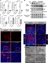
Hepatocyte-specific Sox9 deletion promotes SHP accumulation in nucleus. Sox9 f/f and Alb-CreERT2/+; Sox9 f/f mice pre-treated with tamoxifen were subjected to 1 h of hepatic ischemia. Liver samples were collected 6 h after reperfusion. A qRT-PCR was used to determine Sox9 and SHP mRNA in different cellular fractions in liver samples from Sox9 f/f and H-Sox9 KO mice. N = 3. B Western blot was used to determine SHP protein expression in different cellular fractions in liver samples. C Band intensities were measured by Image J. D Immunofluorescence co-staining for Sox9 and SHP. Scale bar, 50 µm. E The subcellular localization of SHP in hepatocytes was observed using confocal laser scanning microscopy. Scale bars, 50 µm. F Transmission electron microscopy analysis for hepatic mitochondrial morphology in Sox9 f/f and H-Sox9 KO mice induced by IR
Fig. 5
Fig. 5
Loss of Sox9 in hepatocytes inhibits IR-induced cell death associated with SHP. Sox9 f/f and Alb-CreERT2/+; Sox9 f/f mice pre-treated with tamoxifen were subjected to 1 h of hepatic ischemia, liver samples were collected 6 h after reperfusion. A The representative images of TUNEL staining in liver tissue sections from Sox9 f/f and H-Sox9 KO mice with or without IR. Scale bar, 50 µm. B Western blot analysis on the expression levels of apoptosis related proteins Bax, Cyt C, Bid and Caspase-3 p17 (Caspase-3) in liver tissues. GAPDH was used as an internal control. C Quantification analysis of apoptosis related proteins Bax, Cyt C, Bid and Caspase-3. N = 3. D ROS production in frozen liver tissues from Sox9 f/f and H-Sox9 KO mice following hepatic IR. Scale bar, 100 µm. E Quantitative ROS detection in liver samples
Fig. 6
Fig. 6
Overexpression of SOX9 reduces SHP nuclear localization and induces mitochondrial injury. Huh-7 cells were transfected with either SOX9 overexpression plasmid (SOX9 OE) or vehicle plasmid (Control). Then the cells were treated with 400 μM H2O2 for 6 h before harvest. A qRT-PCR was used to determine SOX9 and SHP mRNA expression in different cellular fractions in cell samples from indicated group. N = 3. B The subcellular localization of SHP in Huh-7 cells was observed under a fluorescence microscope. Scale bar, 50 µm. C Mito Tracker Red staining of Huh-7 cells treated with indicated concentration of H2O2. Scale bar, 50 µm. D Ca2+ indicator X-Rhod-1 in Huh-7 cells treated with vehicle or H2O2. Scale bar, 50 µm
Fig. 7
Fig. 7
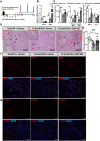
Overexpression of SHP aggravated hepatic ischemia–reperfusion injury in mice with hepatocellular specific knockout of Sox9. Sox9 f/f and Alb-CreERT2/+; Sox9 f/f mice pre-treated with tamoxifen and indicated vectors were subjected to 1 h of hepatic ischemia. Plasma and liver samples were collected at 6 h post reperfusion. A Schematic diagram showing ischemia reperfusion model. B Sox9 and SHP mRNA expression were determined by qRT-PCR in indicated mice. C Plasma ALT level was measured in indicated group. TBA level was detected in livers and plasma in indicated group. TNFα and IL-6 mRNA expression were determined by qRT-PCR in indicated group. D Representative liver sections stained with H&E. Arrows, cell apoptosis; Dashed box, necrotic area. Scale bar, 200 µm. E Quantitative ROS detection in liver samples. F ROS production in frozen liver tissues from Sox9 f/f and H-Sox9 KO mice following IR. Scale bar, 100 μm. G Representative liver sections stained with TUNEL. Scale bar, 100 μm

Similar articles

Cited by

References

    1. Jo A, Denduluri S, Zhang B, et al. The versatile functions of sox9 in development, stem cells, and human diseases. Genes Dis. 2014;1(2):149–161. doi: 10.1016/j.gendis.2014.09.004. - DOI - PMC - PubMed
    1. Wang J, Xu S, Duan J, et al. Invasion of white matter tracts by glioma stem cells is regulated by a notch1-sox2 positive-feedback loop. Nat Neurosci. 2019;22(1):91–105. doi: 10.1038/s41593-018-0285-z. - DOI - PubMed
    1. Riba A, Olier M, Lacroix-Lamandé S, et al. Paneth cell defects induce microbiota dysbiosis in mice and promote visceral hypersensitivity. Gastroenterology. 2017;153(6):1594–1606.e2. doi: 10.1053/j.gastro.2017.08.044. - DOI - PubMed
    1. Chang D, Martinez Alanis D, Miller R, et al. Lung epithelial branching program antagonizes alveolar differentiation. Proc Natl Acad Sci U S A. 2013;110(45):18042–18051. doi: 10.1073/pnas.1311760110. - DOI - PMC - PubMed
    1. Kim J, Bai Y, Jayne L, et al. A kinome-wide screen identifies a cdkl5-sox9 regulatory axis in epithelial cell death and kidney injury. Nat Commun. 2020;11(1):1924. doi: 10.1038/s41467-020-15638-6. - DOI - PMC - PubMed