Single-cell sequencing reveals the existence of fetal vascular endothelial stem cell-like cells in mouse liver
- PMID: 37649114
- PMCID: PMC10468894
- DOI: 10.1186/s13287-023-03460-y
Single-cell sequencing reveals the existence of fetal vascular endothelial stem cell-like cells in mouse liver
Abstract
Background: A resident vascular endothelial stem cell (VESC) population expressing CD157 and CD200 has been identified recently in the adult mouse. However, the origin of this population and how it develops has not been characterized, nor has it been determined whether VESC-like cells are present during the perinatal period. Here, we investigated the presence of perinatal VESC-like cells and their relationship with the adult VESC-like cell population.
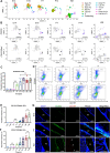
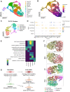
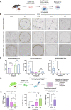
Methods: We applied single-cell RNA sequencing of endothelial cells (ECs) from embryonic day (E) 14, E18, postnatal day (P) 7, P14, and week (W) 8 liver and investigated transcriptomic changes during liver EC development. We performed flow cytometry, immunofluorescence, colony formation assays, and transplantation assays to validate the presence of and to assess the function of CD157+ and CD200+ ECs in the perinatal period.
Results: We identified CD200- expressing VESC-like cells in the perinatal period. These cells formed colonies in vitro and had high proliferative ability. The RNA velocity tool and transplantation assay results indicated that the projected fate of this population was toward adult VESC-like cells expressing CD157 and CD200 1 week after birth.
Conclusion: Our study provides a comprehensive atlas of liver EC development and documents VESC-like cell lineage commitment at single-cell resolution.
Keywords: Development; Endothelial stem cells; Heterogeneity; Mouse liver; RNA velocity; Single-cell transcriptomic sequencing; Trajectory analysis.
© 2023. BioMed Central Ltd., part of Springer Nature.
Conflict of interest statement
The authors declare that they have no conflict of interest.
Figures


Similar articles
-
Discovery of Transcription Factors Involved in the Maintenance of Resident Vascular Endothelial Stem Cell Properties.Mol Cell Biol. 2024 Jan;44(1):17-26. doi: 10.1080/10985549.2023.2297997. Epub 2024 Jan 29. Mol Cell Biol. 2024. PMID: 38247234 Free PMC article.
-
Identification of CD157-Positive Vascular Endothelial Stem Cells in Mouse Retinal and Choroidal Vessels: Fluorescence-Activated Cell Sorting Analysis.Invest Ophthalmol Vis Sci. 2022 Apr 1;63(4):5. doi: 10.1167/iovs.63.4.5. Invest Ophthalmol Vis Sci. 2022. PMID: 35394492 Free PMC article.
-
CD157 Marks Tissue-Resident Endothelial Stem Cells with Homeostatic and Regenerative Properties.Cell Stem Cell. 2018 Mar 1;22(3):384-397.e6. doi: 10.1016/j.stem.2018.01.010. Epub 2018 Feb 8. Cell Stem Cell. 2018. PMID: 29429943
-
Discovery of a Vascular Endothelial Stem Cell (VESC) Population Required for Vascular Regeneration and Tissue Maintenance.Circ J. 2018 Dec 25;83(1):12-17. doi: 10.1253/circj.CJ-18-1180. Epub 2018 Nov 28. Circ J. 2018. PMID: 30487375 Review.
-
Diverse cellular origins of adult blood vascular endothelial cells.Dev Biol. 2021 Sep;477:117-132. doi: 10.1016/j.ydbio.2021.05.010. Epub 2021 May 26. Dev Biol. 2021. PMID: 34048734 Review.
Cited by
-
Single cell transcription revealing key transcription factors in embryonic kidney development.Mol Cell Biochem. 2025 May 20. doi: 10.1007/s11010-025-05307-x. Online ahead of print. Mol Cell Biochem. 2025. PMID: 40392427
-
CD157+ vascular endothelial cells derived from human-induced pluripotent stem cells have high angiogenic potential.Inflamm Regen. 2025 May 14;45(1):14. doi: 10.1186/s41232-025-00379-0. Inflamm Regen. 2025. PMID: 40369638 Free PMC article.
-
Discovery of Transcription Factors Involved in the Maintenance of Resident Vascular Endothelial Stem Cell Properties.Mol Cell Biol. 2024 Jan;44(1):17-26. doi: 10.1080/10985549.2023.2297997. Epub 2024 Jan 29. Mol Cell Biol. 2024. PMID: 38247234 Free PMC article.
-
APJ regulates the balance between self-renewal and differentiation of vascular endothelial stem cells.Inflamm Regen. 2025 Aug 4;45(1):25. doi: 10.1186/s41232-025-00389-y. Inflamm Regen. 2025. PMID: 40760447 Free PMC article.
References
-
- Asahara T, Murohara T, Sullivan A, Silver M, Van Der Zee R, Li T, et al. Isolation of putative progenitor endothelial cells for angiogenesis. Science. 1997;275:964–967. - PubMed
-
- Wakabayashi T, Naito H, Suehiro J, Lin Y, Kawaji H, Iba T, et al. CD157 marks tissue-resident endothelial stem cells with homeostatic and regenerative properties. Cell Stem Cell. 2018;22:384–397. - PubMed
Publication types
MeSH terms
LinkOut - more resources
Full Text Sources
Research Materials

