Review
. 2023 Aug 30;14(1):225.
doi: 10.1186/s13287-023-03456-8.

The characteristics and medical applications of antler stem cells

Affiliations
Review

The characteristics and medical applications of antler stem cells

Qi Liu et al. Stem Cell Res Ther. .

Abstract

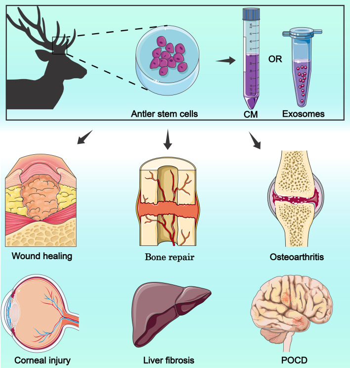
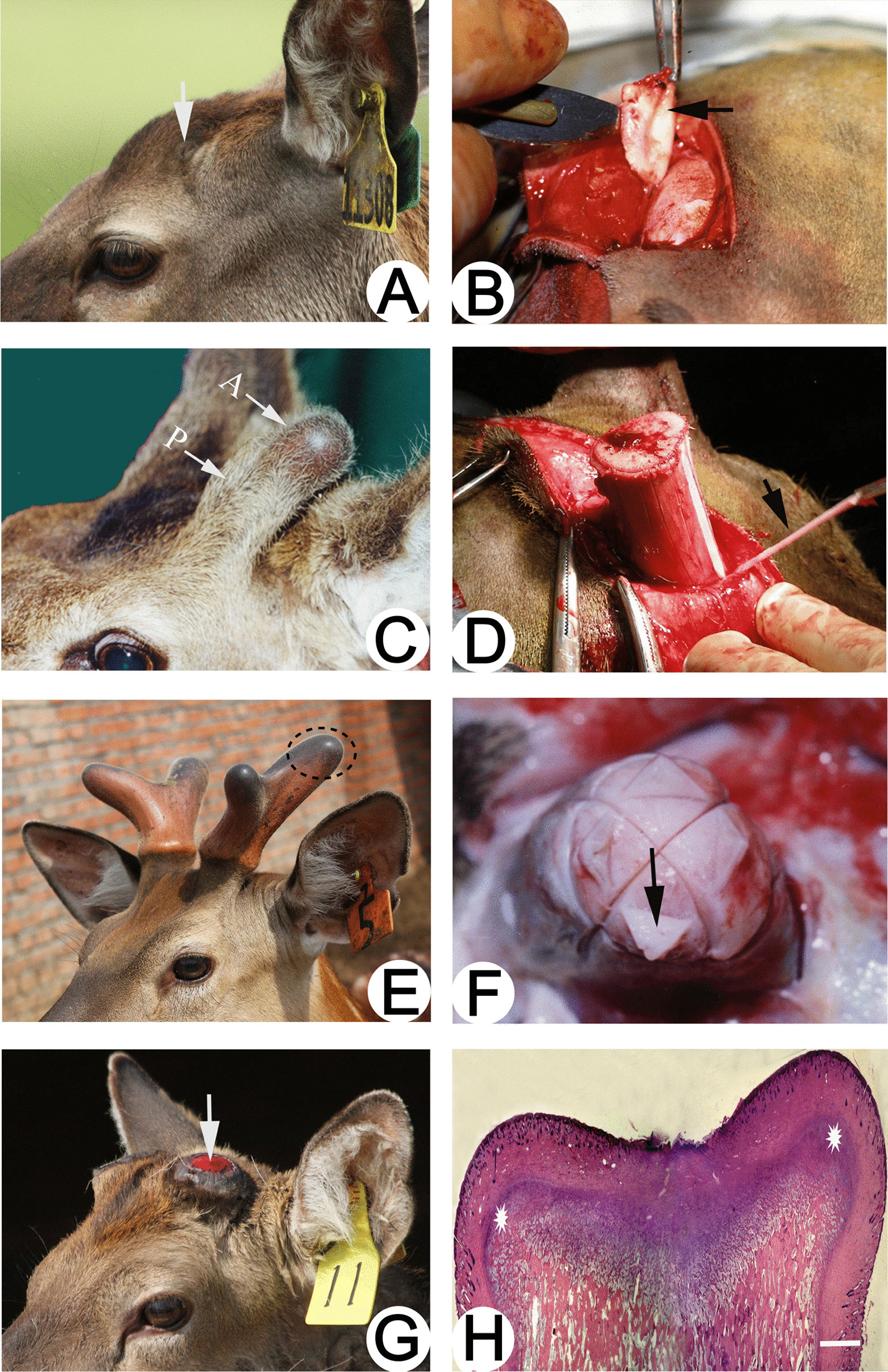
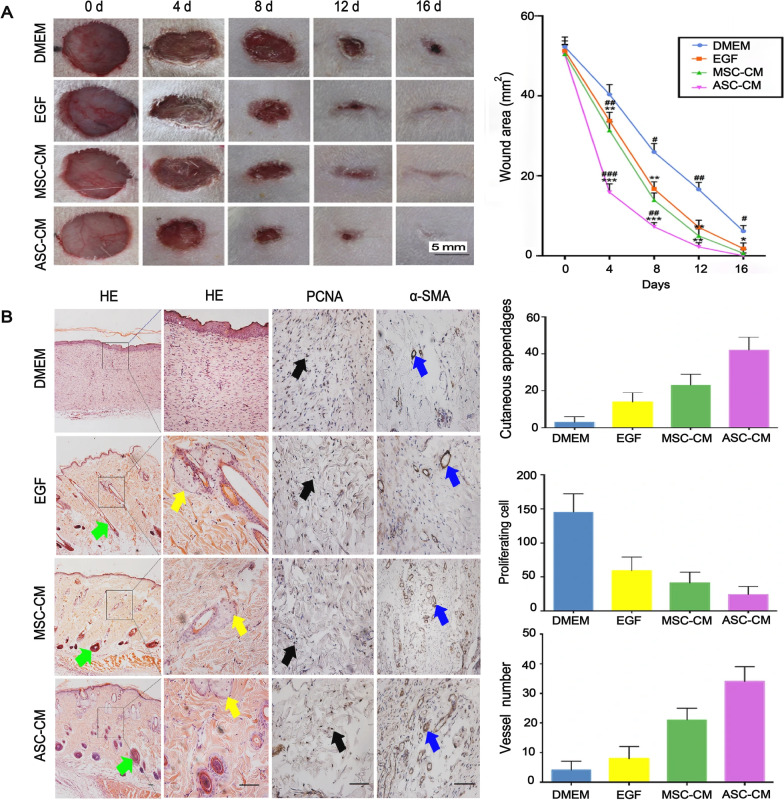
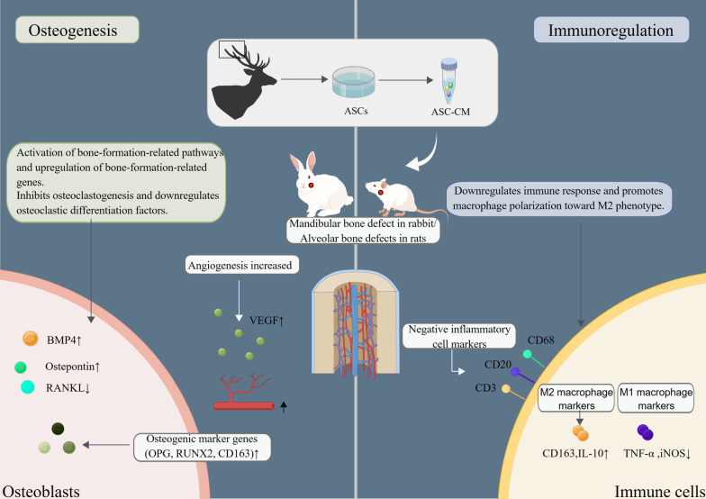
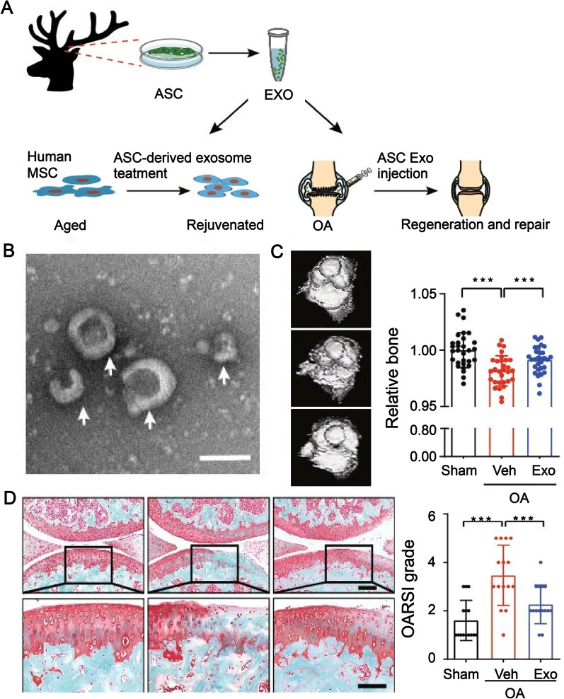
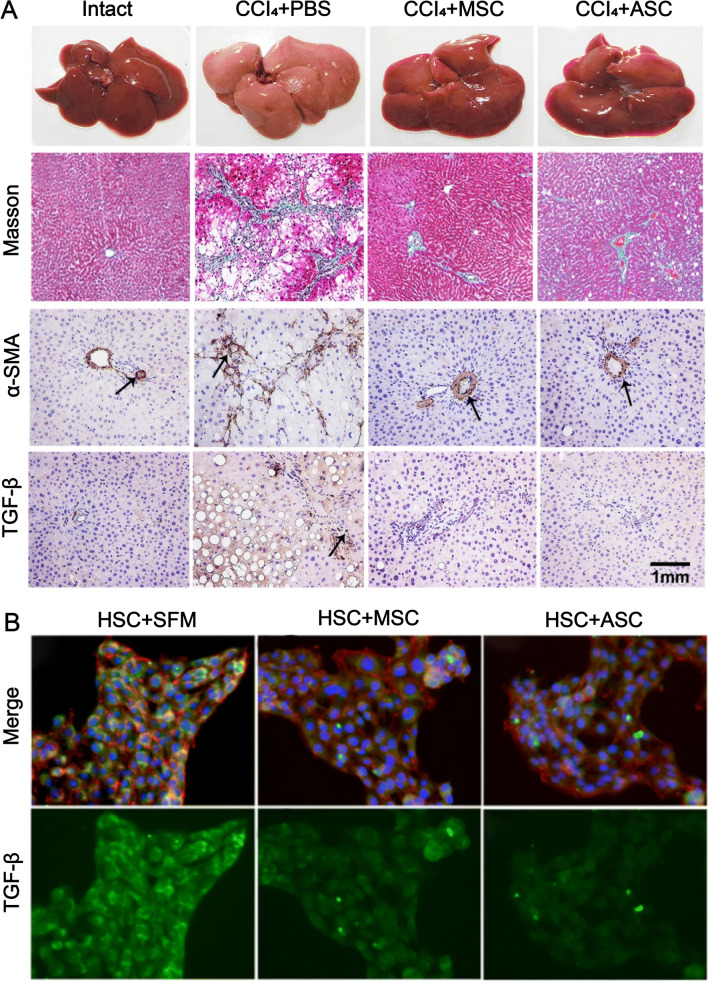
Antlers are the only fully regenerable mammalian appendages whose annual renewal is initiated by antler stem cells (ASCs), defined as a specialized type of mesenchymal stem cells (MSCs) with embryonic stem cell properties. ASCs possess the same biological features as MSCs, including the capacity for self-renewal and multidirectional differentiation, immunomodulatory functions, and the maintenance of stem cell characteristics after multiple passages. Several preclinical studies have shown that ASCs exhibit promising potential in wound healing, bone repair, osteoarthritis, anti-tissue fibrosis, anti-aging, and hair regeneration. Medical applications based on ASCs and ASC-derived molecules provide a new source of stem cells and therapeutic modalities for regenerative medicine. This review begins with a brief description of antler regeneration and the role of ASCs. Then, the properties and advantages of ASCs are described. Finally, medical research advances regarding ASCs are summarized, and the prospects and challenges of ASCs are highlighted.

Keywords: Antler; Medical applications; Stem cells.


Conflict of interest statement

The authors declare that they have no competing interests.

Figures

Fig. 1
Fig. 1
ASCs for medical applications. CM: conditioned medium; POCD: postoperative cognitive impairment. This figure is totally depicted by ourselves
Fig. 2
Fig. 2
Schematic diagram of the origin of antler periosteum and ASCs. A The presumptive antler growth area (arrow) before the occurrence of the first antler; B Antlerogenic periosteum (arrow), where the APCs are the source of the antler pedicle and the first antler; C Antler pedicle (P) and a nascent antler bud (A); D Pedicle periosteum (arrow) (where the PPCs are the source of antler regeneration); E The antler growth center is located at the tip of the antler (ellipse); F The apical perichondrium (arrow) at the distal end of the antler, where the antler growth center and RMCs are located, is associated with rapid antler growth; Reproduced with permission from [45] ©2021 Chunyi Li et al. G Pedicle stump (arrow) formed after spring antler casting; H Newly formed growth centers on the stump of the vertebral arch (indicated by stars). Reproduced with permission from [27] © 2014 Elsevier Ltd
Fig. 3
Fig. 3
Effect of ASC-CM on wound healing. A Effect of ASC-CM versus the control (DMEM, EGF, and MSC-CM) on wound healing speed. The ASC-CM group had the fastest healing rate, which was completed by day 16. B Effect of ASC-CM vs. control (DMEM, EGF, and MSC-CM) on wound healing quality. The group that received ASC-CM had the thickest dermis, the most skin appendages, the most α-SMA positive vessels, and the least number of PCNA positive cells; Reproduced with permission from [76]. © 2019 Xiaoli Rong et al. ASC-CM, antler stem cell-conditioned medium; MSC-CM, mesenchymal stem cell-conditioned medium; EGF, epidermal growth factor; α-SMA, anti-α smooth muscle actin; PCNA, proliferating cell nuclear antigen; DMEM, Dulbecco’s modified eagle medium; HE, Hematoxylin and eosin staining
Fig. 4
Fig. 4
Schematic diagram of the molecular mechanism of ASC-CM treatment of bone injury. ASCs, antler stem cells; ASC-CM, ASC-conditioned medium; BMP, bone morphogenetic proteins; RANKL, Receptor activator of nuclear factor-kB ligand; OPG, osteoprotegerin; iNOS, Inducible nitric oxide synthase; Runx2, Runt-related transcription factor 2; TNF-α, Tumor necrosis factor alpha; IL-10, Interleukin-10. This figure is depicted by ourselves using Figdraw (www.figdraw.com). The Authorization ID which is the permission to use it is PTRWO77989
Fig. 5
Fig. 5
ASC-derived exosomes alleviate the aging of MSCs and treat OA. A Experimental design; B Transmission electron microscopy image showing ASCs-derived exosomes as spherical or cup-shaped; C Microcomputed tomography scan showing bone erosion. The treatment effect was better in the ASC-derived exosome treatment group; D Safranin O and fast green staining images showing that ASC-derived exosome treatment is effective for cartilage regeneration; Reproduced with permission from [69]. © 2021, Oxford University Press. ASC, antler stem cell; MSC, mesenchymal stem cell; Exo, exosome; OA, osteoarthritis; Veh, vehicle
Fig. 6
Fig. 6
Effects of ASCs on liver fibrosis in rats treated with CCI4. A Compared with intact mice, the CCl4 + PBS control group showed increased liver volume and visible nodules on the surface. The CCl4 + ASC-treated group showed smaller liver volume and bright red, smooth surface compared with the CCl4 + PBS-treated group. Masson staining showed that the CCl4 + ASC-treated group had significantly lower collagen fibers compared with the control group. Immunohistochemical staining showed that α-SMA+ and TGF-β+ cells were significantly lower in the CCl4 + ASC treatment group. B Immunofluorescence showed that TGF-β expression was significantly decreased in HSCs co-cultured with ASCs. Reproduced with permission from [96]. © 2019 Xiaoli Rong et al. ASCs, antler stem cells; MSCs, mesenchymal stem cells; α-SMA, anti-α smooth muscle actin; TGF-β, transforming growth factor-β; HSC, hepatic stellate cells; CCl4, carbon tetrachloride; SFM, serum-free medium; PBS, phosphate-buffered saline

Similar articles

Cited by

References

    1. Varani K, Vincenzi F, Pasquini S, Blo I, Salati S, Cadossi M, et al. Pulsed electromagnetic field stimulation in osteogenesis and chondrogenesis: signaling pathways and therapeutic implications. Int J Mol Sci. 2021;22(2):809. - PMC - PubMed
    1. De Luca M, Aiuti A, Cossu G, Parmar M, Pellegrini G, Robey PG. Advances in stem cell research and therapeutic development. Nat Cell Biol. 2019;21(7):801–811. - PubMed
    1. Bacakova L, Zarubova J, Travnickova M, Musilkova J, Pajorova J, Slepicka P, et al. Stem cells: their source, potency and use in regenerative therapies with focus on adipose-derived stem cells—a review. Biotechnol Adv. 2018;36(4):1111–1126. - PubMed
    1. Sid-Otmane C, Perrault LP, Ly HQ. Mesenchymal stem cell mediates cardiac repair through autocrine, paracrine and endocrine axes. J Transl Med. 2020;18(1):336. - PMC - PubMed
    1. Kuo YR, Wang CT, Cheng JT, Kao GS, Chiang YC, Wang CJ. Adipose-derived stem cells accelerate diabetic wound healing through the induction of autocrine and paracrine effects. Cell Transplant. 2016;25(1):71–81. - PubMed

Publication types

LinkOut - more resources