

doi: 10.1080/15548627.2023.2249794.
Epub 2023 Aug 30.
Atl (atlastin) regulates mTor signaling and autophagy in Drosophila muscle through alteration of the lysosomal network
Affiliations
- PMID: 37649246
- PMCID: PMC10761077
- DOI: 10.1080/15548627.2023.2249794
Item in Clipboard
Atl (atlastin) regulates mTor signaling and autophagy in Drosophila muscle through alteration of the lysosomal network
Autophagy.
2024 Jan.
Abstract
atl atlastin; ALR autophagic lysosome reformation; ER endoplasmic reticulum; GFP green fluorescent protein; HSP hereditary spastic paraplegia; Lamp1 lysosomal associated membrane protein 1 PolyUB polyubiquitin; RFP red fluorescent protein; spin spinster; mTor mechanistic Target of rapamycin; VCP valosin containing protein.
Keywords: ATG8; Hereditary Spastic Paraplegia; LAMP1; Neurodegeneration; endosome; protein aggregates; rab7.
Conflict of interest statement
No potential conflict of interest was reported by the authors.
Figures
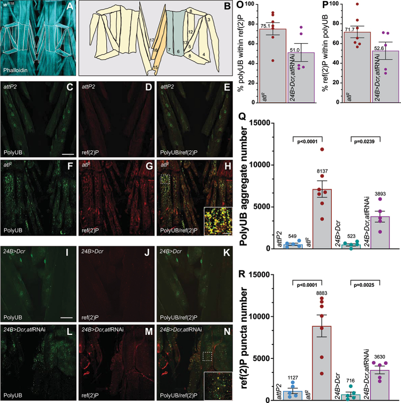
Loss of atl causes polyUB accumulation that is marked for degradation by autophagy with ref(2)P. (A) Phalloidin stain of a wild-type (w1118) third instar Drosophila larva. (B) Schematic representation of one segment (indicated by a white dashed line in A) of larval musculature with muscles labeled in the right hemisegment. The body wall muscles studied in this work include the ventral oblique muscles (VO, also called chevrons, labeled 15,16, and 17, colored tan) and ventral lateral longitudinal muscles (VL, labeled 6, and 7, colored light blue). (C-N) confocal imaging (maximum intensity z-projections) of fixed third instar larval musculature. Scale bar: 50 µm. (C-H) antibodies against polyubiquitin (polyUB, green, C, F, I, and L) and ref(2)P (red, D, G, J, and M) in wild-type control larva (attP2, C-E) and the atl null mutant (atl2, F-H). (I-N) muscle RNAi knockdown of atl (L-N). Panels E, H, K, and N represent overlayed images of polyUB (green) and ref(2)P (red). The inset in H and N are enlarged images of the region indicated by the dashed white box. (O-P). Quantification of ref(2)P and polyUB colocalization. (O) Imaris-generated surfaces of polyUB that lies within surfaced generated by ref(2)P, similar to Manders M1 coefficient. (P) Imaris-generated surfaces of ref(2)P that lies within surfaced generated by polyUB, similar to Manders M2 coefficient. (Q). Quantification of total cytoplasmic polyUB aggregates within the volume examined in panels C, F, I and L. (R). Quantification of total ref(2)P puncta number within the volume examined in panels D, G, J and M. Imaging data was quantified using Imaris v9.7.2. Quantitative data are presented as mean ± S.E.M with each individual sample represented in the scatter plot. Data shown are representative of at least five different experiments (n = 5).
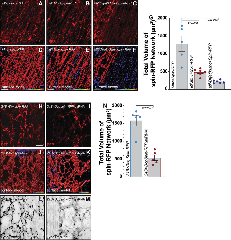
Loss of atl decreases the volume and complexity of the apical lysosomal network marked by spin-RFP. (A-F) Live imaging (maximum intensity z-projections) of larval body wall muscle 6 expressing the lysosomal/endosomal marker spin-RFP in wild-type control animals (Mhc>spin-GFP, A), atl2 (B), and an atl deficiency (atl2 /Df(atl), C). Scale bar: 5 µm. Imaris surface modeling of the spin-RFP surface of the indicated genotypes are shown in (D, E, and G) Discontinuous surfaces are represented as different colors. (H-M) Live imaging (maximum intensity z-projections) of larval body wall muscle 6 expressing the lysosomal/endosomal marker spin-RFP in wild-type control animals (24B>spin-RFP, H) and muscle RNAi knockdown of atl (24B>spin-RFP, atlRNAi, I). (J and K) Imaris surface modeling of the spin-RFP surface of the indicated genotypes are shown in (H, and I). (L and M) LysoTracker staining in wild-type (L) and atl knockdown larvae (M). Quantification of endolysosomal volume marked by spin-RFP in wild type, atl2, and an atl deficiency (atl2 /Df(atl), (G) and muscle RNAi knockdown (N).
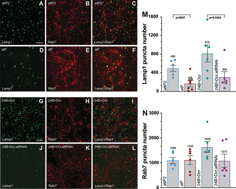
Loss of atl decreases conventional lysosome numbers.

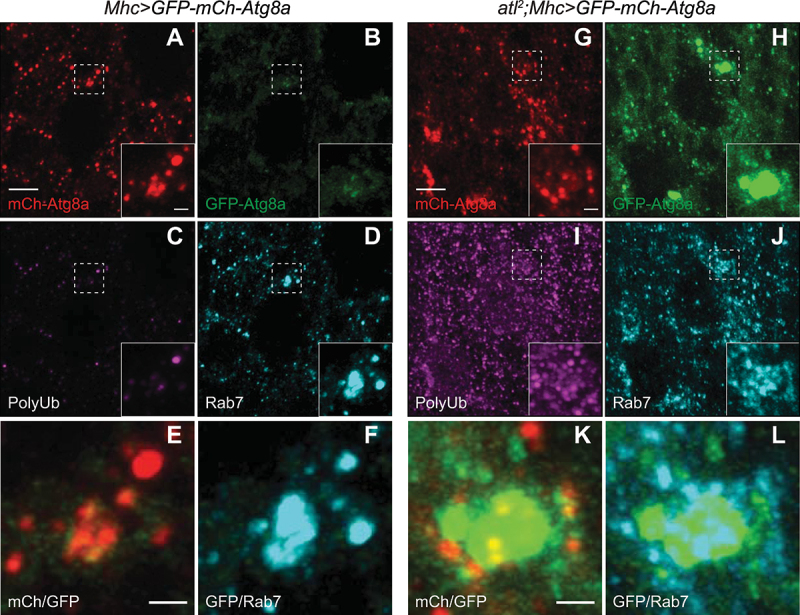
Loss of atl results in a blockage of autophagic flux.
Loss of atl results in the accumulation of undegraded cargo in Rab7 positive compartments.
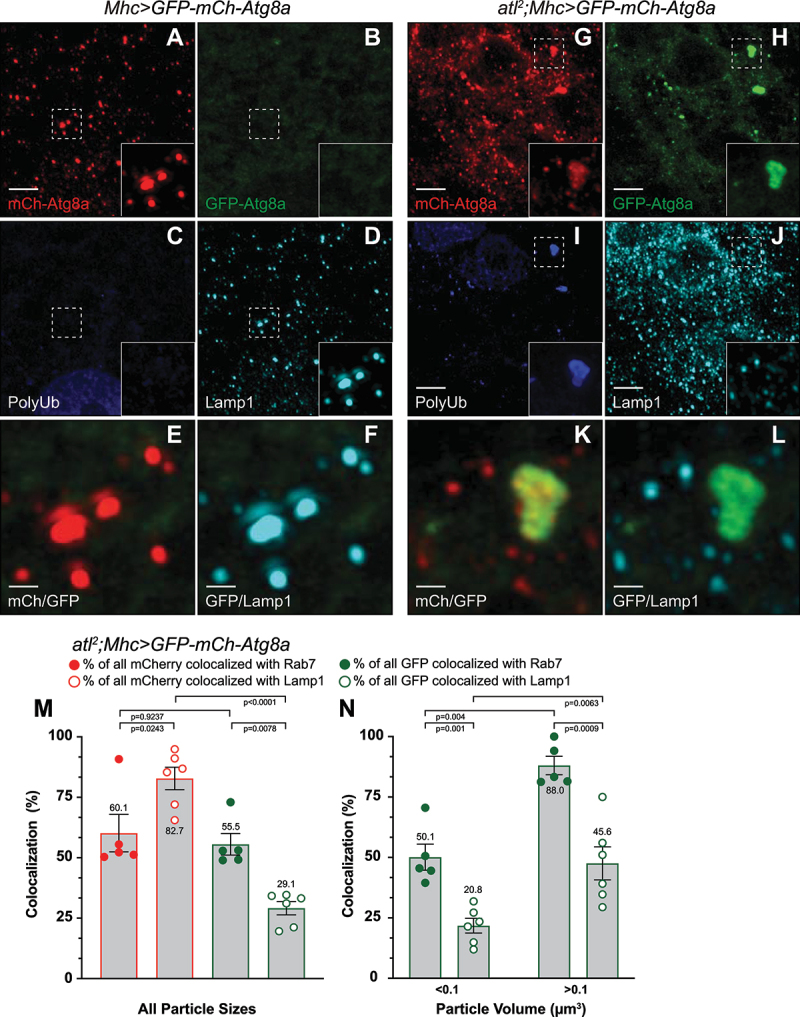
Autophagic cargo does not reach Lamp1 positive lysosomes without atl.

Visualization of autophagic structures with electron microscopy.

Loss of atl results in decrease mTor activity in muscle.

Reduction in atl, but not mTor, increases polyubiquitin aggregate accumulation.

Models of mTor signaling and basal autophagy.
Similar articles
-
Mouse models for hereditary spastic paraplegia uncover a role of PI4K2A in autophagic lysosome reformation.Autophagy. 2021 Nov;17(11):3690-3706. doi: 10.1080/15548627.2021.1891848. Epub 2021 Mar 9. Autophagy. 2021. PMID: 33618608 Free PMC article.
-
Spinster is required for autophagic lysosome reformation and mTOR reactivation following starvation.Proc Natl Acad Sci U S A. 2011 May 10;108(19):7826-31. doi: 10.1073/pnas.1013800108. Epub 2011 Apr 25. Proc Natl Acad Sci U S A. 2011. PMID: 21518918 Free PMC article.
-
ZFYVE26/SPASTIZIN and SPG11/SPATACSIN mutations in hereditary spastic paraplegia types AR-SPG15 and AR-SPG11 have different effects on autophagy and endocytosis.Autophagy. 2019 Jan;15(1):34-57. doi: 10.1080/15548627.2018.1507438. Epub 2018 Sep 13. Autophagy. 2019. PMID: 30081747 Free PMC article.
-
Recent progress in autophagic lysosome reformation.Traffic. 2017 Jun;18(6):358-361. doi: 10.1111/tra.12484. Epub 2017 May 5. Traffic. 2017. PMID: 28371052 Review.
-
MTOR, PIK3C3, and autophagy: Signaling the beginning from the end.Autophagy. 2015;11(12):2375-6. doi: 10.1080/15548627.2015.1106668. Autophagy. 2015. PMID: 26565689 Free PMC article. Review.
Cited by
-
Atlastin-1 regulates endosomal tubulation and lysosomal proteolysis in human cortical neurons.Neurobiol Dis. 2024 Sep;199:106556. doi: 10.1016/j.nbd.2024.106556. Epub 2024 Jun 6. Neurobiol Dis. 2024. PMID: 38851544 Free PMC article.
-
Molecular mechanism on autophagy associated cardiovascular dysfunction in Drosophila melanogaster.Front Cell Dev Biol. 2025 Mar 3;13:1512341. doi: 10.3389/fcell.2025.1512341. eCollection 2025. Front Cell Dev Biol. 2025. PMID: 40099194 Free PMC article. Review.
-
Motor neuron activity enhances the proteomic stress caused by autophagy defects in the target muscle.PLoS One. 2024 Jan 2;19(1):e0291477. doi: 10.1371/journal.pone.0291477. eCollection 2024. PLoS One. 2024. PMID: 38166124 Free PMC article.
-
Loss of Fic causes progressive neurodegeneration in a Drosophila model of hereditary spastic paraplegia.Biochim Biophys Acta Mol Basis Dis. 2024 Oct;1870(7):167348. doi: 10.1016/j.bbadis.2024.167348. Epub 2024 Jul 8. Biochim Biophys Acta Mol Basis Dis. 2024. PMID: 38986817
-
The Drosophila Nesprin-1 homolog MSP300 is required for muscle autophagy and proteostasis.J Cell Sci. 2024 Jun 1;137(11):jcs262096. doi: 10.1242/jcs.262096. Epub 2024 Jun 10. J Cell Sci. 2024. PMID: 38757366 Free PMC article.
References
Publication types
MeSH terms
Substances
Grants and funding
LinkOut - more resources
Full Text Sources
Other Literature Sources
Molecular Biology Databases
Research Materials
Miscellaneous