Case Reports
. 2023 Nov;50(11):963-970.
doi: 10.1111/cup.14524. Epub 2023 Aug 30.

Spatial transcriptomics of a giant pilomatricoma

Affiliations
Case Reports

Spatial transcriptomics of a giant pilomatricoma

Apoorva T Patil et al. J Cutan Pathol. 2023 Nov.

Abstract

Pilomatricomas (PMs) are common benign adnexal tumors that show a predilection for the head and neck region and are characterized at the molecular level by activating mutations in the beta-catenin (CTNNB1) gene. Giant PMs are a rare histopathological variant, according to the World Health Organization, which are defined by a size greater than 4 cm and are reported to show upregulation of yes-associated protein compared to PMs of typical 1-3 cm size. We describe the case of a 67-year-old man with an 8 cm giant PM involving his temporal scalp, whose PM we characterized by 10X spatial gene expression analysis. This revealed five total transcriptomic clusters, including four distinct clusters within the giant PM, each with a unique transcriptional pattern of hair follicle-related factors, keratin gene expression, and beta-catenin pathway activity.

Keywords: adnexal and skin; beta-catenin; gene expression profiling; neoplasms; pilomatricoma; spatial transcriptomics.


Conflict of interest statement

Conflicts of Interest Disclosure: The authors declare no conflicts of interest.

Figures

Figure 1.
Figure 1.. Histopathology and beta catenin immunohistochemistry (IHC) of giant pilomatricoma.
(A-B) Low and high power images showing sections of pilomatricoma demonstrating characteristic non-invasive basal cell layer with an abrupt transition to enucleated keratin layers. H&E (A) 200X and (B) 400X. (C-D) Low and high power images showing staining pattern of beta catenin IHC, confirming strong and diffuse staining throughout which is mostly localized to the nucleus, and to a lesser extent the plasma membrane. DAB (C) 200X and (D) 600X.
Figure 2.
Figure 2.. Localization of unique gene expression clusters within giant pilomatricoma.
(A) Image of region selected for spatial gene expression profiling. H&E 400X. (B) Overlay of tissue regions corresponding to clusters. (C) t-distributed stochastic neighbor embedding (t-SNE) visualization of pilomatricoma spatial gene expression clusters. The transcriptomic data within each of the circular regions overlaid onto the tissue in panel (B) is visualized as a single point in two dimensions. Points with similar features tend to cluster near each other. (D) Solid-fill visualization of points from panel (C) with labels indicating select tissue types and arrow showing maturation direction from basal layer to superficial ghosted keratin.
Figure 3.
Figure 3.. Gene enrichment within spatial gene expression clusters.
(A) The most significantly enriched genes within each cluster. (B) Total number of genes showing significantly higher or lower transcript number in each clusters when compared to all other clusters. Box indicates clusters used for all remaining analyses of tumor spatial transcriptomics, in which Cluster 5 representing stromal elements was excluded. (C-E) Gene ontology (GO) enrichment analysis for biological processes applied to the top 250 most significantly highly expressed genes from each cluster for which enough significantly expressed transcripts were available to analyze.
Figure 4.
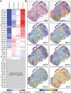
Figure 4.. Expression patterns of genes implicated in hair follicle biology and pilomatricoma pathogenesis.
(A) Expression level of select transcripts related to hair follicle biology and pilomatricoma pathogenesis across pilomatricoma spatial transcriptomic clusters. Transcripts from a subset of genes were not detected in sufficient number to enable spatial comparisons. (B) Spatial overlays of relative transcript quantity for select genes plotted in panel (A). N.S. = not statistically significant.
Figure 5.
Figure 5.. Expression patterns of all detected keratin gene transcripts.
(A) Expression level of all detectable transcripts coding for keratin family genes across pilomatricoma spatial transcriptomic clusters. Transcripts from a subset of keratin genes were detected, but not in sufficient number to enable spatial comparisons. (B) Spatial overlays of relative transcript quantity for select genes plotted in panel (A). N.S. = not statistically significant.

References

    1. Jones CD, Ho W, Robertson BF, Gunn E, Morley S. Pilomatrixoma: a comprehensive review of the literature. Am J Dermatopathol. 2018;40(9):631–641. doi:10.1097/DAD.0000000000001118. - DOI - PubMed
    1. Guinot-Moya R, Valmaseda-Castellon E, Berini-Aytes L, Gay-Escoda C. Pilomatrixoma. Review of 205 cases. Med Oral Patol Oral Cir Bucal. 2011;16(4):e552–555. doi:10.4317/medoral.16.e552. - DOI - PubMed
    1. Hernández-Núñez A, Nájera Botello L, Romero Maté A, et al. Retrospective study of pilomatricoma: 261 tumors in 239 patients. Actas Dermosifiliogr. 2014;105(7):699–705. doi:10.1016/j.ad.2014.01.003. - DOI - PubMed
    1. Miura T, Yamamoto T. Perforating pilomatricoma with anetodermic epidermis in an adolescent with lymphoma. Pediatr Dermatol. 2013;30(4):e68–69. doi:10.1111/j.1525-1470.2012.01825.x. - DOI - PubMed
    1. Li L, Zeng Y, Fang K, et al. Anetodermic pilomatricoma: molecular characteristics and trauma in the development of its bullous appearance. Am J Dermatopathol. 2012;34(4):e41–45. doi:10.1097/DAD.0b013e31823583f5. - DOI - PMC - PubMed

Publication types