

Marsdenia tenacissima enhances immune response of tumor infiltrating T lymphocytes to colorectal cancer
- PMID: 37649480
- PMCID: PMC10465246
- DOI: 10.3389/fimmu.2023.1238694
Marsdenia tenacissima enhances immune response of tumor infiltrating T lymphocytes to colorectal cancer
Abstract
Introduction: Tumor-infiltrating T lymphocytes in the tumor microenvironment are critical factors influencing the prognosis and chemotherapy outcomes. As a Chinese herbal medicine, Marsdenia tenacissima extract (MTE) has been widely used to treat cancer in China. Its immunoregulatory effects on tumor-associated macrophages is well known, but whether it regulates tumor-infiltrating T-cell functions remains unclear.
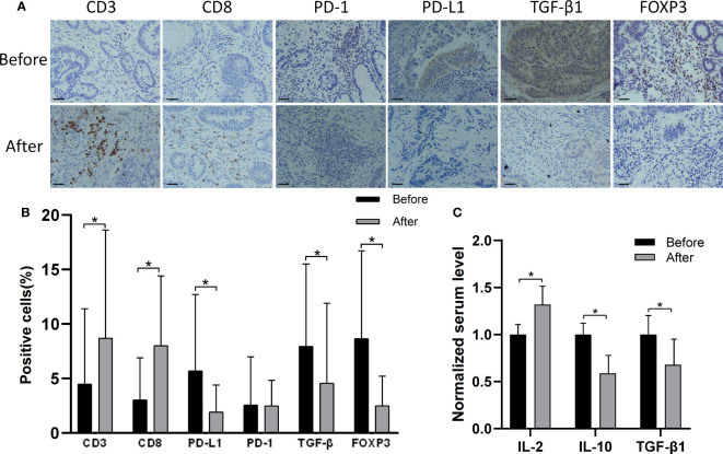
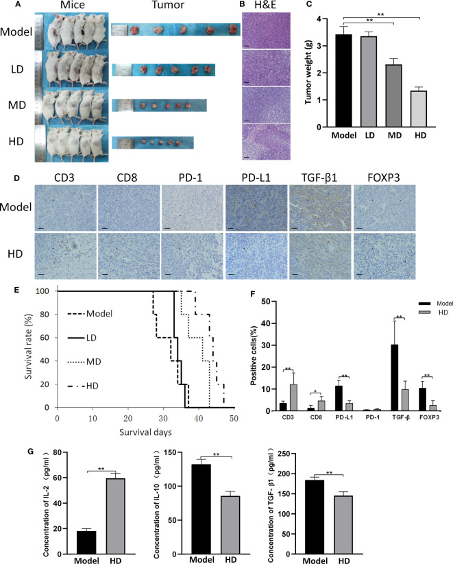
Method: We collected 17 tumor samples from MTE-administered colorectal cancer patients, 13 of which showed upregulation of CD3+/CD8+ tumor-infiltrating T cells. Further in vitro and in vivo experiments were performed to investigate the regulatory effects of MTE on tumor-infiltrating T cells and immune escape of tumors.
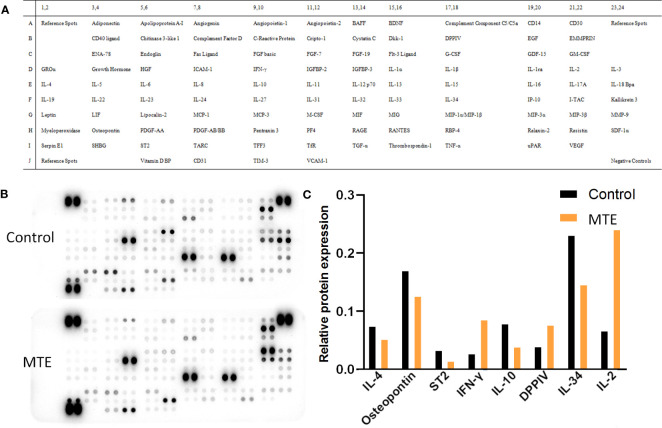
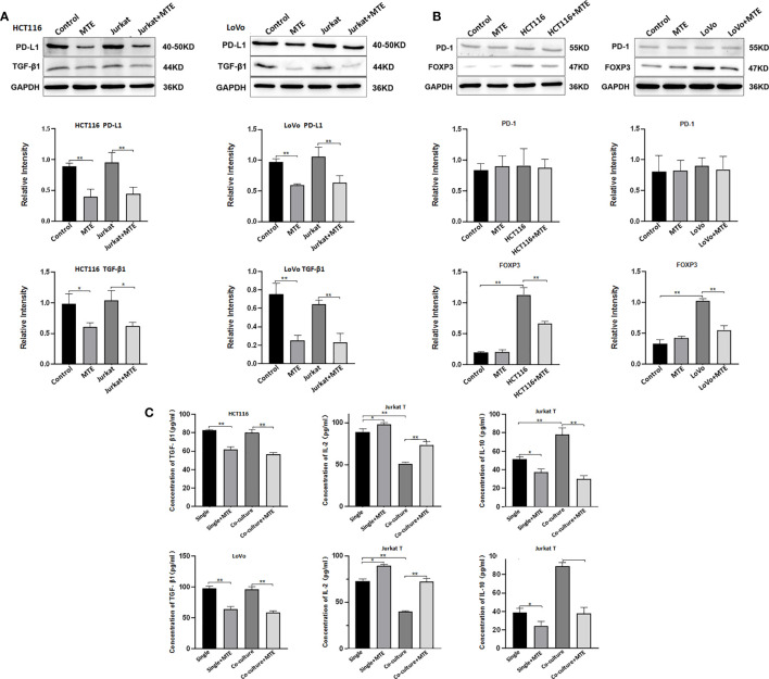
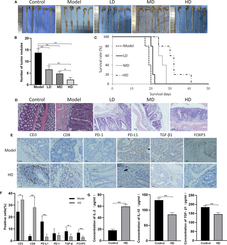
Results: Under single and co-culture conditions, MTE inhibited TGF-β1 and PD-L1 expression in the colorectal cancer (CRC) cell lines HCT116 and LoVo. In Jurkat cells, MTE inhibited FOXP3 and IL-10 expression, increased IL-2 expression, but had no effect on PD-1 expression. These findings were confirmed in vitro using subcutaneous and colitis-associated CRC mouse models. MTE also increased the density of CD3+/CD8+ tumor-infiltrating T cells and exhibited considerable tumor-suppressive effects in these two tumor mouse models.
Conclusions: Our findings suggested that MTE inhibits the immune escape of cancer cells, a precipitating factor increasing the immune response of T lymphocytes.
Keywords: CD8; colorectal cancer; marsdenia tenacissima extraction; tumor infiltrating T cells; tumor microenvironment.
Copyright © 2023 Yi, Zhang, Yan, Liu, Feng, Chu, Liu, Wang, Xue, Zhang and Wang.
Conflict of interest statement
The authors declare that the research was conducted in the absence of any commercial or financial relationships that could be construed as a potential conflict of interest.
Figures
Similar articles
-
Marsdenia tenacissima extract disturbs the interaction between tumor-associated macrophages and non-small cell lung cancer cells by targeting HDGF.J Ethnopharmacol. 2022 Nov 15;298:115607. doi: 10.1016/j.jep.2022.115607. Epub 2022 Aug 13. J Ethnopharmacol. 2022. PMID: 35973634
-
Nitric oxide, a communicator between tumor cells and endothelial cells, mediates the anti-tumor effects of Marsdenia Tenacissima Extract (MTE).J Ethnopharmacol. 2020 Mar 25;250:112524. doi: 10.1016/j.jep.2019.112524. Epub 2019 Dec 26. J Ethnopharmacol. 2020. PMID: 31884032
-
Marsdenia tenacissima extract inhibits gefitinib metabolism in vitro by interfering with human hepatic CYP3A4 and CYP2D6 enzymes.J Ethnopharmacol. 2014;151(1):210-7. doi: 10.1016/j.jep.2013.10.021. Epub 2013 Oct 21. J Ethnopharmacol. 2014. PMID: 24157377
-
Oral and injectable Marsdenia tenacissima extract (MTE) as adjuvant therapy to chemotherapy for gastric cancer: a systematic review.BMC Complement Altern Med. 2019 Dec 12;19(1):366. doi: 10.1186/s12906-019-2779-y. BMC Complement Altern Med. 2019. PMID: 31830977 Free PMC article.
-
A Systematic Review of the Tumor-Infiltrating CD8+ T-Cells/PD-L1 Axis in High-Grade Glial Tumors: Toward Personalized Immuno-Oncology.Front Immunol. 2021 Sep 17;12:734956. doi: 10.3389/fimmu.2021.734956. eCollection 2021. Front Immunol. 2021. PMID: 34603316 Free PMC article.
Cited by
-
Effect of colorectal cancer stem cells on the development and metastasis of colorectal cancer.World J Gastrointest Oncol. 2024 Nov 15;16(11):4354-4368. doi: 10.4251/wjgo.v16.i11.4354. World J Gastrointest Oncol. 2024. PMID: 39554751 Free PMC article. Review.
-
Targeting the mTOR Pathway in Hepatocellular Carcinoma: The Therapeutic Potential of Natural Products.J Inflamm Res. 2024 Dec 6;17:10421-10440. doi: 10.2147/JIR.S501270. eCollection 2024. J Inflamm Res. 2024. PMID: 39659752 Free PMC article. Review.
-
Research progress of tumor-associated macrophages in immune checkpoint inhibitor tolerance in colorectal cancer.World J Gastrointest Oncol. 2024 Oct 15;16(10):4064-4079. doi: 10.4251/wjgo.v16.i10.4064. World J Gastrointest Oncol. 2024. PMID: 39473964 Free PMC article. Review.
-
Phillygenin regulates the colorectal cancer tumor microenvironment by inhibiting hypoxia-inducible factor 1 alpha.Cytotechnology. 2025 Feb;77(1):17. doi: 10.1007/s10616-024-00679-2. Epub 2024 Dec 10. Cytotechnology. 2025. PMID: 39669690
References
-
- Yothers G, Venook AP, Oki E, Niedzwiecki D, Lin Y, Crager MR, et al. . Patient-specific meta-analysis of 12-gene colon cancer recurrence score validation studies for recurrence risk assessment after surgery with or without 5FU and oxaliplatin. J Gastrointest Oncol (2022) 13(1):126–36. doi: 10.21037/jgo-21-620 - DOI - PMC - PubMed
-
- Wu ZL, Chen Y, Qu Z, Wu GY, He XF, Huang JW, et al. . An ester derivative of tenacigenin B from Marsdenia tenacissima (Roxb.) Wight et Arn reversed paclitaxel-induced MDR in vitro and in vivo by inhibiting both P-gp and MRP2. J Ethnopharmacol (2022) 294:115353. doi: 10.1016/j.jep.2022.115353 - DOI - PubMed
Publication types
MeSH terms
LinkOut - more resources
Full Text Sources
Research Materials