. 2023 Aug 15:14:1238321.
doi: 10.3389/fimmu.2023.1238321. eCollection 2023.

Evolution of T cell receptor beta loci in salmonids

Affiliations

Evolution of T cell receptor beta loci in salmonids

Pierre Boudinot et al. Front Immunol. .

Abstract

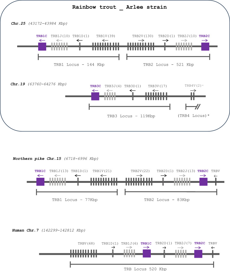
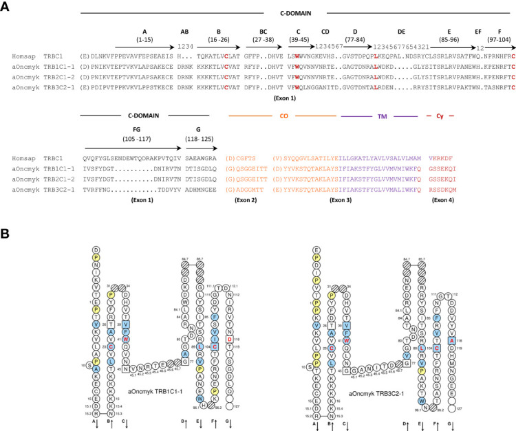
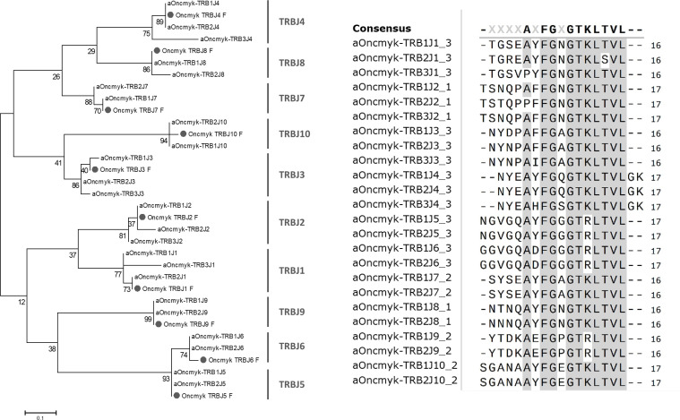
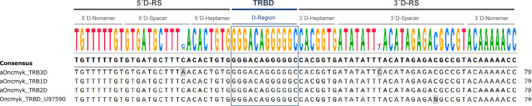
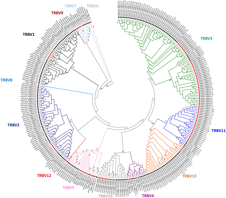
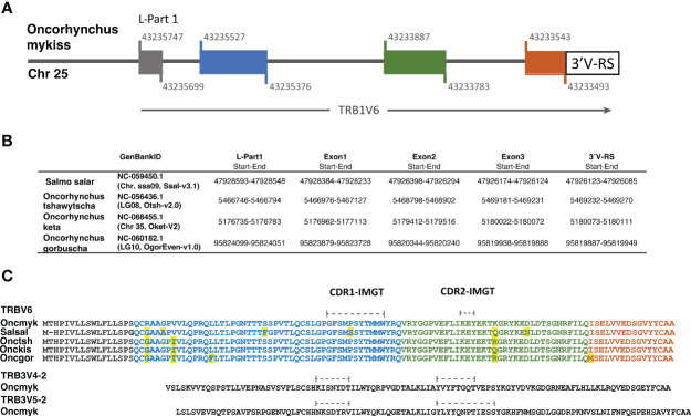
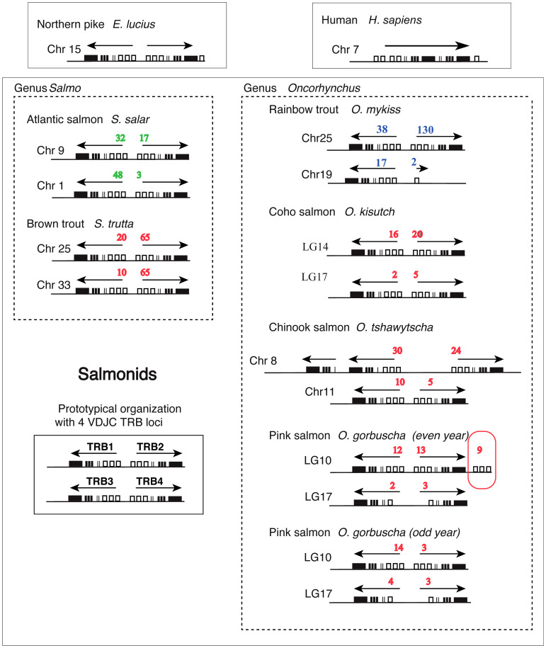
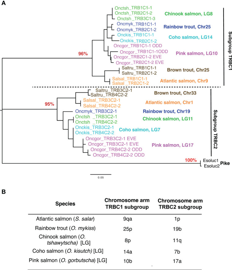
T-cell mediated immunity relies on a vast array of antigen specific T cell receptors (TR). Characterizing the structure of TR loci is essential to study the diversity and composition of T cell responses in vertebrate species. The lack of good-quality genome assemblies, and the difficulty to perform a reliably mapping of multiple highly similar TR sequences, have hindered the study of these loci in non-model organisms. High-quality genome assemblies are now available for the two main genera of Salmonids, Salmo and Oncorhynchus. We present here a full description and annotation of the TRB loci located on chromosomes 19 and 25 of rainbow trout (Oncorhynchus mykiss). To get insight about variations of the structure and composition of TRB locus across salmonids, we compared rainbow trout TRB loci with other salmonid species and confirmed that the basic structure of salmonid TRB locus is a double set of two TRBV-D-J-C loci in opposite orientation on two different chromosomes. Our data shed light on the evolution of TRB loci in Salmonids after their whole genome duplication (WGD). We established a coherent nomenclature of salmonid TRB loci based on comprehensive annotation. Our work provides a fundamental basis for monitoring salmonid T cell responses by TRB repertoire sequencing.

Keywords: T cell receptor; TRB locus; adaptive immunity; evolution; rainbow trout; repertoire; salmonid fish.


Conflict of interest statement

The authors declare that the research was conducted in the absence of any commercial or financial relationships that could be construed as a potential conflict of interest.

Figures

Figure 1
Figure 1
Comparative genomic organization of TRB loci. Gene organization of TRB loci in rainbow trout (Oncorhynchus mykiss, Arlee strain) compared to those of Northern pike (Esox lucius) and human (Homo sapiens). TRBC genes as shown as purple boxes, TRBJ and TRBV genes as light and dark grey lines, respectively; and TRBD genes as white lines. The number of corresponding genes is between brackets. The arrow indicates the transcriptional orientation. The symbols representing the genes are not to scale. See Supplementary File 2 for exact location of each gene. * TRBV genes located approximately 390 Kbp downstream to the previous TRBV gene. TRB4 in parenthesis indicates the absence of D-J-C cluster.
Figure 2
Figure 2
Deduced amino acid sequences from rainbow trout TRBC genes annotated in the USDA_OmykA_1.1 genome (GenBank GCA_013265735.3, isolate Arlee). (A) Alignment of the TRBC amino acid sequences from human (Homsap) and rainbow trout (aOncmyk). The domains and regions are indicated as follow: C-DOMAIN (constant domain), CO (connecting region), TM (transmembrane region) and CY (cytoplasmic region). The four conserved amino acids of the C-DOMAIN: 1st-CYS 23 and 2nd-CYS 104 (cysteins of the disulfide bridge), CONSERVED-TRP 41 (tryptophan, W) and the hydrophobic amino acid at position 89 are in red. Description of the C domain strands (A, B, C, D, E, F, G), turns (AB, DE, EF), loops (BC, FG), and their IMGT positions are according to the IMGT unique numbering for C-DOMAIN (29). Missing positions 32 and 33 in the BC loop are not shown. The amino acids between parentheses at the beginning of EX1, EX2 and EX3 corresponds to the first codon resulting from a splicing frame 1. The splicing between EX3 and EX4 is a splicing frame 0. (B) IMGT Collier de Perles of the rainbow trout TRB1C1-1 and TRB3C2-1 genes as derived from the alignment with the human TRBC proteins.
Figure 3
Figure 3
Rainbow trout TRBJ sequences. Phylogenetic tree of rainbow trout TRBJ nucleotide sequences. It includes the 24 TRBJ genes annotated in the USDA_OmykA_1.1 genome and the ten TRBJ gene sequences (labelled with a grey circle) previously identified in rainbow trout (21). The tree was inferred using the Neighbor joining method and JTT matrix based model. A bootstrap of 1000 replicates was used. Alignment of deduced amino acid sequences of rainbow trout TRBJ genes annotated in rainbow trout Arlee genome. Amino acids are in single letter code and conserved positions are grey highlighted. A consensus sequences is shown above the alignment.
Figure 4
Figure 4
Alignment of the D-GENE-UNIT sequences of the TRBD (diversity) genes annotated in the genome of Oncorhynchus mykiss, USDA_OmykA_1.1 (GenBank GCA_013265735.3, isolate Arlee). The consensus sequence is provided at the top of the figure. It includes the 5’ and 3’ recombination signal (RS) sequences (5´D-RS and 3´D-RS) and the TRBD region. Oncmyk_TRBD_U97590 corresponds to the TRBD gene previously identified in rainbow trout (20).
Figure 5
Figure 5
Salmonids TRBV subgroups. Phylogenetic tree of TRBV nucleotide sequences from Atlantic salmon (red triangles) and rainbow trout (black triangles). The tree was inferred using the Neighbor joining method and JTT matrix based model. A bootstrap of 1000 replicates was used. The tree with highest likelihood is shown, and is drawn to scale, with branch lengths corresponding to the number of substitutions per site. The percentage of trees in which the associated taxa cluster together is indicated when >50. The analysis has been performed with all annotated TRBV genes, functional, with open reading frame and pseudogenes (P). The subgroup branches are represented in different colors.
Figure 6
Figure 6
The functional TRBV6 gene comprises 4 exons and is conserved across Salmonids. (A) Structure of the functional TRB1V6-1 gene in the rainbow trout genome (Arlee strain; USDA OmykA_1.1, GenBank GCA_013265735.3). Colored boxes indicate exons and black line indicates introns. Nucleotide position of the exon start and exon end at the Chr. 25 is shown above and below of each box. (B) The localization of TRBV6 exons and 3´V-RS sequences in the genome of 4 additional salmonid species is shown in a table. In Supplementary file 3 the corresponding nucleotide sequences are available. (C) Alignment of TRBV6 amino acid sequences of several salmonid species: rainbow trout, Oncmyk AAN15758; Atlantic salmon, SalsalTRBV6 Genome Ssal_3.1 reference release 102. Chr09, join:47916622‐47916678; 47916813‐47916971; 47926449‐47926282; 47926180‐47926127); Chinook salmon, OnctshTRBV6: XP_042181049; Chum salmon, OnckisTRBV6: XP_052352121; Pink salmon, OncgorTRBV6: XP_046223527). The amino acid sequence encoded by each exon is shown in the corresponding color. Differences between species are yellow highlighted.
Figure 7
Figure 7
Structure of TRB loci across salmonid species. Genomic location and organization of TRB loci was determined based on last available genome assemblies at NCBI and Ensembl. Loci from rainbow trout (this work) and Atlantic salmon (22) are represented besides those from brown trout (Salmo trutta), Coho salmon (Oncorhynchus kisutch), Chinook salmon (Oncorhynchus tshawytscha), and Pink salmon (Oncorhynchus gorbuscha) (even and odd year). Northern pike (Esox lucius) is shown above as a representative from a sister group of Salmonids, which has not been subjected to WGD. Human (Homo sapiens) is shown above as a representative from mammals. White rectangles represent TRBV genes, thin lines TRBD genes, thick lines TRBJ genes and black rectangles TRBC genes. Numbers of TRBV genes (functional, ORF and pseudogenes) detected in our analysis are indicated in blue for rainbow trout, in green for Atlantic salmon and in red for the other species. Transcriptional orientation (determined by TRBC genes) is indicated above each locus.
Figure 8
Figure 8
TRBC subgroups. (A) Phylogenetic analysis of TRBC genes reveals two distinct types located on different chromosomes in each salmonid species. These types have been shown to correspond to two TRBC subgroups, C1 and C2. The TRBC1 subgroup includes TRB1C1-1 and TRB2C1-2 and the TRBC2 subgroup includes TRB3C2-1 and TRBC4C2-2. Phylogenetic reconstruction was inferred by using the Maximum Likelihood method and JTT matrix-based model. The tree with the highest log likelihood (-1537.90) is shown. The percentage of trees in which the associated taxa clustered together is shown next to the key nodes corresponding to types A and B as a percentage (in red). Evolutionary analyses were conducted in MEGA X. The tree was rooted by TRBC sequences from the Northern pike. (B) TRB loci are found on distinct chromosome arms generated by the ssWGD. The location of TRB loci is consistent with chromosomal orthology relationships between species.

Similar articles

Cited by

References

    1. Flajnik M, Du Pasquier L. Evolution of the immune system. In: Paul W, editor. Fundam. Immunol., 7th edn. (2012) New York: Wolters Kluwer & Lippincott Willians & Wilkins. p. 67–128.
    1. Flajnik MF. A cold-blooded view of adaptive immunity. Nat Rev Immunol (2018) 18:438–53. doi: 10.1038/S41577-018-0003-9 - DOI - PMC - PubMed
    1. Danilova N. The evolution of adaptive immunity. Adv Exp Med Biol (2012) 738:218–35. doi: 10.1007/978-1-4614-1680-7_13 - DOI - PubMed
    1. Lefranc M-P, Lefranc G. The immunoglobulin FactsBook. London (UK): Academic Press; (2001).
    1. Lefranc M-P, Lefranc G. Immunoglobulins or antibodies: IMGT® Bridging genes, structures and functions. Biomedicines (2020) 8(9):E319. doi: 10.3390/BIOMEDICINES8090319 - DOI - PMC - PubMed

Publication types

LinkOut - more resources