Review
. 2023 Aug 15:14:1226038.
doi: 10.3389/fimmu.2023.1226038. eCollection 2023.

What role does PDL1 play in EMT changes in tumors and fibrosis?

Affiliations
Review

What role does PDL1 play in EMT changes in tumors and fibrosis?

Yun-Chao Zhang et al. Front Immunol. .

Abstract

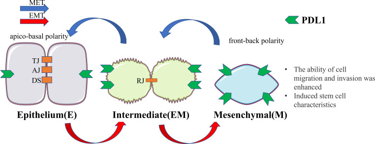
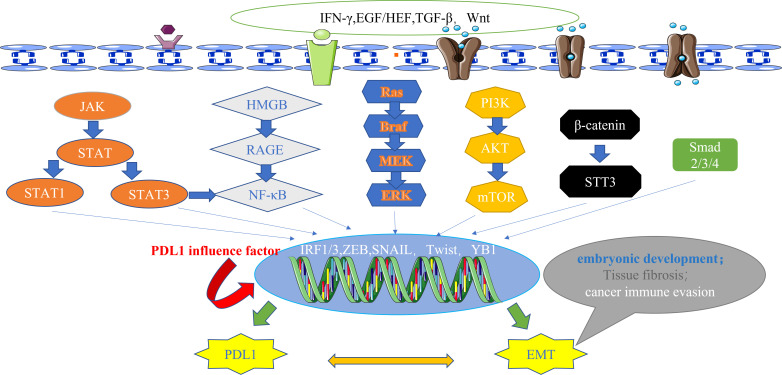
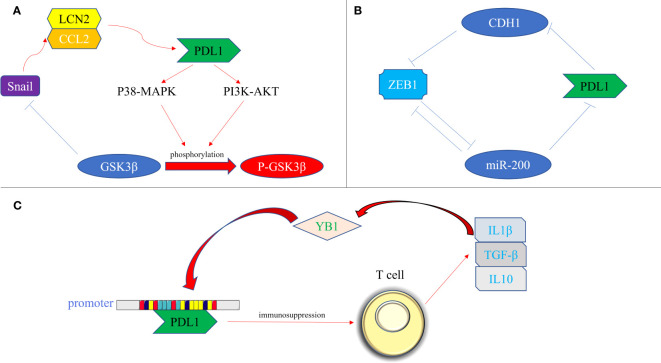
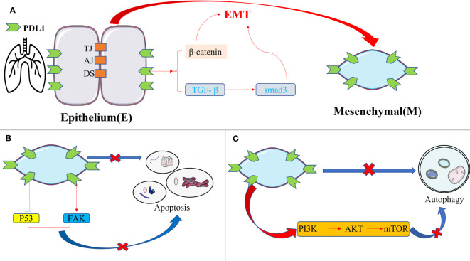
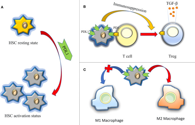
Epithelial-mesenchymal transformation (EMT) plays a pivotal role in embryonic development, tissue fibrosis, repair, and tumor invasiveness. Emerging studies have highlighted the close association between EMT and immune checkpoint molecules, particularly programmed cell death ligand 1 (PDL1). PDL1 exerts its influence on EMT through bidirectional regulation. EMT-associated factors, such as YB1, enhance PDL1 expression by directly binding to its promoter. Conversely, PDL1 signaling triggers downstream pathways like PI3K/AKT and MAPK, promoting EMT and facilitating cancer cell migration and invasion. Targeting PDL1 holds promise as a therapeutic strategy for EMT-related diseases, including cancer and fibrosis. Indeed, PDL1 inhibitors, such as pembrolizumab and nivolumab, have shown promising results in clinical trials for various cancers. Recent research has also indicated their potential benefit in fibrosis treatment in reducing fibroblast activation and extracellular matrix deposition, thereby addressing fibrosis. In this review, we examine the multifaceted role of PDL1 in immunomodulation, growth, and fibrosis promotion. We discuss the challenges, mechanisms, and clinical observations related to PDL1, including the limitations of the PD1/PDL1 axis in treatment and PD1-independent intrinsic PDL1 signaling. Our study highlights the dynamic changes in PDL1 expression during the EMT process across various tumor types. Through interplay between PDL1 and EMT, we uncover co-directional alterations, regulatory pathways, and diverse changes resulting from PDL1 intervention in oncology. Additionally, our findings emphasize the dual role of PDL1 in promoting fibrosis and modulating immune responses across multiple diseases, with potential implications for therapeutic approaches. We particularly investigate the therapeutic potential of targeting PDL1 in type II EMT fibrosis: strike balance between fibrosis modulation and immune response regulation. This analysis provides valuable insights into the multifaceted functions of PDL1 and contributes to our understanding of its complex mechanisms and therapeutic implications.

Keywords: EMT-related disease; epithelial-mesenchymal transformation; fibrosis; immune escape; programmed death-ligand 1.


Conflict of interest statement

The authors declare that the research was conducted in the absence of any commercial or financial relationships that could be construed as a potential conflict of interest.

Figures

Figure 1
Figure 1
Cellular changes during EMT and MET. Upon undergoing EMT, cellular adhesion is diminished, leading to the loss of the epithelial phenotype and the gradual acquisition of a mesenchymal phenotype. In this transitional state, there is an upregulation of PDL1 expression. This transformation endows cells with the capacity for distant metastasis and redifferentiation. Conversely, MET induces the reversal of these alterations. EMT, epithelial-mesenchymal transformation; MET, mesenchymal - epithelial transformation; TJ, tight junction; AJ, adhesive junctions; DS, desmosome; RJ, residual junction.
Figure 2
Figure 2
The main signal pathways that affect the expression of PDL1.These pathways also have downstream signals, including ZEB and Snail, which act as transcription factors for EMT and regulate the expression of PDL1, highlighting the bidirectional regulatory relationship between EMT and PDL1 at the signaling pathway level. IFN-γ, interferon-γ; EGF, epidermal growth factor; HGF, hepatocyte growth factor; TGF-β, transforming growth factor–β; Wnt, wingless; JAK, janus kinase; STAT, signal transducer and activator of transcription; HMGB1, high mobility group protein 1; RAGE, receptor for advanced glycation end products; Ras, rasopathes; Braf, RAF family serine/threonine protein kinases; MEK, MAPK kinase; ERK, extracellular-signal regulated protein kinase; PI3K, phosphoinositide 3-kinase; AKT, protein kinase B; mTOR, mammalian target of rapamycin; STT3, N-oligosaccharide transferase complex; Smad, skin growth factor; IRF, interferon regulatory factor; ZEB, zinc finger E-box binding homeobox; PDL1, programmed death ligand 1; EMT, epithelial-mesenchymal transformation.
Figure 3
Figure 3
Bidirectional regulation of EMT transcription factors and PDL1. (A) At the protein level. PDL1 activates the p38-MAPK or AKT pathways, leading to the phosphorylation of GSK3β and subsequent upregulation of Snail expression. Snail, in turn, promotes the generation of the CCL2/LCN2 complex, inducing an immunosuppressive state in cells and further enhancing PDL1 expression; (B) At the miRNA level, ZEB1 acts as a transcriptional repressor of miR-200, alleviating the inhibitory effect of miR-200 on PDL1 expression. Additionally, PDL1 can influence ZEB1 through the generation of CDH1; (C) At the transcriptional level, YB1 directly binds to the promoter sequence of PDL1, promoting PDL1 production. The excessive presence of PDL1 induces the release of fibrosis-promoting factors, such as TGF-β, from T cells, thereby promoting the upregulation of YB1. PDL1, programmed death ligand 1; P38-MAPK, p38 mitogen activated protein kinase; PI3K, phosphoinositide 3-kinase; AKT, protein kinase B; GSK3β, glycogen synthase kinase 3β; P- GSK3β, phosphorylated glycogen synthetase kinase 3β; CCL2, C-C motif chemokine ligand 2; LCN2, lipocalin 2; ZEB, zinc finger E-box binding homeobox; miRNA-200, micro RNA 200 family; YB1, Y-box binding protein 1; IL1β, interleukin 1; IL10, interleukin 10; TGF-β, transforming growth factor–β.
Figure 4
Figure 4
The intrinsic pro-fibrotic effect of PDL1 in pulmonary fibrosis. (A) PDL1 promotes fibrosis by mediating fibroblast-to-myofibroblast transition and EMT via TGF-β/Smad3 and β-catenin signaling pathways; (B) Overexpression of PDL1 leads to myoblasts’ resistance to apoptosis and escape from macrophage phagocytosis through inhibition of p53 pathway or activation of FAK pathway, ultimately leading to excessive proliferation of myoblasts and triggering IPF; (C) PDL1 can inhibit autophagy of fibrosis-related cells through the PI3K/AKT/mTOR signal pathway. PDL1, programmed death ligand 1; EMT, epithelial-mesenchymal transformation; TJ, tight junction; AJ, adhesive junctions; DS, desmosome; TGF-β, transforming growth factor–β; Smad, skin growth factor; FAK, focal adhesion kinase; PI3K, phosphoinositide 3-kinase; AKT, protein kinase B; mTOR, mammalian target of rapamycin.
Figure 5
Figure 5
The immune regulation of PDL1 in hepatic fibrosis, thus promoting the role of fibrosis. (A) PDL1 activates HSCs (HSCs are considered to be the main effector cells of liver fibrosis); (B) PDL1 induces the production of Tregs and releases exocrine bodies showing TGF-β; (C) PDL1 stimulates the polarization of M2 macrophages. HSCs, hepatic stellate cells; PDL1, programmed death ligand 1; PD1, programmed cell death protein 1; TGF-β, transforming growth factor–β.

Similar articles

Cited by

References

    1. Aroeira LS, Aguilera A, Sanchez-Tomero Jose´A. Epithelial to mesenchymal transition and peritoneal membrane failure in peritoneal dialysis patients:Pathologic significance and potential therapeutic interventions. J Am Soc Nephrol (2007) 18:2004–13. doi: 10.1681/ASN.2006111292 - DOI - PubMed
    1. Nieto Ma, Huang Ry, Jackson Ra, Thiery Jp. Emt: 2016. Cell (2016) 166(1):21–45. doi: 10.1016/j.cell.2016.06.028 - DOI - PubMed
    1. Grassmann a, Gioberge S, Moeller S, Brown G. Esrd patients in 2004: global overview of patient numbers, treatment modalities and associated trends. Nephrol Dial Transplant (2005) 20:2587–93. doi: 10.1093/ndt/gfi159 - DOI - PubMed
    1. Strippoli R, Moreno-Vicente R, Battistelli C, Cicchini C, Noce V, Amicone L, et al. . Molecular mechanisms underlying peritoneal emt and fibrosis. Stem Cells Int (2016) 2016:3543678. doi: 10.1155/2016/3543678 - DOI - PMC - PubMed
    1. Jiang Y, Zhan H. Communication between Emt and Pd-L1 Signaling: New Insights into Tumor Immune Evasion. Cancer Lett (2020) 468:72–81. doi: 10.1016/J.Canlet.2019.10.013 - DOI - PubMed

Publication types