Review
. 2023 Aug 29:9:107.
doi: 10.1038/s41378-023-00579-z. eCollection 2023.

Polymeric and biological membranes for organ-on-a-chip devices

Affiliations
Review

Polymeric and biological membranes for organ-on-a-chip devices

Kendra Corral-Nájera et al. Microsyst Nanoeng. .

Abstract

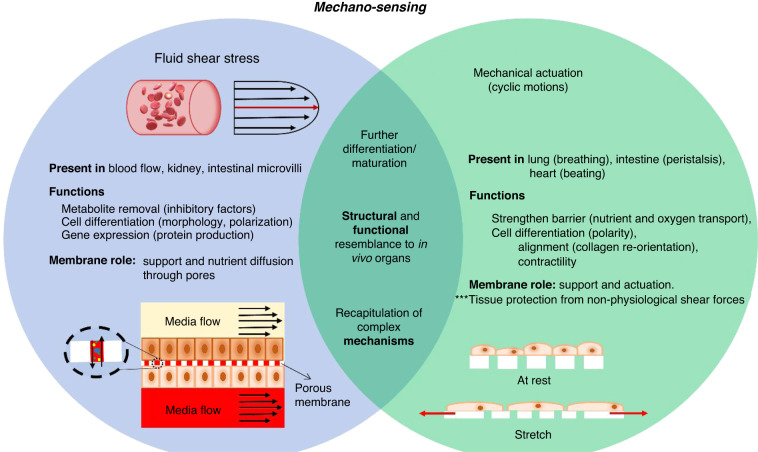
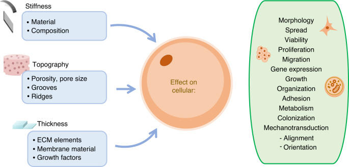
Membranes are fundamental elements within organ-on-a-chip (OOC) platforms, as they provide adherent cells with support, allow nutrients (and other relevant molecules) to permeate/exchange through membrane pores, and enable the delivery of mechanical or chemical stimuli. Through OOC platforms, physiological processes can be studied in vitro, whereas OOC membranes broaden knowledge of how mechanical and chemical cues affect cells and organs. OOCs with membranes are in vitro microfluidic models that are used to replace animal testing for various applications, such as drug discovery and disease modeling. In this review, the relevance of OOCs with membranes is discussed as well as their scaffold and actuation roles, properties (physical and material), and fabrication methods in different organ models. The purpose was to aid readers with membrane selection for the development of OOCs with specific applications in the fields of mechanistic, pathological, and drug testing studies. Mechanical stimulation from liquid flow and cyclic strain, as well as their effects on the cell's increased physiological relevance (IPR), are described in the first section. The review also contains methods to fabricate synthetic and ECM (extracellular matrix) protein membranes, their characteristics (e.g., thickness and porosity, which can be adjusted depending on the application, as shown in the graphical abstract), and the biological materials used for their coatings. The discussion section joins and describes the roles of membranes for different research purposes and their advantages and challenges.

Keywords: Materials science; Microfluidics.


Conflict of interest statement

Competing interestsThe authors declare no competing interests.

Figures

Fig. 1
Fig. 1. Membranes mediate cell mechanosensing under fluid shear stress and mechanical actuation.
The combination of fluid shear stress and mechanical actuation results in an increased cell differentiation, achieves a closer resemblance to in vivo organs and allows the recapitulation of complex biological mechanisms, as opposed to the mechanical stimulation methods individually
Fig. 2
Fig. 2. Fluidic flow relevance in microfluidic devices.
a OOC developed by Delon et al. (2019) to investigate the effect of fluid shear stress on Caco-2 intestinal epithelial cells. The device is sectioned to deliver differential shear stress (top), with the highest shear stress on the left and the lowest, on the right (Image adapted from Figs. 1 and 6 from reference). The formation of tight junctions is monitored via immunofluorescence staining (bottom) of ZO-1, a tight junction protein stained in green. b, c Hybrid insertable fluidic devices have been proposed by Shin et al. (2019) to introduce fluid flow to Transwell ® inserts (Images adapted from Figure S5 from reference Supplemental Information)
Fig. 3
Fig. 3. OOC mechanical actuation.
a A gut-on-a-chip proposed by Kim et al. (2012). b, c The device has a basal‒apical conformation, channels are separated by a porous PDMS membrane. d The lateral vacuum chambers stretch the membrane, delivering mechanical stimulus to cells to mimic peristaltic motions. Image adapted from Fig. 1 from reference
Fig. 4
Fig. 4. Membrane characteristics can be adjusted to mimic in vivo surfaces and promote cellular specialization.
Stiffness, topography and thickness impact cellular structure, function and activity
Fig. 5
Fig. 5. Porous membrane fabrication processes can be divided into the following main steps: mold fabrication, treatment to reduce adhesion, and membrane fabrication.
Left: a widely implemented technique is photolitographic mold fabrication and posterior replica molding. Right: laser machining mold fabrication to create dissolvable molds is a novel fabrication approach
Fig. 6
Fig. 6. Porous PDMS membrane fabrication via high-pressure saturated steam method proposed by Jang et al. (2019).
Schematic illustration of 4 stages: 1st—initial heating process to 100 °C, 2nd—temperature increase from 100 °C to 120 °C for pressurization, 3rd—pressurization at 0.12 MPa for 20 min, 4th—pressure release for 40 min. Image from Fig. 1 reference
Fig. 7
Fig. 7. PDMS 3D printing improves cell adhesion and spreading due to uneven surfaces.
Immuno-images of adhered cells on cast and 3D-printed surfaces. Image from Fig. 6 reference
Fig. 8
Fig. 8. Protein and cell patterning.
a Schematic of the patterning process with reusable parylene stencil. b, c Fluorescent protein pattern. d, e Cell patterning (NIH-3T3 fibroblasts) on PDMS, substrate initially coated with FN to increase adhesion. Image adapted from Figs. 2, 4, and 6 from reference
Fig. 9
Fig. 9. Tibbe et al. (2018) in situ chitosan membrane fabrication.
a Chip design where V1 is the inlet for a basic solution (pH 9.6), V2 the inlet for the chitosan solution, and V3 the inlet for the neutral (pH 7) solution. The chitosan membrane forms at 4, whereas 5 is the outlet for the chitosan solution. b Bright field image of the basic solution (red), chitosan solution (transparent), and neutral solution (blue). The experiment is depicted in ce. After membrane polymerization, astrocytes are seeded in the bottom chamber (c), the chitosan membrane is later removed (d) and last, endothelial cells are seeded in direct contact with astrocytes for coculture (e). Image adapted from Figs. 1 and 3 from reference

Similar articles

Cited by

References

    1. Yang X, et al. Nanofiber membrane supported lung-on-a-chip microdevice for anti-cancer drug testing. Lab Chip. 2018;18:486–495. - PubMed
    1. Bein A, et al. Microfluidic organ-on-a-chip models of human intestine. Cell. Mol. Gastroenterol. Hepatol. 2018;5:659–668. - PMC - PubMed
    1. Zamprogno P, et al. Second-generation lung-on-a-chip with an array of stretchable alveoli made with a biological membrane. Commun. Biol. 2021;4:168. - PMC - PubMed
    1. Felder M, et al. Impaired wound healing of alveolar lung epithelial cells in a breathing lung-on-a-chip. Front. Bioeng. Biotechnol. 2019;7:3. - PMC - PubMed
    1. Novak R, et al. Robotic fluidic coupling and interrogation of multiple vascularized organ chips. Nat. Biomed. Eng. 2020;4:407–420. - PMC - PubMed