. 2023 Aug 31;18(8):e0290359.
doi: 10.1371/journal.pone.0290359. eCollection 2023.

Descending interneurons of the stick insect connecting brain neuropiles with the prothoracic ganglion

Affiliations

Descending interneurons of the stick insect connecting brain neuropiles with the prothoracic ganglion

Jens Goldammer et al. PLoS One. .

Abstract

Stick insects respond to visual or tactile stimuli with whole-body turning or directed reach-to-grasp movements. Such sensory-induced turning and reaching behaviour requires interneurons to convey information from sensory neuropils of the head ganglia to motor neuropils of the thoracic ganglia. To date, descending interneurons are largely unknown in stick insects. In particular, it is unclear whether the special role of the front legs in sensory-induced turning and reaching has a neuroanatomical correlate in terms of descending interneuron numbers. Here, we describe the population of descending interneurons with somata in the brain or gnathal ganglion in the stick insect Carausius morosus, providing a first map of soma cluster counts and locations. By comparison of interneuron populations with projections to the pro- and mesothoracic ganglia, we then estimate the fraction of descending interneurons that terminate in the prothoracic ganglion. With regard to short-latency, touch-mediated reach-to-grasp movements, we also locate likely sites of synaptic interactions between antennal proprioceptive afferents to the deutocerebrum and gnathal ganglion with descending or ascending interneuron fibres. To this end, we combine fluorescent dye stainings of thoracic connectives with stainings of antennal hair field sensilla. Backfills of neck connectives revealed up to 410 descending interneuron somata (brain: 205 in 19 clusters; gnathal ganglion: 205). In comparison, backfills of the prothorax-mesothorax connectives stained only up to 173 somata (brain: 83 in 16 clusters; gnathal ganglion: 90), suggesting that up to 60% of all descending interneurons may terminate in the prothoracic ganglion (estimated upper bound). Double stainings of connectives and antennal hair field sensilla revealed that ascending or descending fibres arborise in close proximity of afferent terminals in the deutocerebrum and in the middle part of the gnathal ganglia. We conclude that two cephalothoracic pathways may convey cues about antennal movement and pointing direction to thoracic motor centres via two synapses only.


Conflict of interest statement

The authors have declared that no competing interests exist.

Figures

Fig 1
Fig 1. Descending interneurons (DN) in the CRG after staining a neck connective.
Stick insects possess prognathous mouth parts, which is why the cerebral ganglion (CRG) is tilted backwards in the adult head capsule. As a consequence, the neuraxis (n-) is pointing rearward, and n-ventral CRG neuropils are facing dorsally inside the head. a: Depth colour-coded whole-mount of the CRG, combined with anti-synapsin immunostaining (gray). The CRG outline was captured by scanning the autofluorescence of the tissue (dark gray). Depth is encoded from n-dorsal to n-ventral as blue to red. Brackets indicate approximate borders of the tritocerebrum (TR), deutocerebrum (DE), and protocerebrum (PR). Arrowheads mark two thick fibre bundles of potential axons of ascending interneurons (AN) or primary sensory neurons projecting from the gnathal ganglion and the ventral nerve cord into the lateral PR. b: Schematic drawing of the soma cluster distribution in the CRG, ipsi- (i) and contralaterally (c) to the side of the dyed connective (arrow). pi: pars intercerebralis cluster. Shades of gray indicate n-dorsoventral location of the clusters: n-dorsal = white with black outline; intermediate = gray; n-ventral = black. The two unlabelled clusters in the n-posterior mid region of the PR were found rarely. Scale bar = 100 μm.
Fig 2
Fig 2. Thick optical CRG sections with DNs stained through a neck connective.
Connective backfill (magenta) combined with anti-synapsin immunostaining (gray). a-f: Optical sections (same whole-mount shown in Fig 1A) from n-dorsal (a) to n-ventral CRG parts (f). For an explanation of the term neuraxis (n-) please see Fig 1. Arrow in (a) marks the site of the dyed neck connective. Labels indicate soma clusters and prominent CRG neuropils. AL: antennal lobe; CA: calyces; CX: central complex; DL: dorsal lobe; PB: protocerebral bridge; VFA: ventral area of flagellar afferents. Arborisations of DNs occur in the posterior lateral protocerebrum (PLP), the posterior ventrolateral protocerebrum (PVLP), the anterior ventrolateral protocerebrum (AVLP), the posterior slope (PS), the lateral accessory lobe (LAL), the ipsi- and contralateral DLs and VFAs, and in the medial contralateral protocerebrum (b-e). Arrowheads in (b) and (c) mark two thick fibre bundles of potential axons of ascending interneurons (AN) or primary sensory neurons projecting from the gnathal ganglion and the ventral nerve cord into the lateral PR. Asterisks mark clusters with undefined borders. Question marks indicate solitary neurons. The dark CRG outline, e.g. the cell body rind, was captured by scanning the autofluorescence of the tissue. Scale bars = 100 μm.
Fig 3
Fig 3. GNG whole-mount, showing DNs stained through a neck connective.
a: Depth-colour coded specimen. Depth is encoded from dorsal to ventral as blue to red. Outline (gray) was captured by scanning the autofluorescence (also in a1-a3). About 200 cell bodies were dyed ipsi- and contralaterally to the stained connective and along the GNG midline. Asterisk marks the position of DUM cell bodies. a1-a3: Thick optical GNG sections of specimen shown in (a) from dorsal (a1) to ventral regions (a3). Contralateral cell bodies are located mainly dorsolaterally, but also in intermediate and ventral regions (a1-a3). Ipsilateral cell bodies were stained posterolaterally in intermediate and ventral parts (Fig 3A2–3A3). Along the GNG midline, the majority of stained cell bodies are located ventrally in the cell body rind (a2-a3). Dense arborisations of branches occur in the core GNG neuropil (a1-a2). Asterisks mark the position of stained DUM cell bodies. Scale bars = 100 μm.
Fig 4
Fig 4. DNs in the CRG and GNG after staining the prothorax-mesothorax connective.
a: Depth colour-coded CRG with minor distortion along z-axis. The CRG outline was captured by scanning the autofluorescence of the tissue (gray). Depth is encoded from n-dorsal to n-ventral as blue to red. Arrowheads mark two thick fibre bundles of potential ascending interneuron (AN) axons projecting from the gnathal ganglion and the ventral nerve cord into the lateral PR. b: Schematic drawing of the soma cluster distribution in the CRG, ipsi- (i) and contralaterally (c) to the side of the dyed connective (arrow). pi: pars intercerebralis cluster. Gray scale indicates the n-dorso-ventral location of the clusters; n-dorsal: white with black outline; intermediate: gray; n-ventral: black. For an explanation of the term neuraxis (n-) please see Fig 1. c: DNs in the GNG. The outline (gray) was captured by scanning autofluorescence. Depth coding as in (a). Approximately 80 cell bodies were dyed in different regions and clusters, the majority in ventral parts of the ganglion. d: Intersegmental interneurons in the prothoracic ganglion. Outline and depth coding as in (a) and (c). Approximately 100 cell bodies with axons to the mesothoracic ganglion were stained in ipsi- and contralateral ganglion regions. Arrowhead marks branches in the VAC, a prominent sensory neuropil. Scale bars = 100 μm.
Fig 5
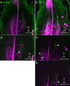
Fig 5. Potential contact sites between scapal HF afferents and DN branches in the DE of CRG, contralateral to the dyed prothorax-mesothorax connective.
a: Longitudinal projection of sHPd afferents (green) and DN branches (magenta). a1-a3: Single images of (a) from n-dorsal to more n-ventral parts of the DL and tract T6I. For an explanation of the term neuraxis (n-) please see Fig 1. a1: Few fine DN branches enter the DL. Arrowheads indicate regions with overlap. Lower arrowhead marks vicinity of boutons in tract T6II with DN branches. a2-a3: DN branches entering the VFA (medial). Arrowheads label vicinity of sHPd afferents and DN branches in the medial-n-anterior tract 6I. b: Longitudinal projection of sHPv afferents (green) and DN branches (magenta). b1-b3: Single images of (b) from dorsal to more ventral parts of the DL. b1: Arrowheads mark DN branches in vicinity to sHPv afferents in tract T6II. b2-b3: Arrowheads indicate vicinity of DN branches with sHPv afferents in the anterior DL and along T6I. Scale bars = 100 μm.
Fig 6
Fig 6. Potential contact sites between scapal HF afferents and of DN branches.
a: CRG, ipsilateral to the dyed prothorax-mesothorax connective. Longitudinal projection of sHPv afferents (green) and DN branches (magenta). a1-a3: Single images of (a) from n-dorsal to more n-ventral parts of the DL. a1-a2: Few fine DN branches enter the DL. Arrowheads indicate possible overlap with HF afferent terminals. a3: Possible overlap of sHPv afferents and DN branches in the medial-n-anterior tract 6I (arrowheads). b: Deutocerebrum, contralaterally to a dyed neck connective. Longitudinal projection of sHPv afferents (green) and DN branches (magenta). b1-b3: Single images of (a), from n-dorsal to more n-ventral parts of the DL. For an explanation of the term neuraxis (n-) please see Fig 1. b1-b2: Arrowheads indicate DN branches in vicinity to sHPv afferents along T6I. b3: Arrowheads indicate vicinity of DNs with sHPv afferents in the anterior DL and along T6I. Scale bars = 100 μm.
Fig 7
Fig 7. Potential contact sites between hair field afferents and DNs within the GNG.
Double stainings of scapal HF afferents (green) and backfilled from the prothorax-mesothorax connective (magenta). a: Overview of GNG whole-mount with sHPd afferents (green) stained bilaterally (only the HF afferents on the contralateral side with respect to the stained connective were stained sufficiently). a1: Single image of a again reveals potential contact regions between afferents and DN branches near the terminal inverted-“V” structure of the afferents (arrowhead). Scale bars = 100 μm. b: Overview of another GNG whole-mount with sHPv afferents (green) stained bilaterally. b1-b2: Single images of (b), revealing regions where DN branches and HF afferents are in close vicinity, i.e., potential contact sites between them (arrowheads).

References

    1. Photohorotaxis Kalmus H., eine neue Reaktionsart, gefunden an den Eilarven von Dixippus morosus. Z vergl Physiol. 1937; 24, pp. 644–655. doi: 10.1007/BF00592302 - DOI
    1. Jander R, Volk-Heinrichs I. Das Strauch-spezifische Perceptor-System der Stabheuschrecke (Carausius morosus). Z vergl Physiol. 1970; 70, pp. 425–447. doi: 10.1007/bf00298197 - DOI
    1. Frantsevich I, Frantsevich L. Space constancy in form perception by the stick insect. Naturwiss. 1996; 83 (7), pp. 323–324. doi: 10.1007/BF01152214 - DOI
    1. Rosano H, Webb B. A dynamic model of thoracic differentiation for the control of turning in the stick insect. Biol Cybern. 2007; 97 (3), pp. 229–246. doi: 10.1007/s00422-007-0170-4 - DOI - PubMed
    1. Dürr V, Ebeling W. The behavioural transition from straight to curve walking: kinetics of leg movement parameters and the initiation of turning. J Exp Biol. 2005; 208 (12), pp. 2237–2252. doi: 10.1242/jeb.01637 - DOI - PubMed

Publication types