. 2023 Aug 31;19(8):e1011614.
doi: 10.1371/journal.ppat.1011614. eCollection 2023 Aug.

A single inactivating amino acid change in the SARS-CoV-2 NSP3 Mac1 domain attenuates viral replication in vivo

Affiliations

A single inactivating amino acid change in the SARS-CoV-2 NSP3 Mac1 domain attenuates viral replication in vivo

Taha Y Taha et al. PLoS Pathog. .

Abstract

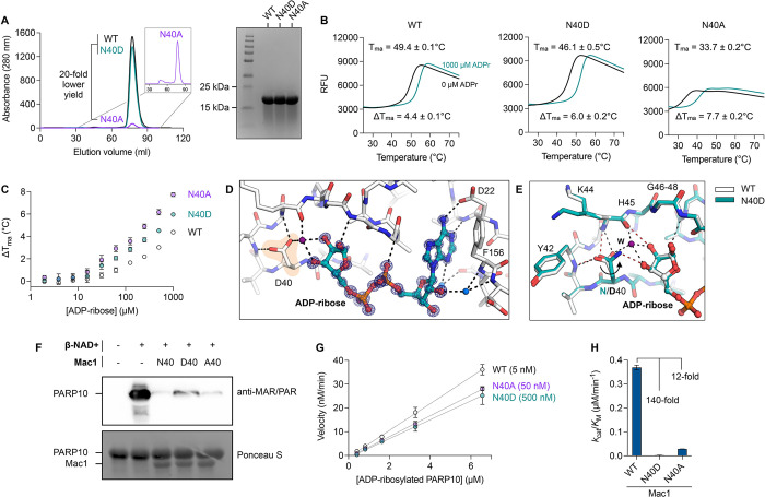
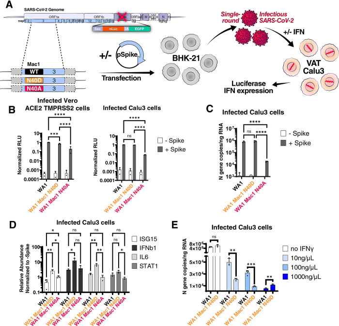
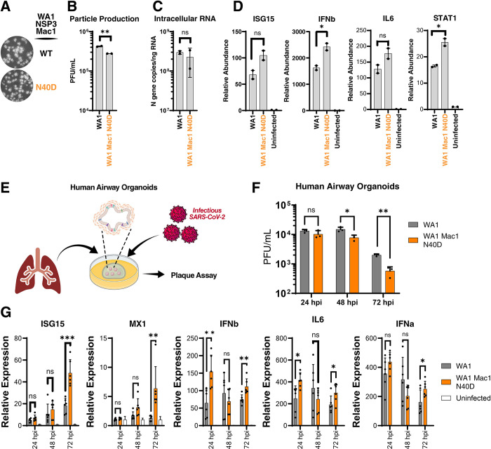
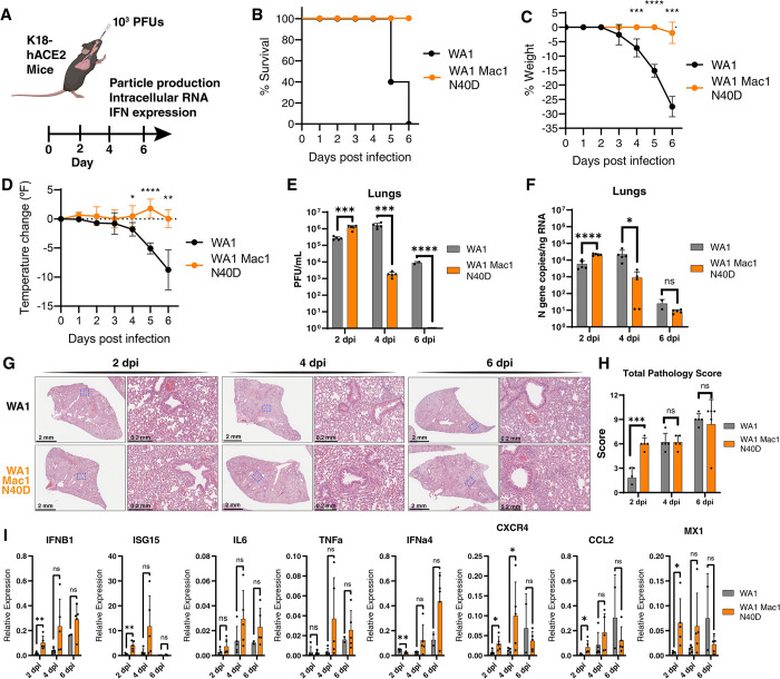
Despite unprecedented efforts, our therapeutic arsenal against SARS-CoV-2 remains limited. The conserved macrodomain 1 (Mac1) in NSP3 is an enzyme exhibiting ADP-ribosylhydrolase activity and a possible drug target. To determine the role of Mac1 catalytic activity in viral replication, we generated recombinant viruses and replicons encoding a catalytically inactive NSP3 Mac1 domain by mutating a critical asparagine in the active site. While substitution to alanine (N40A) reduced catalytic activity by ~10-fold, mutations to aspartic acid (N40D) reduced activity by ~100-fold relative to wild-type. Importantly, the N40A mutation rendered Mac1 unstable in vitro and lowered expression levels in bacterial and mammalian cells. When incorporated into SARS-CoV-2 molecular clones, the N40D mutant only modestly affected viral fitness in immortalized cell lines, but reduced viral replication in human airway organoids by 10-fold. In mice, the N40D mutant replicated at >1000-fold lower levels compared to the wild-type virus while inducing a robust interferon response; all animals infected with the mutant virus survived infection. Our data validate the critical role of SARS-CoV-2 NSP3 Mac1 catalytic activity in viral replication and as a promising therapeutic target to develop antivirals.


Conflict of interest statement

T.Y.T. and M.O. are inventors on a patent application filed by the Gladstone Institutes that covers the use of pGLUE to generate SARS-CoV-2 infectious clones and replicons. A.A. is a co-founder of Tango Therapeutics, Azkarra Therapeutics, Ovibio Corporation and Kytarro, a member of the board of Cytomx and Cambridge Science Corporation, a member of the scientific advisory board of Genentech, GLAdiator, Circle, Bluestar, Earli, Ambagon, Phoenix Molecular Designs and Trial Library, a consultant for SPARC, ProLynx, GSK and Novartis, receives grant or research support from SPARC and AstraZeneca, and holds patents on the use of PARP inhibitors held jointly with AstraZeneca from which he has benefited financially (and may do so in the future). The Krogan Laboratory has received research support from Vir Biotechnology, F. Hoffmann-La Roche, and Rezo Therapeutics. N.J.K has financially compensated consulting agreements with Maze Therapeutics, Interline Therapeutics, Rezo Therapeutics, and GEn1E Lifesciences, Inc.. N.J.K is on the Board of Directors of Rezo Therapeutics and is a shareholder in Tenaya Therapeutics, Maze Therapeutics, Rezo Therapeutics, and Interline Therapeutics. All other authors declare no competing interests.

Figures

Fig 1
Fig 1. Expression and characterization of Mac1 N40A and N40D mutants in vitro.
A) Analysis of WT, Asn40Ala (N40A) and Asn40Asp (N40D) soluble expression in BL21(DE3) E. coli by size exclusion chromatography (HiLoad 16/600 Superdex 75 pg column) and SDS-PAGE (Coomassie stained). B) Thermostability of Mac1 mutants determined by differential scanning fluorimetry (DSF). Protein (2 μM) was incubated ± 1 mM ADP-ribose with SYPRO orange (10 μM). Apparent melting temperatures were calculated by DSFworld [66] using fitting model 1. Data are presented as the mean of four replicates. C) Difference in Tma for Mac1 incubated with ADP-ribose from 2 μM to 1 mM. Data are presented as the mean ± SD of four technical replicates. D) X-ray crystal structure of ADP-ribose bound to N40D. Difference electron density (mFO-DFC map) is shown prior to modeling ADP-ribose (blue mesh contoured at 8 σ). E) ADP-ribose binding is conserved between WT (teal sticks) and N40D (white sticks). Selected hydrogen bonds are shown for WT (red dashed lines) and N40D (black dashed lines). The D40 side chain is rotated ~80° relative to N40 and therefore does not form a hydrogen bond with the terminal ribose, however, a water molecule (purple sphere labeled W) creates a new hydrogen bond network. F) Western blot analysis of Mac1 activity with auto-MARylated PARP10 (residues 819–1007). MARylated PARP10 (0.5 μM) was incubated with Mac1 variants (20 μM) for 1 hour at room temperature prior to SDS-PAGE and transfer to nitrocellulose membrane and blotting with anti-MAR/PAR antibody (1:1000 dilution, Cell Signaling, 83732). G) Kinetic measurements of MARylated PARP10 hydrolysis by Mac1 variants (concentration indicated between parentheses) using NudT5/AMP-Glo to detect ADP-ribose [51,59]. H) Catalytic efficiency (kcat/KM) was determined by linear regression of data in G using GraphPad Prism. Data are presented as the mean ± SD of three technical replicates.
Fig 2
Fig 2. SARS-CoV-2 replicon with Mac1 N40D mutation fails to suppress innate immune responses in cells.
A) Experimental workflow of the SARS-CoV-2 replicon system. SARS-CoV-2 WA1 and Mac1 domain mutant replicons were transfected along with a S and N expression vectors into BHK-21 cells. At 72 hours post-transfection, the supernatant containing single-round infectious particles is used to infect VAT and Calu3 cells in the presence or absence of exogenous IFN. At 72 hours post-infection, luciferase activity in the supernatants was used as a readout for viral RNA replication and innate immune response gene expression is measured by RT-qPCR. B) Luciferase readout of infected VAT and Calu3 cells with indicated replicons. Data are presented as mean +/- SD of three biological replicates conducted in triplicate. C) Intracellular viral RNA (N gene copies) of Calu3 cells in B was measured by RT-qPCR using a standard curve. Data are presented as mean +/- SD of three biological replicates conducted in triplicate. D) Relative expression of indicated innate immune response genes relative to GAPDH control and normalized to the -Spike control of each replicon in infected cells in B by RT-qPCR. Data are presented as mean +/- SD of three biological replicates conducted in triplicate. E) Calu3 cells were infected with indicated replicons in the presence of indicated IFNγ concentrations. Intracellular viral RNA (N gene copies) was measured by RT-qPCR using a standard curve. Data are presented as mean +/- SD of one biological replicate conducted in triplicate. *, p<0.05; **, p<0.01; ***, p<0.001; ****, p<0.0001 by two-sided Student’s T-test.
Fig 3
Fig 3. SARS-CoV-2 Mac1 domain N40D mutant is attenuated in human airway organoids.
A) Plaque morphology of indicated viruses in VAT cells. The images were pseudocolored in grayscale to optimize plaque visualization. B) Viral particle release of infected Calu3 cells (MOI 0.01) with indicated viruses was measured at 48 hours post-infection by plaque assay on VAT cells. Data are presented as mean +/- SD of two biological replicates conducted in duplicate. C) Intracellular viral RNA (N gene copies) of Calu3 cells in B was measured by RT-qPCR using a standard curve. Data are presented as mean +/- SD of two biological replicates conducted in duplicate. D) Relative levels of ISG15, IFNb, IL6, and STAT1 in Calu3 cells in B normalized to uninfected cells were determined by RT-qPCR. Data are presented as mean +/- SD of two biological replicates conducted in duplicate. E) Experimental workflow of the generation of primary human lung organoids and infection with SARS-CoV-2. Clip art was created with BioRender.com with permission. F) Viral particle release of infected primary human lung organoids with indicated viruses was measured at indicated times post-infection by plaque assay on VAT cells. Data are presented as mean +/- SD of one biological replicate conducted in triplicate. G) Relative levels of ISG15, MX1, IFNb, IL6, and IFNa to 18s RNA in infected human airway organoids in F normalized to uninfected organoids were determined by RT-qPCR. Data are presented as mean +/- SD of one biological replicate conducted in triplicate. *, p<0.05; **, p<0.01; ***, p<0.001 by two-sided Student’s T-test.
Fig 4
Fig 4. SARS-CoV-2 Mac1 N40D mutant is attenuated in K18-hACE2 mice.
A) K18-hACE2 mice (5 mice per group per timepoint) were infected with 103 PFUs of WA1 and WA1 Mac1 N40D viruses intranasally. At 2, 4, and 6 days post-infection, the lung tissue was collected for viral particle abundance measurement by plaque assay (VAT) and viral RNA and innate immune response gene expression analysis by RT-qPCR. Clip art was created with BioRender.com with permission. B) Survival curve of mice infected in A. C) Change in body weight of mice infected in A. Data are shown as mean ± SD. D) Change in body temperature of mice infected in A. Data are shown as mean ± SD. E) Viral particle abundance in the lungs was measured by plaque assay (VAT). Data are presented as mean +/- SD. F) Viral RNA levels in the lungs were measured by RT-qPCR using a standard curve. Data are presented as mean +/- SD. G) Hematoxylin and eosin (H&E) staining of lungs of mice infected in A. H) Total pathology scores of all infected mice lungs in G. Breakdown of scores is available in S7 Fig. I) Relative levels of IFNB1, ISG15, IL6, TNFa, IFNa4, CXCR4, CCL2, and MX1 to GAPDH were determined by RT-qPCR. Data are presented as mean +/- SD. *, p<0.05; **, p<0.01; ***, p<0.001; ****, p<0.0001 by two-sided Student’s T-test.

Update of

References

    1. Carabelli AM, Peacock TP, Thorne LG, Harvey WT, Hughes J, Consortium C-GU, et al.. SARS-CoV-2 variant biology: immune escape, transmission and fitness. Nat Rev Microbiol. 2023:1–16. Epub 2023/01/19. doi: 10.1038/s41579-022-00841-7 ; PubMed Central PMCID: PMC9847462. - DOI - PMC - PubMed
    1. WHO. WHO Coronavirus (COVID-19) Dashboard: World Health Organization; 2022. [cited 2022 9/17/2022]. Available from: https://covid19.who.int/.
    1. Beigel JH, Tomashek KM, Dodd LE, Mehta AK, Zingman BS, Kalil AC, et al.. Remdesivir for the Treatment of Covid-19—Final Report. N Engl J Med. 2020;383(19):1813–26. Epub 2020/05/24. doi: 10.1056/NEJMoa2007764 ; PubMed Central PMCID: PMC7262788. - DOI - PMC - PubMed
    1. Jayk Bernal A, Gomes da Silva MM, Musungaie DB, Kovalchuk E, Gonzalez A, Delos Reyes V, et al.. Molnupiravir for Oral Treatment of Covid-19 in Nonhospitalized Patients. N Engl J Med. 2022;386(6):509–20. Epub 2021/12/17. doi: 10.1056/NEJMoa2116044 ; PubMed Central PMCID: PMC8693688. - DOI - PMC - PubMed
    1. Hammond J, Leister-Tebbe H, Gardner A, Abreu P, Bao W, Wisemandle W, et al.. Oral Nirmatrelvir for High-Risk, Nonhospitalized Adults with Covid-19. N Engl J Med. 2022;386(15):1397–408. Epub 2022/02/17. doi: 10.1056/NEJMoa2118542 ; PubMed Central PMCID: PMC8908851. - DOI - PMC - PubMed

Publication types