. 2023 Sep 20;15(714):eadi1145.
doi: 10.1126/scitranslmed.adi1145. Epub 2023 Sep 20.

Epitope base editing CD45 in hematopoietic cells enables universal blood cancer immune therapy

Affiliations

Epitope base editing CD45 in hematopoietic cells enables universal blood cancer immune therapy

Nils Wellhausen et al. Sci Transl Med. .

Abstract

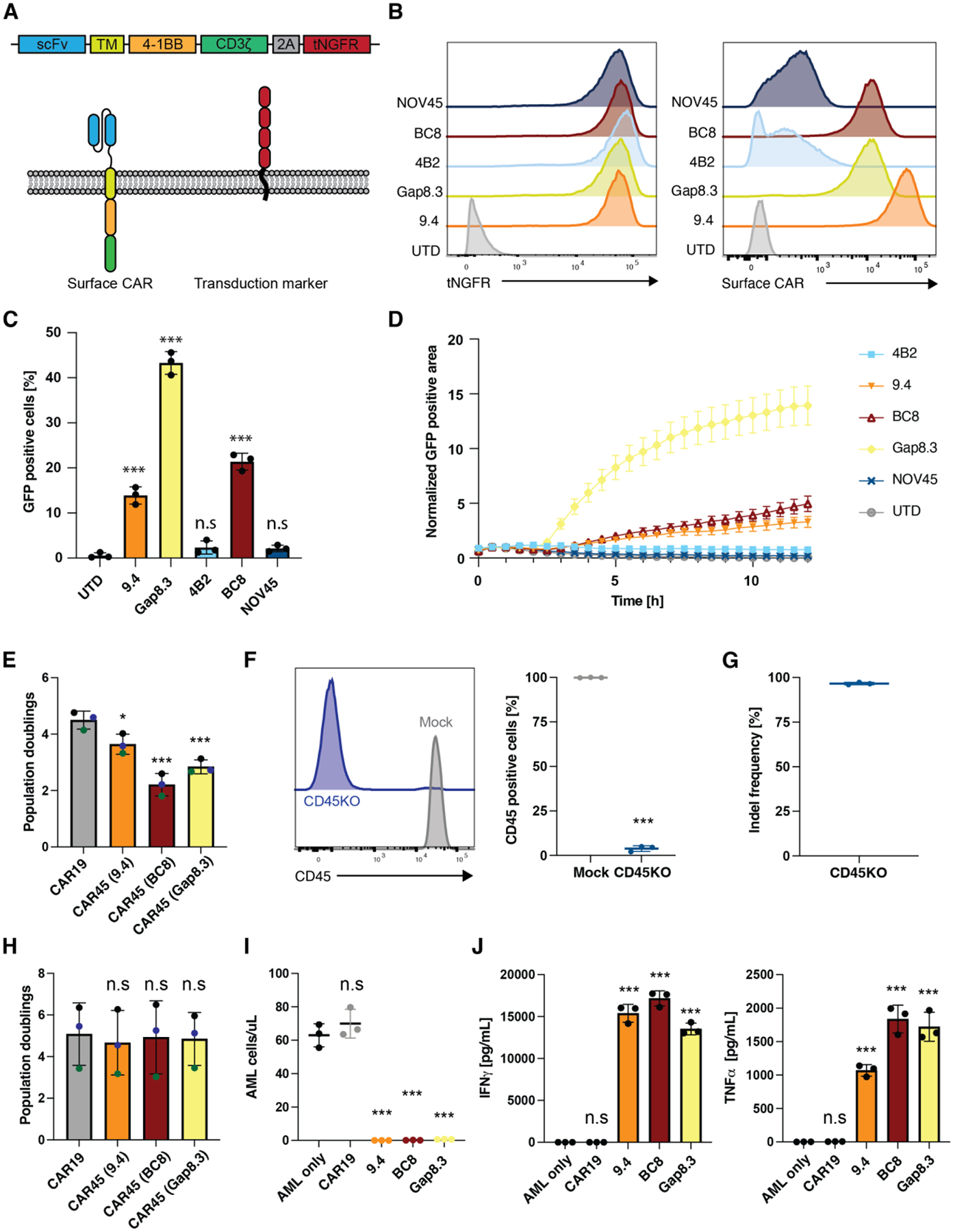
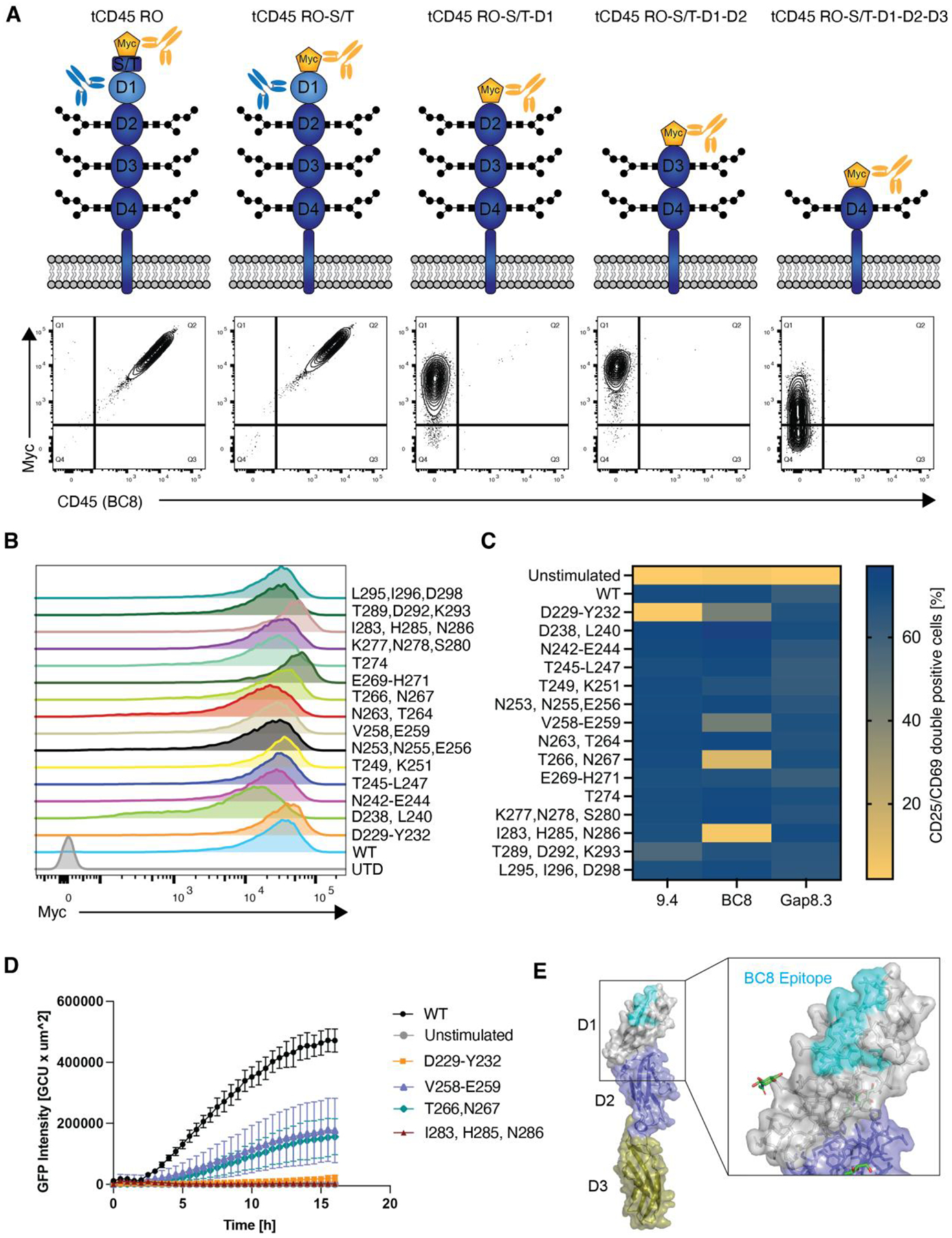
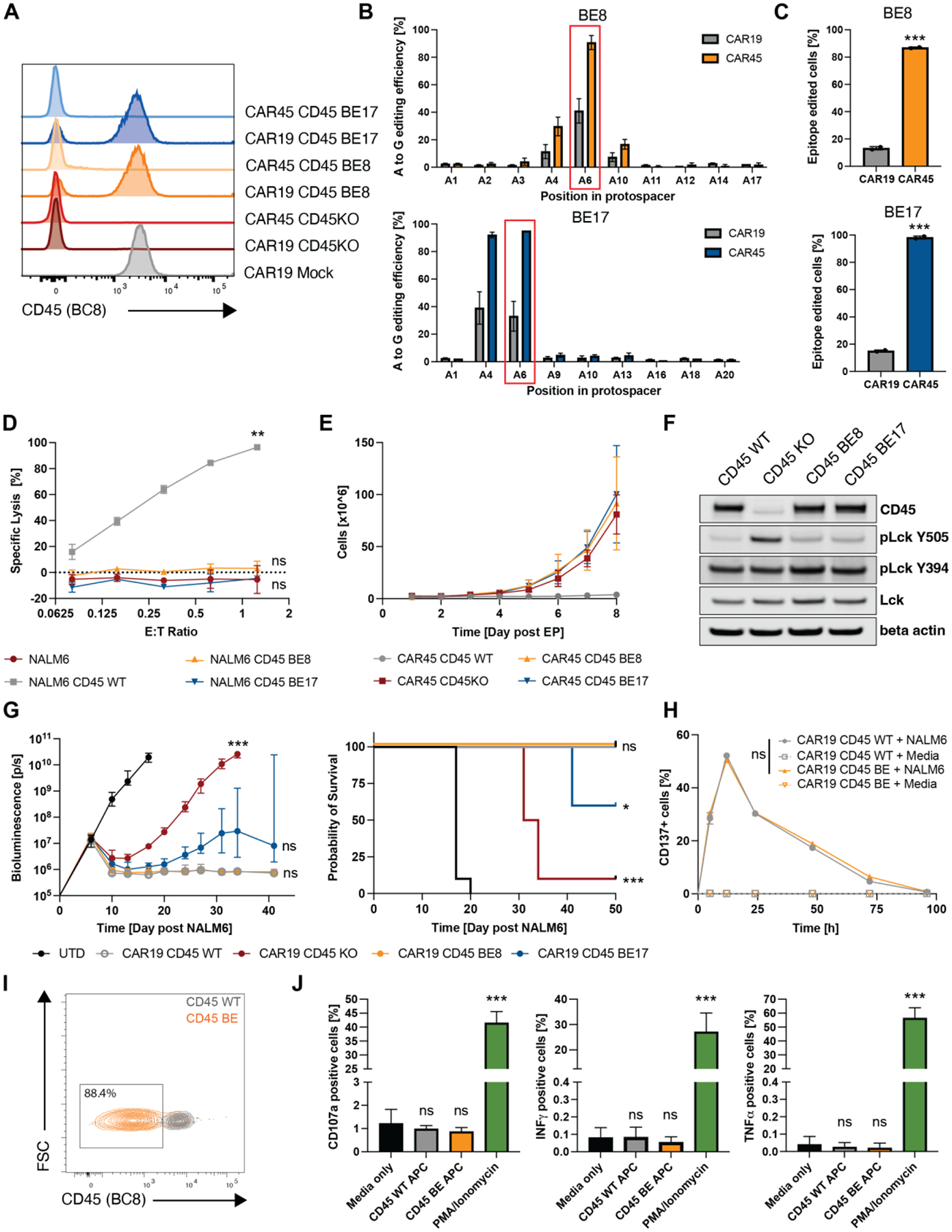
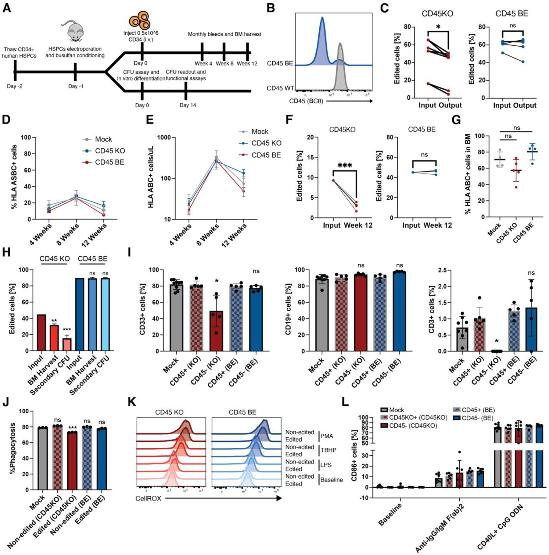
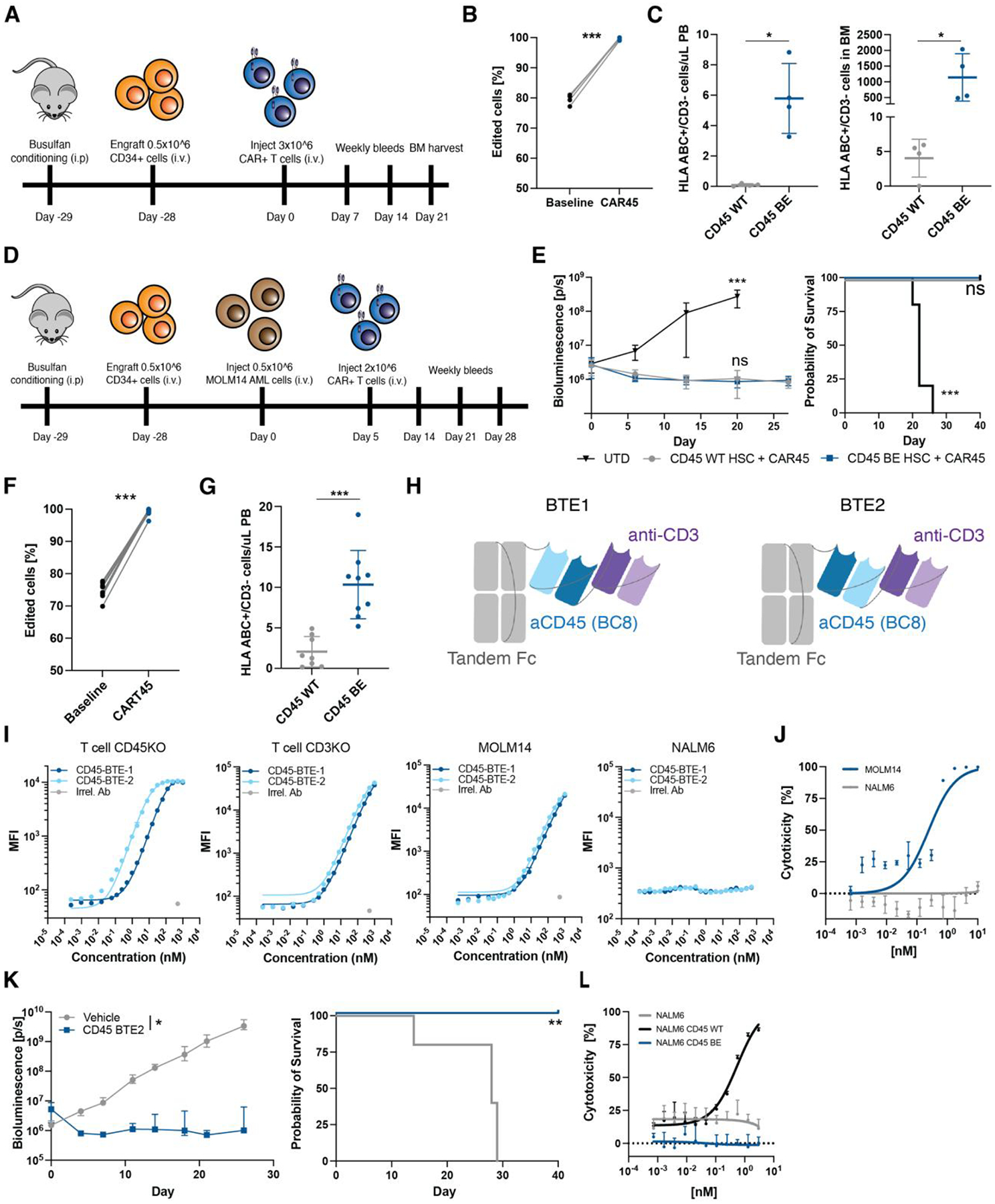
In the absence of cell surface cancer-specific antigens, immunotherapies such as chimeric antigen receptor (CAR) T cells, monoclonal antibodies, or bispecific T cell engagers typically target lineage antigens. Currently, such immunotherapies are individually designed and tested for each disease. This approach is inefficient and limited to a few lineage antigens for which the on-target/off-tumor toxicities are clinically tolerated. Here, we sought to develop a universal CAR T cell therapy for blood cancers directed against the pan-leukocyte marker CD45. To protect healthy hematopoietic cells, including CAR T cells, from CD45-directed on-target/off-tumor toxicity while preserving the essential functions of CD45, we mapped the epitope on CD45 that is targeted by the CAR and used CRISPR adenine base editing to install a function-preserving mutation sufficient to evade CAR T cell recognition. Epitope-edited CD45 CAR T cells were fratricide resistant and effective against patient-derived acute myeloid leukemia, B cell lymphoma, and acute T cell leukemia. Epitope-edited hematopoietic stem cells (HSCs) were protected from CAR T cells and, unlike CD45 knockout cells, could engraft, persist, and differentiate in vivo. Ex vivo epitope editing in HSCs and T cells enables the safe and effective use of CD45-directed CAR T cells and bispecific T cell engagers for the universal treatment of hematologic malignancies and might be exploited for other diseases requiring intensive hematopoietic ablation.


Conflict of interest statement

Authors (NW, KCG, CHJ, SIG) have filed an invention disclosure with the University of Pennsylvania and Stanford University based on this work.

C.H.J. has patents related to CAR therapy with royalties paid from Novartis to the University of Pennsylvania. C.H.J. is a scientific co-founder and holds equity in Capstan Therapeutics and Tmunity Therapeutics. C.H.J. serves on the board of AC Immune and is a scientific advisor to Alaunos, BluesphereBio, Cabaletta, Carisma, Cartography, Cellares, Cellcarta, Celldex, Danaher, Decheng, ImmuneSensor, Kite, Poseida, Verismo, Viracta, and WIRB-Copernicus group.

S.G. has patents related to CAR therapy with royalties paid from Novartis to the University of Pennsylvania. S.G. is a scientific co-founder and holds equity in Interius Biotherapeutics and Carisma Therapeutics. S.G. is a scientific advisor to Carisma, Cartography, Currus, Interius, Kite, NKILT, Mission Bio, and Vor Bio.

DW has received grant funding from industry for sponsored research collaborations, he has received speaking honoraria, and received fees for consulting or serving on scientific review committees. Remunerations received by DW include direct payments and equity/options. DW also discloses the following associations with commercial partners: Geneos consultant/advisory board, AstraZeneca advisory board, speaker, Inovio board of directors, consultant, Sanofi advisory board, BBI advisory board, Pfizer advisory Board, Flagship consultant, and Advaccine consultant.

R.M.Y. is an inventor on patents and/or patent applications licensed to Novartis Institutes of Biomedical Research and Tmunity Therapeutics and receives license revenue from such licenses.

Figures

Fig. 1.
Fig. 1.. CD45 is a universal blood cancer antigen that can be targeted with CD45 knockout CAR-T cells.
(A) Schematic overview of the tested CAR45 constructs. The heavy and light chains of different anti-CD45 antibody clones were cloned into a second-generation CAR containing a 4–1BB costimulatory domain and truncated NGFR (tNGFR) as a transduction marker. (B) CD45KO Jurkat reporter cells were transduced with CAR45 constructs and sorted for purity based on expression of tNGFR. Sorted cells were incubated with His-tagged recombinant CD45 followed by secondary staining with an anti-His antibody showing that CARs derived from clones 9.4, Gap8.3, and BC8 show highest expression of surface CAR. (C) CD45KO CAR45 Jurkat reporter cells were incubated with wild type Jurkat cells (CD45+) for 4hrs and NFAT-mediated GFP expression was measured by flow cytometry. CARs derived from antibody clones Gap8.3, BC8, and 9.4 show significant upregulation of GFP compared to untransduced reporter cells (n=3, One-way ANOVA compared to UTD, ***p<0.001). Data are represented as the mean ± SD. (D) CD45KO CAR45 Jurkat reporter cells were incubated with wild type Jurkat cells (CD45+) for 12hrs and NFAT-mediated GFP expression was measured by time lapse microscopy and the GFP positive area (normalized to t=0) was quantified. Data are represented as the mean ± SEM. (E) Population doublings of CD3/CD28 activated T cells transduced with either CAR19 or CAR45 after 13 days of ex vivo expansion. CAR45 transduced cells have significantly lower T population doublings due to fratricide (n=3 independent donors, one-ANOVA compared to CAR19, ***p<0.001; *p<0.05). Data are represented as the mean ± SD. (F) Human T cells were electroporated with SpCas9 protein and CD45 targeting gRNA (screening of CD45-specific gRNA not shown). Surface CD45 protein expression was assessed by flow cytometry (n=3 independent donors, unpaired t-test, ***p<0.001. Data are represented as the mean ± SD. (G) INDEL frequency in CD45KO T cells were quantified by TIDE following 9 days of in vitro culture. (H) T cells were electroporated with SpCas9 protein pre-complexed with CD45 gRNA followed by CD3/CD28 activation and transduction with either CAR19 or CAR45 and expansion for 13 days. CD45 deletion enables the expansion of CAR45 transduced cells similarly to CART19 cells (n=3 independent donors, one-ANOVA compared to CAR19, n.s=p>0.05). Data are represented as the mean ± SD. (I) CD45KO CART45 or CART19 cells were incubated with patient AML cells for 24hrs at a 1:4 E:T ratio and AML cells were quantified by flow cytometry. CART45 efficiently eliminates AML cells compared to CART19 (n=3 technical replicates, one way ANOVA compared to AML only, ***p<0.001; n.s=p>0.05). Data are represented as the mean ± SD. (J) CD45KO CART45 or CART19 control cells were incubated with patient AML cells for 24hrs at a 1:1 E:T ratio and cytokines in the supernatant were quantified by cytometric bead array (n=3 technical replicates, one way ANOVA compared to AML only, ***p<0.001; n.s=p>0.05). Data are represented as the mean ± SD.
Fig. 2.
Fig. 2.. Sequential epitope mapping identifies CAR45 epitopes on human CD45.
(A) Truncated human CD45 constructs that sequentially lack CD45’s extracellular subdomains were expressed in CD45 negative NALM6 cells. WT CD45RO was expressed as a positive control. Cells were stained with anti-myc to verify surface expression and with clone BC8 to determine CD45 binding. BC8 was mapped to the D1 domain of the CD45 ECD as the BC8 clone no longer bound to the CD45 constructs that lacked this domain. (B) Myc-tagged alanine mutants of the D1 domain were expressed in NALM6 cells. Alanine mutagenesis did not affect protein expression or transport to the cell surface. (C) NALM6 cells expressing either WT or alanine-mutated CD45 were co-cultured with CD45KO CART45 for 24h. Activation of CART45 cells was measured by surface expression of CD25 and CD69. Data are represented as the mean of n=2 technical replicates. (D) Jurkat NFAT-GFP reporter cells expressing the BC8-based CAR were co-cultured with NALM6 cells expressing CD45 alanine mutants and activation was measured by time lapse fluorescence microscopy (n=4 technical replicates). Data are represented as the mean ± SD. (E) Amino acid mutations that decreased BC8 CAR activation compared to CD45WT recognition were superimposed onto CD45 protein structure (PDB ID: 5FMV). BC8 recognizes a conformational epitope on the N-terminal portion of the CD45 D1 domain.
Fig. 3.
Fig. 3.. Epitope base editing CD45 enables CAR45 T cells expansion while preserving CD45 expression and CAR-T cell function.
(A) Edited human T cells were transduced with either CAR19 or CAR45 and stained for CD45 expression using the BC8 antibody clone 7 days post transduction. (B) Sequencing the targeted PTPRC loci shows that the edited cells are positively selected when transduced with CAR45 compared to CAR19 at the end of CAR-T cell expansion. (C) Cells with the intended edit are positively selected over time (7 days) when transduced with CAR45 compared to CAR19 (***p<0.001, unpaired t-test, n=2 independent donors). (D) Luciferase+ NALM6 expressing WT or base edited CD45 mutants were co-cultured with CART45 for 24hrs. CART45 was able to lyse cells expressing CD45WT but not CD45BE, suggesting that cells expressing base-edited CD45 are protected from CART45. (n=3 technical replicates, two-way ANOVA, **p<0.002; ns=p>0.05). Data are represented as mean ± SD. (E) Ex vivo expansion of unedited, CD45KO, and CD45BE CART45 cells demonstrates that epitope editing and CD45 knockout prevents CART45 fratricide (n=3 independent donors). Data are represented as mean ± SD. (F) Western blots of cell lysates from CD45WT, CD45KO and CD45BE T cells show that epitope-edited cells maintain CD45 expression and a higher frequency of dephosphorylated Lck Y505 (the substrate of CD45) compared to CD45KO cells. (G) NSG mice were injected with 0.5 × 10⁶ Luc+ NALM6 cells followed by injection of 3 × 10⁶ CAR19+ T cells. Tumor burden was measured by bioluminescent imaging and survival was monitored. Mice that were treated with CD45KO CART19 cells rapidly relapsed and succumbed to their tumor whereas mice that received CD45BE CART19 cells cleared their tumors comparable to unedited CART19 cells (n=10 per group BLI=ANOVA, survival = log-rank test, ***p<0.001; **p<0.0021; *p<0.033; n.s=p>0.12). (H) Unedited and base edited CART19 cells were briefly stimulated with NALM6 tumor cells at a 1:1 E:T ratio and expression of the activation marker CD137 was measured over 96hrs. (n=3 technical replicates, two-way ANOVA, ns=p>0.05). Data are represented as mean ± SD. (I) M-CSF stimulated monocytes were electroporated with ABE8e mRNA + BE8 sgRNA and cultured for 5–7 days prior to staining with anti-CD45 (clone BC8). Efficient epitope base editing (>85%) was observed by flow cytometry. (J) T cells were co-cultured with unedited, or epitope edited autologous monocytes for 5hrs to assess T cell reactivity against the mutant peptide arising from base editing. PMA/Ionomycin and media only conditions served as controls. T cell reactivity was measured by degranulation (CD107a+) and intracellular cytokine production. (n=2 independent donors of different HLA type, one way ANOVA compared to media only, ***p<0.001; ns=p>0.05). Data are represented as the mean ± SD.
Fig. 4.
Fig. 4.. CART45 shows potent activity against multiple hematologic cancer cell lines and primary AML xenografts.
(A) Luc+ MOLM14 AML cells were incubated with CART45 cells at multiple effector:target ratios for 24hrs. CART45 efficiently lysed MOLM14 cells whereas CART19 control cells did not (n=2 independent donors, unpaired t-test, **p<0.002). Data are represented as mean ± SD. (B) MOLM14 AML cells were incubated with CellTrace labeled CART45 cells at a 1:1 E:T ratio for 5 days. CART45 cells proliferated extensively compared to CART19 control cells as measured by dye dilution. (C) Schematic overview of experimental timeline for AML patient-derived xenograft model. (D) Number of AML cells/uL peripheral blood after CART45 treatment. CART45 cells rapidly clear the primary patient tumor cells in NSG mice compared to UTD control cells. Data are represented as mean ± SEM. (E) NSGS mice that were treated with CART45 cells show improved survival compared to UTD T cells (**p<0.002, log-rank test, n=4 per group). (F) Number of CD3+ T cells in the peripheral blood. CART45 cells undergo initial expansion followed by contraction after the tumor cleared. (G) Number of AML cells/uL peripheral blood after tumor rechallenge and initial CAR45 treatment (***p<0.001, unpaired t-test, n=7 from 2 independent donors). Data are represented as mean ± SEM. Days since initial CAR45 treatment adjusted to one donor with the second donor cohort offset by 22 days. (H) Blood samples from 22 patients with nine different hematologic malignancies were stained for CD45. CD45 is expressed on all patient cells except one multiple myeloma case. Grey histograms represent fluorescence intensity of unstained controls. (I) In vitro cytotoxicity assay demonstrates that CART45 can target multiple tumor cell lineages simultaneously, whereas CAR19 and CAR33 can only target B cell or myeloid cell leukemia/lymphoma cells respectively (n=3 technical replicates). Data are represented as mean ± SD. (J) T cells (CD3+/CD45WT+) engrafted from AML patient apheresis get eliminated and replaced by CD45BE CART45 cells. (K) 48hr co-culture of unedited CD34+ HSCs with autologous CART45 cells demonstrates susceptibility of normal HSCs to CART45 on-target/off-tumor toxicity.
Fig. 5.
Fig. 5.. Epitope edited hematopoietic system remains functional.
(A) Schematic overview. Human CD34+ HSCs were edited with gRNA and either ABE8e mRNA or Cas9 RNP’s. After recovery, CD34+ cells were injected into NSG mice to measure engraftment and differentiation in peripheral blood. A subset of edited CD34+ cells were plated in semi-solid methocult media for colony formation or differentiated into myeloid cells for functional assays. (B) Edited and control CD34+ HSCs were stained with BC8 antibody clone. (C) INDEL frequency in CD45KO colonies as quantified by TIDE(43) decreased after 14 days in methocult media whereas the frequency of base-edited alleles as measured by EditR remained stable (*p<0.05, paired t-test, n=6 or 7 from 4 independent donors). (D) Mice engrafted with CD45BE HSCs show similar frequency of human engrafted cells at early, mid, and late timepoints (ns=p>0.05; *p<0.05, two-way ANOVA, n=5–8). Data are represented as mean ± SD. (E) Mice engrafted with CD45BE HSCs show similar numbers of human engrafted cells at early, mid, and late timepoints (ns=p>0.05*p<0.05, two-way ANOVA, n=5–8). Data are represented as mean ± SD. (F) Longitudinal analysis of human engrafted cells shows a decrease in the frequency of CD45KO cells, while epitope edited cells show stable engraftment over 12 weeks (***p<0.001, paired t-test, n=5). (G) Mice engrafted with CD45BE HSCs show similar frequency of human engrafted in the bone marrow (ns=p>0.05, one way ANOVA, n=4–5). Data are represented as mean ± SD. (H) Mice engrafted with CD45KO HSCs show a decline in the frequency of edited cells in the BM and after secondary in vitro colony formation whereas the frequency of CD45BE edited cells remains unchanged compared to the injection input. (ns=p>0.05, **=p<0.01, ***=p<0.001, one way ANOVA compared to input, n=4–5). (I) Epitope edited HSCs show comparable myeloid, B cell, and T cell differentiation compared to unedited HSCs whereas CD45KO HSCs have a decreased frequency of myeloid cells and undetectable levels of T cells in the peripheral blood. Myeloid, B-cell, and T-cell differentiation was assessed at 4 weeks, 8 weeks, and 12 weeks respectively when peak differentiation into the corresponding lineage occurs in the NSG xenograft model. (ns=p>0.05; *=p<0.05, one way ANOVA, n=5–8). Data are represented as mean ± SD. (J) In vitro-differentiated CD45KO and CD45BE myeloid cells retain phagocytosis ability as measured by internalization of pHrodo deep red E. coli bioparticles. (ns=p>0.05; ***p<0.01, one way ANOVA, n=3 technical replicates). Data are represented as mean ± SD. (K) Levels of reactive oxygen species (ROS) production after lipopolysaccharide (LPS), tert-butyl hydroperoxide (TBHP), or phorbol myristate acetate (PMA) stimulation are similar among unedited and CD45 edited cells. ROS production was measured by fluorescence of CellROX deep red reagent. (L) In vivo differentiated B cells were harvested from peripheral blood of mice 10 weeks post-transplant and activated by either CD40L+CpG ODN or anti-IgG/IgM (Fab)2 for 48hrs. Activation as measured by surface level expression of CD86 did not differ between edited and unedited B cells and was significantly above baseline (n=5–8 mice per group). Data are represented as mean ± SD.
Fig. 6.
Fig. 6.. Epitope engineered hematopoietic system is shielded from CD45 targeted CAR-T cells and BTEs.
(A) Schematic overview of experimental timeline for in vivo protection of epitope edited HSC and progeny cells from CART45. (B) Frequency of edited cells in peripheral blood of NSG mice engrafted with CD45BE HSCs before and after treatment with CART45 cells (***p<0.001, paired t-test, n=4). (C) Number of human cells in peripheral blood and bone marrow of mice engrafted with CD45WT or CD45BE HSCs after CART45 injection (gated on mouse CD45-/human CD3-). n=4 mice per group, ***p<0.001, Mann-Whitney test. Data are represented as mean ± SD (D) Schematic overview of experimental timeline for in vivo protection of epitope edited HSC and progeny cells from CART45 in the presence of AML tumor cells. (E) Mice treated with CART45 clear AML cells resulting in extended survival. (n=9 for CART45 and n=4 for UTD, one-way ANOVA compared to CD45WT+CART45, ***p<0.001). Data are represented as mean ± SD. (F) Frequency of edited cells in peripheral blood of NSG mice engrafted with CD45BE HSCs before and after treatment with CART45 cells (***p<0.001, paired t-test, n=9). (G) Number of human cells in peripheral blood engrafted with CD45WT or CD45BE HSCs 3 weeks after CART45 injection (gated on mouse CD45-/human CD3-). n=9 mice per group, ***p<0.001, Mann-Whitney test. Data are represented as mean ± SD. (H) Schematic overview of BTE design. Anti-CD45 (clone BC8) and anti-CD3 (clone UCHT1) were fused to a silenced tandem Fc domain in LHHL (BTE1) and HLHL (BTE2) configurations. (I) Flow cytometry binding assay shows specific binding of BTE1 and BTE2 to CD3 and CD45 respectively. NALM6 cells (CD3 and CD45 negative) were used as a negative control. Irrelevant antibody (IgG1) was used as a staining control. (J) Dose-dependent killing of MOLM14 AML cells by T cells in the presence of BTEs. (K) Mice treated with anti-CD45 BTE clear MOLM14 AML cells resulting in extended survival. (left panel: *=p<0.05, two-way ANOVA, right panel: **=p<0.01, log-rank test, n=5). Data are represented as mean ± SD. (L) Dose-dependent killing of CD45WT expressing NALM6 cells but not CD45BE expressing NALM6 cells by T cells in the presence of BTEs suggests that CD45BE expressing cells are protected from BTE mediated cytotoxicity.

References

    1. Zammarchi F et al. , ADCT-402, a PBD dimer–containing antibody drug conjugate targeting CD19-expressing malignancies. Blood, The Journal of the American Society of Hematology 131, 1094–1105 (2018). - PubMed
    1. Kantarjian H et al. , Blinatumomab versus chemotherapy for advanced acute lymphoblastic leukemia. New England Journal of Medicine 376, 836–847 (2017). - PMC - PubMed
    1. Maude SL et al. , Chimeric antigen receptor T cells for sustained remissions in leukemia. New England Journal of Medicine 371, 1507–1517 (2014). - PMC - PubMed
    1. Kantarjian HM et al. , Inotuzumab ozogamicin versus standard therapy for acute lymphoblastic leukemia. New England Journal of Medicine 375, 740–753 (2016). - PMC - PubMed
    1. Falchi L, Vardhana SA, Salles GA, Bispecific antibodies for the treatment of B-cell lymphoma: promises, unknowns, and opportunities. Blood, The Journal of the American Society of Hematology 141, 467–480 (2023). - PMC - PubMed

Publication types