. 2023 Sep 13;15(36):42293-42303.
doi: 10.1021/acsami.3c09627. Epub 2023 Aug 31.

Goldilocks Energy Minimum: Peptide-Based Reversible Aggregation and Biosensing

Affiliations

Goldilocks Energy Minimum: Peptide-Based Reversible Aggregation and Biosensing

Wonjun Yim et al. ACS Appl Mater Interfaces. .

Erratum in

Abstract

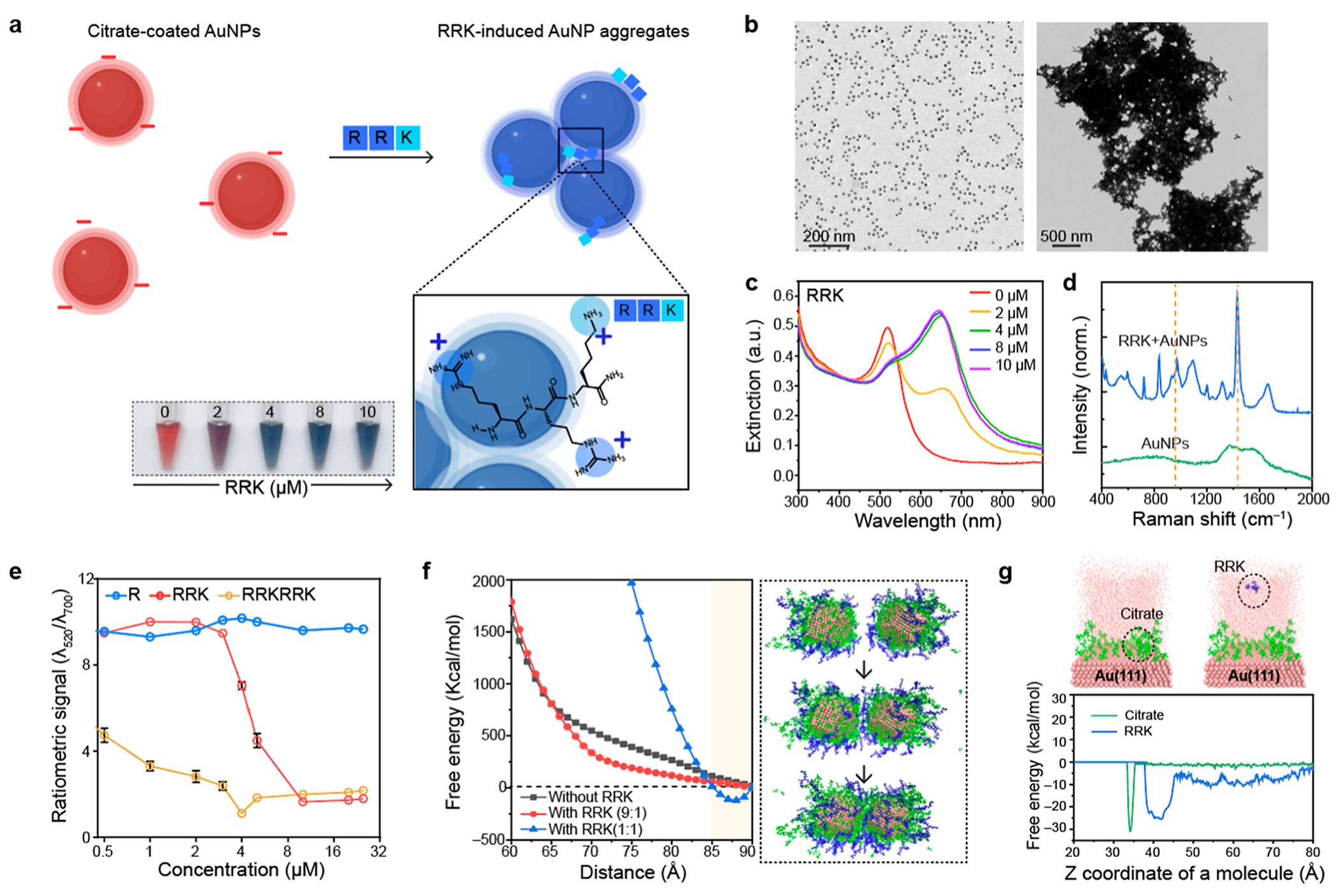
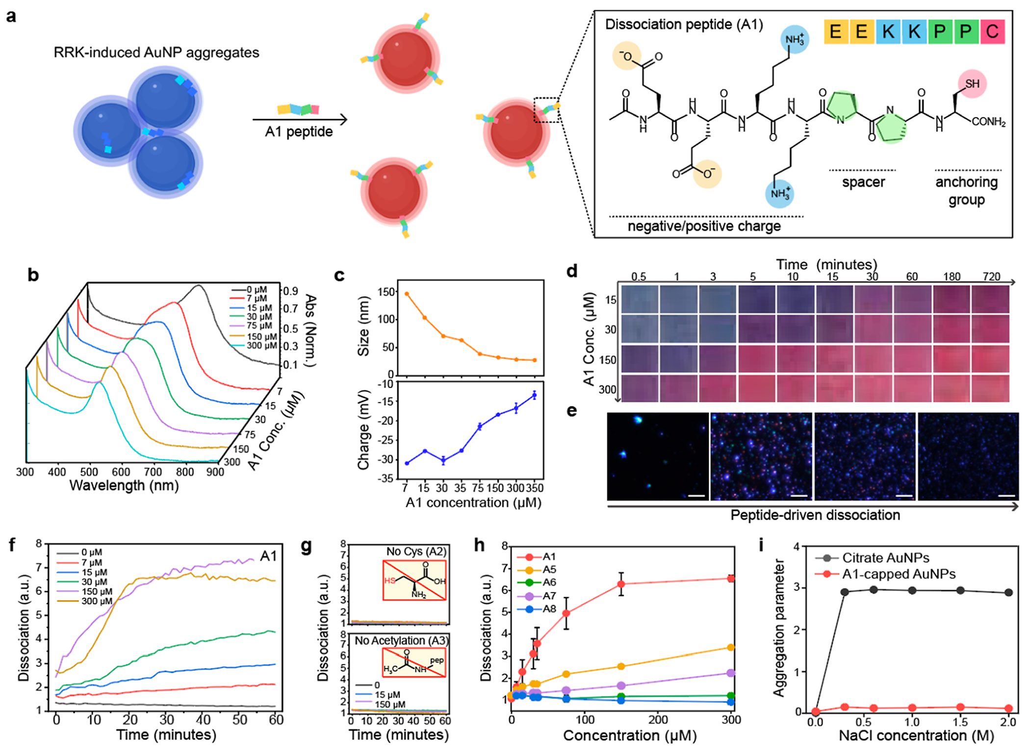
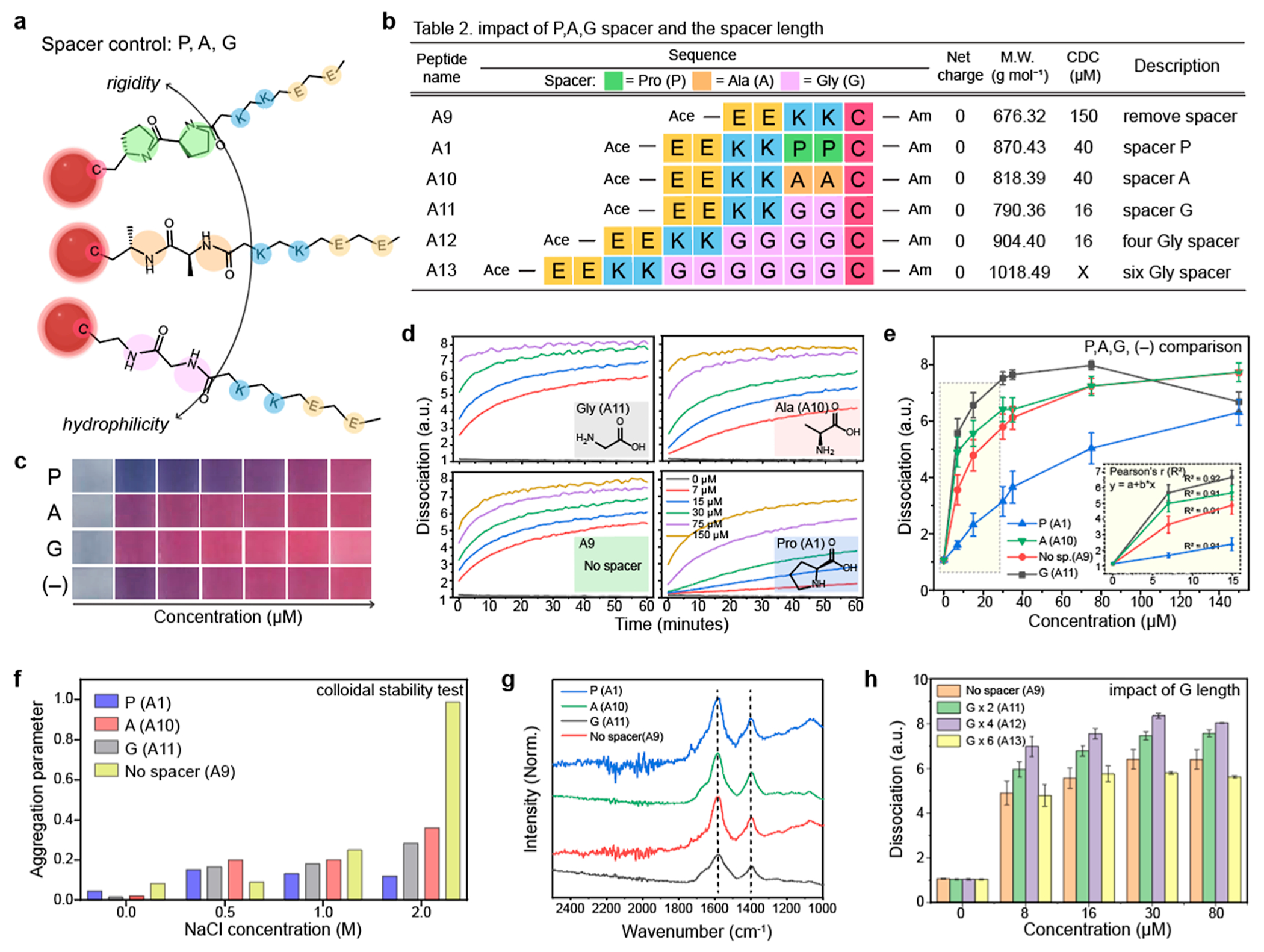
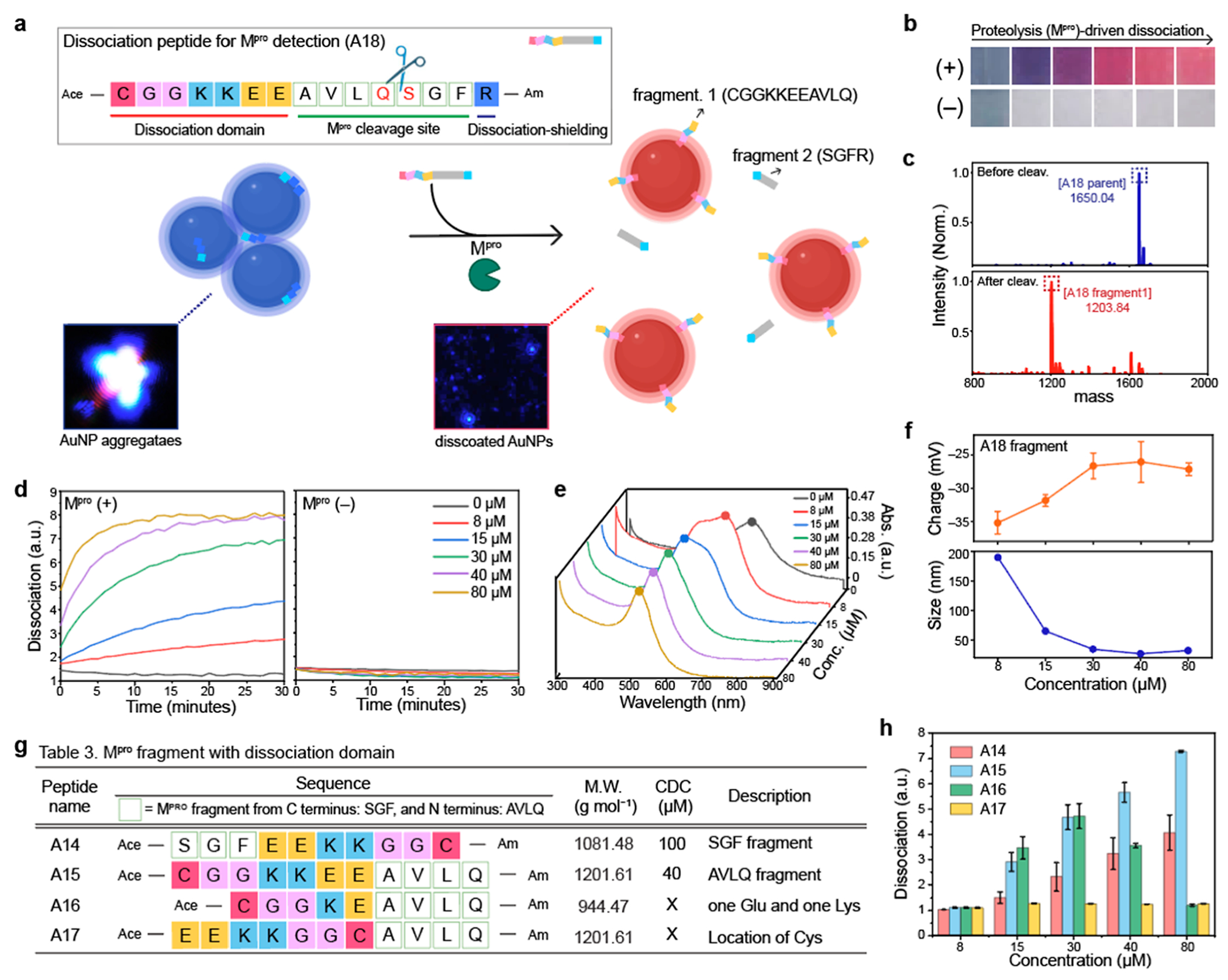
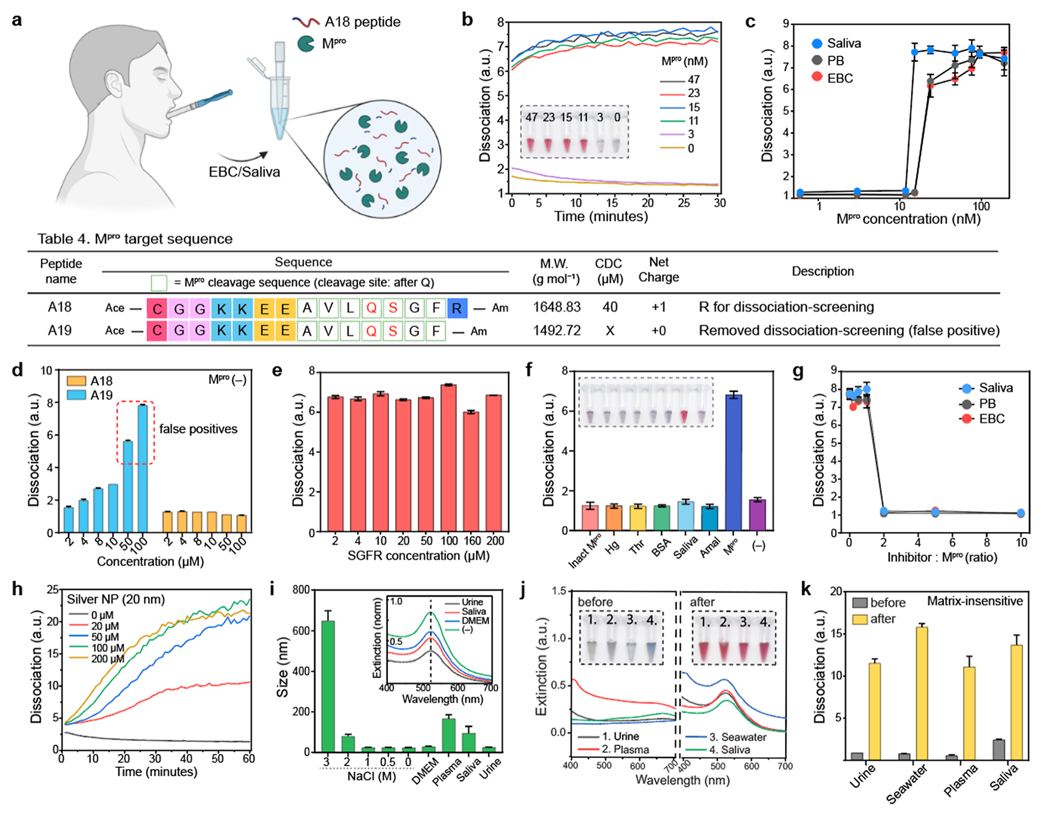
Colorimetric biosensors based on gold nanoparticle (AuNP) aggregation are often challenged by matrix interference in biofluids, poor specificity, and limited utility with clinical samples. Here, we propose a peptide-driven nanoscale disassembly approach, where AuNP aggregates induced by electrostatic attractions are dissociated in response to proteolytic cleavage. Initially, citrate-coated AuNPs were assembled via a short cationic peptide (RRK) and characterized by experiments and simulations. The dissociation peptides were then used to reversibly dissociate the AuNP aggregates as a function of target protease detection, i.e., main protease (Mpro), a biomarker for severe acute respiratory syndrome coronavirus 2. The dissociation propensity depends on peptide length, hydrophilicity, charge, and ligand architecture. Finally, our dissociation strategy provides a rapid and distinct optical signal through Mpro cleavage with a detection limit of 12.3 nM in saliva. Our dissociation peptide effectively dissociates plasmonic assemblies in diverse matrices including 100% human saliva, urine, plasma, and seawater, as well as other types of plasmonic nanoparticles such as silver. Our peptide-enabled dissociation platform provides a simple, matrix-insensitive, and versatile method for protease sensing.

Keywords: DLVO theory; SARS-CoV-19; colorimetric biosensor; dissociation peptide; matrix-insensitive; reversible aggregation.


Conflict of interest statement

The authors declare no competing financial interest.

Figures

Figure 1.
Figure 1.
Short cationic peptides for reversible aggregation. (a) Schematic illustration of RRK-based particle aggregation. AuNPs were aggregated by electrostatic attractions between negatively charged citrate on AuNPs and positively charged RRK peptides. The inset photograph shows color change from red to blue as a function of the RRK peptide (2–10 μM). (b) TEM images of citrate-coated AuNPs (left) and RRK-induced AuNP aggregates (right). (c) UV–vis spectrum of RRK-induced AuNP aggregates. The plasmonic resonance peak of AuNPs was redshifted due to the plasmonic coupling. (d) Raman shifts before and after adding RRK peptides into citrate-coated AuNPs. The Raman peak at 1443 cm−1 was attributed to the C–N stretching in the Arg residue. (e) Ratiometric signal (λ520/λ700) of AuNPs after adding R, RRK, and RRKRRK peptides with different concentrations 0.5–32 μM, respectively. The error bars represent the standard deviation of three independent samples. (f) SMD simulations for free energy investigation as a function of AuNP distance at 298 K and 1 atm. Energy minimum point was observed after adding RRK peptides. Black, red, and blue lines indicate citrate to RRK molar ratios of 1:0, 9:1, and 1:1, respectively. The inset images indicate the simulation stages along a trajectory of the citrate-coated AuNPs with the RRK (9:1) system. (g) MTD free energy investigation for a system with 1 RRK on the Au(111) surface (right) and a system with 1 citrate on the Au(111) surface (left). The Z coordinate value was calculated based on the center mass of RRK and citrate molecule as shown in the inset images. The upper surface of the Au(111) slab was located at 31 Å. The MTD results observed no surface ligand exchange on Au(111) during the electrostatic interactions between RRK and citrate molecules.
Figure 2.
Figure 2.
Peptide-enabled dissociation of AuNP aggregates. (a) Schematic illustration of peptide-based particle dissociations. AuNP aggregates induced by RRK peptides were reversibly dissociated by the A1 peptides. The structural component of the A1 peptide contains charge, spacer, and anchoring group. (b) UV–vis spectrum shows that the plasmonic resonance peak of AuNP aggregates blueshifted upon addition of the A1 peptide (7–300 μM). (c) Hydrodynamic diameter and the surface charge after adding the A1 peptide. (d) Time-dependent photographs show 150 μM of the A1 peptide required to dissociate AuNP aggregates. x and y axis indicate time and the A1 concentration, respectively. (e) Darkfield images of AuNP aggregates (left) and the dissociated AuNPs (right). The scale bar indicates 10 μm. Blue dots represent actual AuNPs dissociated by the A1 peptide. (f) Time-dependent particle dissociation driven by the A1 peptide. The ratiometric signal (λ520/λ700) was referred to as dissociation (y axis). (g) Particle dissociation was quenched without Cys (A2) and acetylation (A3). (h) Dissociation capacity of the A1, A5, A6, A7, and A8 peptides. (i) Dissociated AuNPs by the A1 (red) showed higher colloidal stability than citrate-coated AuNPs (black). Panel (c,h,i) repeated three independent times and showed similar results.
Figure 3.
Figure 3.
Impact of hydrophilicity and steric bulk on particle dissociation. (a) Different Pro-, Ala-, and Gly-spacers have different nature of rigidity and hydrophilicity which can impact on the dissociation capacity. Table 2 in (b) describes peptide sequences that are designed to investigate the impact of spacers. (c) Photographs of the dissociated AuNPs by the PP, AA, and GG spacers and without spacer (−) as a negative control. (d) Time-dependent particle dissociations driven by the A1, A9, A10, and A11 peptides, respectively. (e) Gly spacer showed a higher dissociation capacity than the Pro- and Ala- spacers. (f) Aggregation parameter of the dissociated AuNPs driven by the A1, A9, A10, and A11 peptides. The results showed that the peptide with spacer can provide higher colloidal stability for AuNPs than the peptide without spacer. (g) FTIR data of the dissociated AuNPs by the A1, A9, A10, and A11 peptides. The peaks at 1400 and 1600 cm−1 were attributed to the carboxyl group in the Glu amino acid. (h) Impact of the spacer length on the particle dissociation. Increasing the length of the spacer (from two to four) improved dissociation capacity, while the spacer with six Glu (i.e., A13) showed lower dissociation capacity than the A12 peptide. The panel (e,f,h) repeated three independent times and showed similar results.
Figure 4.
Figure 4.
Mpro detection using dissociation strategy. (a) Schematic illustrates that Mpro cleavage releases dissociation domains, changing the color from blue to red. Our dissociation peptide (i.e., A18) consists of three parts: dissociation domain (CGGKKEE), cleavage site (AVLQ↓.SGF), and dissociation shielding site (R). The inset images are before and after particle dissociation obtained by darkfield microscopy. Blue dots indicate actual AuNP aggregates (left) and the dissociated AuNPs (right). (b) Color changes with (+) and without (−) Mpro in PB buffer. The released A18 fragment (CGGKKEEAVLQ) dissociated AuNP aggregates, changing the color from blue to red. (c) MALDI-TOF MS data before and after Mpro cleavage, confirming the mass peaks of the A18 parent and its fragment. (d) Time-dependent particle dissociation by the A18 fragments. The results showed that at least 40 μM of the A18 fragment was required for particle dissociations. (e) UV–vis spectrum before and after particle dissociation by the A18 fragments with different concentrations (8–80 μM). (f) Changes in the size and surface charge after the dissociation induced by Mpro cleavage. Table 3 in (g) describes peptide sequences that are designed to confirm the best location and order of the dissociation domain for Mpro detection. (h) Particle dissociations driven by the A14, A15, A16, and A17 peptides. The results show that the dissociation domain located at C-terminus showed the highest dissociation capacity. In addition, the thiol group at the tail showed higher dissociation affinity than the thiol group in the middle. The panel (f,h) repeated three independent times and showed similar results.
Figure 5.
Figure 5.
Matrix-insensitive Mpro detection (a) Schematic illustration of Mpro detection in EBC or saliva. The released A18 fragment by Mpro cleavage was used for colorimetric biosensing in saliva or EBC. (b) Time-dependent Mpro detection from 0 to 47 nM in saliva. The inset photograph shows that our dissociation strategy can provide a clear readout of the positive Mpro sample above 11 nM in saliva. (c) Detection limits of Mpro in saliva, EBC, and PB buffer, respectively. Table 4 describes peptide sequences that are designed to verify the role of the dissociation screening domain. (d) One Arg at the C-terminus can prevent false positives. False positives occurred in A19 when the peptide concentration was over 50 μM, while A18 showed no false positive in the absence of Mpro. (e) A18 fragment from C terminus (i.e., SGFR) had negligible impact on the dissociation process. (f) Specificity test using different biological proteins [e.g., inactivated Mpro (inact Mpro), hemoglobin (Hg), thrombin (Thr), BSA, saliva, and amalyase (Amal)]. (g) GC376 inhibitor assay test in saliva, EBC, and PB buffer, respectively. (h) The released A18 fragments by Mpro cleavage can dissociate other types of plasmonic assemblies such as AgNP aggregates. (i) After particle dissociation, the A18-capping AuNPs maintained high colloidal stability in different biological media (e.g., urine, saliva, plasma, DMEM) and extreme conditions (e.g., 2 M NaCl). (j) Plasmonic resonance peaks of the AuNP aggregates blueshifted after particle dissociation in 100% of (1) human urine, (2) plasma, (3) seawater, and (4) saliva. The inset photographs show before (left) and after (right) adding dissociation peptides. (k) Ratiometric signal (λ520/λ700) of the dissociated AuNPs in diverse matrixes, indicating that our dissociation strategy is less affected by the sample matrix. The panel (c–g,i–k) repeated three independent times and showed similar results.

References

    1. Liu J; Lu Y A Colorimetric Lead Biosensor Using DNAzyme-Directed Assembly of Gold Nanoparticles. J. Am. Chem. Soc 2003, 125, 6642–6643. - PubMed
    1. Jin Z; Li Y; Li K; Zhou J; Yeung J; Ling C; Yim W; He T; Cheng Y; Xu M; et al. Peptide Amphiphile Mediated Co-assembly for Nanoplasmonic Sensing. Angew. Chem., Int. Ed 2023, 62, No. e202214394. - PMC - PubMed
    1. Guo L; Xu Y; Ferhan AR; Chen G; Kim D-H Oriented Gold Nanoparticle Aggregation for Colorimetric Sensors with Surprisingly High Analytical Figures of Merit. J. Am. Chem. Soc. 2013, 135, 12338–12345. - PubMed
    1. Liu J; Lu Y Accelerated Color Change of Gold Nanoparticles Assembled by DNAzymes for Simple and Fast Colorimetric Pb2+ Detection. J. Am. Chem. Soc 2004, 126, 12298–12305. - PubMed
    1. Ghosh SK; Pal T Interparticle Coupling Effect on the Surface Plasmon Resonance of Gold Nanoparticles: from Theory to Applications. Chem. Rev 2007, 107, 4797–4862. - PubMed