Review
. 2023 Oct:130:103563.
doi: 10.1016/j.dnarep.2023.103563. Epub 2023 Aug 19.

Finding significance: New perspectives in variant classification of the RAD51 regulators, BRCA2 and beyond

Affiliations
Review

Finding significance: New perspectives in variant classification of the RAD51 regulators, BRCA2 and beyond

Hayley L Rein et al. DNA Repair (Amst). 2023 Oct.

Abstract

For many individuals harboring a variant of uncertain functional significance (VUS) in a homologous recombination (HR) gene, their risk of developing breast and ovarian cancer is unknown. Integral to the process of HR are BRCA1 and regulators of the central HR protein, RAD51, including BRCA2, PALB2, RAD51C and RAD51D. Due to advancements in sequencing technology and the continued expansion of cancer screening panels, the number of VUS identified in these genes has risen significantly. Standard practices for variant classification utilize different types of predictive, population, phenotypic, allelic and functional evidence. While variant analysis is improving, there remains a struggle to keep up with demand. Understanding the effects of an HR variant can aid in preventative care and is critical for developing an effective cancer treatment plan. In this review, we discuss current perspectives in the classification of variants in the breast and ovarian cancer genes BRCA1, BRCA2, PALB2, RAD51C and RAD51D.

Keywords: BRCA1; BRCA2; Breast cancer; Homologous recombination; Homologous recombination deficient tumors; Ovarian cancer; PALB2; RAD51; RAD51C; RAD51D; Variant of unknown/uncertain significance.


Conflict of interest statement

Declaration of Competing Interest The authors declare that they have no known conflicts of interest that would influence the contents of this manuscript.

Figures
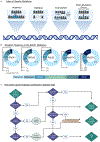
Fig. 1.
Fig. 1.. Types of genetic alterations in the RAD51 regulators and a classification schematic
(A) Simplified types of genetic alterations including insertions, deletions, frameshifts and point mutations. The inserted or mutated residues are in blue and deleted residues in light grey. (B) Mutation distribution represented as a pie chart in the HR genes; BRCA1, BRCA2, PALB2, RAD51C, RAD51D reported in COSMIC [69]. Missense mutations (sky blue), nonsense mutations (grey/blue), silent point mutations (medium blue) along with in frame insertion/deletion mutations (light green) and frameshift mutations (dark green). Mutations within the “other” category (navy) include promoter mutations, mutations in intronic regions and combinations of mutations. Percentages indicated are the number of samples where the mutation type is present divided by total number of samples analyzed. In some cases, the total is not 100% due to the addition of multiple uncategorized mutations and multiple mutations found in a single sample. (C) The flow chart determines disease risk contribution of five types of point mutations; silent, nonsense, missense benign, missense pathogenic and missense VUS. The prior four mutations have a known contribution to disease risk, while it is unknown how missense VUS contribute to disease risk. Created with BioRender.com
Fig. 2.
Fig. 2.. The roles of the RAD51 regulators during HR and replication
(A1-5) The steps of homologous recombination are shown from the perspective of BRCA1 and the RAD51 mediator proteins BRCA2, PALB2, RAD51C and RAD51D. Additional proteins required for each step are listed on the left of the arrows. 1. After a DSB occurs, the DNA ends are resected through the activities of MRN complex, CtIP, DNA2, WRN, BLM, EXO1 with BRCA1 (blue oval) and BARD1 (purple oval). The ssDNA is coated with the ssDNA binding complex RPA, which is a heterotrimer (beige ovals) 2. BRCA2 (blue oval) with BRCA1 and PALB2 (light teal oval) bring RAD51 to ssDNA and enable RAD51 filament nucleation (green circles). 3. RAD51 presynaptic filament formation is promoted by the CX3 complex (RAD51C (navy oval) and XRCC3 (light green oval) and the BCDX2 complex (RAD51B (blue oval); RAD51C (navy oval); RAD51D (green oval); XRCC2 (dark green oval). Additional factors include RAD54, RAD54B etc. 4. RAD51 filaments perform the homology search and strand invasion steps of a homologous sequence (light blue DNA; a sister chromatid or a homologous chromosome) with the assistance of RAD54, BRCA1-BARD1, among others. 5. The catentated DNA Holliday junctions are either resolved or dissoluted by many additional factors including helicases and topoisomerases (not shown). (B) The effects of functional and non-functional RAD51 mediator proteins during replication. When DNA polymerase (purple) is halted by a fork blocking lesion (green starburst), the RAD51 regulators are required for fork protection and replication restart (right side). RAD51 binding to the ssDNA gap protects the fork from nucleolytic attack and enables replication fork reversal that promotes replication fork restart. Dysfunction of the RAD51 mediator proteins results in nucleolytic degradation and replication fork collapse. Created with BioRender.com
Fig. 3.
Fig. 3.. Variant classification pyramid for benign and pathogenicity and evidence combination for classification key
Adapted from Richards et al. [63] (A) Evidence for variant classification is organized into three benign groups (left side of triangle) and four pathogenic groups (right side of triangle), with evidence decreasing in strength down to the bottom of the pyramid. Each evidence grouping is given a specific label (i.e. BA, BS, BP, PVS, PS, PM and PP) and each type of evidence in the grouping is given a numbered label as well (1-7). PP1 evidence classification is shown as supporting but this base classification can increase to moderate or strong with increasing segregation data (white arrow). The described variant classification pyramid is based off of the general ACMG guidelines. Variant classification pyramids should be edited to fit VCEP criteria for specific genes. (B) Evidence combinations needed for variant classification as likely benign, benign, likely pathogenic and pathogenic classification are listed. Each colored box represents a piece of evidence from the specified category in (A). The number of times a specific box is listed represents different pieces of evidence required under the same category. For example, there are two ways for a variant to be classified as likely benign; 1) one piece of evidence from BS1-3 and one from BP1-7 (i.e. BS3 WT functional results and BP4 computational WT prediction) or two pieces of evidence from BP1-7 (i.e. BP4 computational WT prediction and BP5 found in patients with alternative cause). Created with BioRender.com
Fig. 4.
Fig. 4.. Functional assays for variant classification.
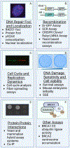
Functional Assays can be utilized to examine how variants change the activities of the RAD51 mediator proteins. These activities include (A) formation of DNA repair foci (including RAD51 and the protein of interest), colocalization with gamma-H2AX (γH2AX; shown with DAPI colocalization) and nuclear localization), (B) recombination capabilities, which can be measured using the DR-GFP assay (shown), SCR assay, CRISPR Clover/Ruby-LMNA Assay, and yeast-based recombination assays, (C) perturbed cell cycle progression and replication which can be measured by cell cycle analysis and fiber spreading assays (shown), (D) contribution to cell viability and sensitivity to DNA damaging agents, measured by survival assays (plates shown), early mouse development studies, and SGE, (E) protein-protein interaction capabilities as measured by yeast and mammalian hybrid assay systems (yeast shown) and co-immunoprecipitation (co-IP), in vitro pull downs (F) and other non-recombination or replication-based functions like the E3 ubiquitin ligase activity of BRCA1, examined by western blotting and centrosome accumulation in the absence of functional BRCA2, visualized by microscopy. Created with BioRender.com

Similar articles

Cited by

References

    1. Varol U, Kucukzeybek Y, Alacacioglu A, Somali I, Altun Z, Aktas S, Oktay Tarhan M, BRCA genes: BRCA 1 and BRCA 2, J BUON, 23 (2018) 862–866. - PubMed
    1. Miki Y, Swensen J, Shattuck-Eidens D, Futreal PA, Harshman K, Tavtigian S, Liu Q, Cochran C, Bennett LM, Ding W, et al., A strong candidate for the breast and ovarian cancer susceptibility gene BRCA1, Science, 266 (1994) 66–71. - PubMed
    1. Hall JM, Lee MK, Newman B, Morrow JE, Anderson LA, Huey B, King MC, Linkage of early-onset familial breast cancer to chromosome 17q21, Science, 250 (1990) 1684–1689. - PubMed
    1. Wooster R, Neuhausen SL, Mangion J, Quirk Y, Ford D, Collins N, Nguyen K, Seal S, Tran T, Averill D, et al., Localization of a breast cancer susceptibility gene, BRCA2, to chromosome 13q12-13, Science, 265 (1994) 2088–2090. - PubMed
    1. Fanale D, Pivetti A, Cancelliere D, Spera A, Bono M, Fiorino A, Pedone E, Barraco N, Brando C, Perez A, Guarneri MF, Russo TDB, Vieni S, Guarneri G, Russo A, Bazan V, BRCA1/2 variants of unknown significance in hereditary breast and ovarian cancer (HBOC) syndrome: Looking for the hidden meaning, Crit Rev Oncol Hematol, 172 (2022) 103626. - PubMed

Publication types