. 2023 Oct 4;31(10):2861-2871.
doi: 10.1016/j.ymthe.2023.08.010. Epub 2023 Aug 30.

Inhalable Nanofitin demonstrates high neutralization of SARS-CoV-2 virus via direct application in respiratory tract

Affiliations

Inhalable Nanofitin demonstrates high neutralization of SARS-CoV-2 virus via direct application in respiratory tract

Sébastien Viollet et al. Mol Ther. .

Abstract

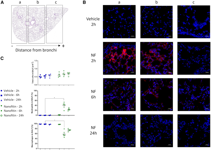
Nanofitins are small and hyperthermostable alternative protein scaffolds that display physicochemical properties making them suitable for the development of topical therapeutics, notably for the treatment of pulmonary infectious diseases. Local administration of biologics to the lungs involves a particularly stressful step of nebulization that is poorly tolerated by most antibodies, which limits their application by this delivery route. During the COVID-19 pandemic, we generated anti-SARS-CoV-2 monomeric Nanofitins of high specificity for the spike protein. Hit Nanofitin candidates were identified based on their binding properties with punctual spike mutants and assembled into a linear multimeric construction constituting of four different Nanofitins, allowing the generation of a highly potent anti-SARS-CoV-2 molecule. The therapeutic efficacy of the multimeric assembly was demonstrated both in in vitro and in vivo models. Interestingly, the neutralization mechanism of the multimeric construction seems to involve a particular conformation switch of the spike trimer. In addition, we reported the stability and the conserved activity of the tetrameric construction after nebulization. This advantageous developability feature for pulmonary administration associated with the ease of assembly, as well as the fast generation process position the Nanofitin technology as a potential therapeutic solution for emerging infectious diseases.

Keywords: Nanofitin; antibody mimetic; infectious disease; inhalable; nebulization; protein scaffold; pulmonary delivery.


Conflict of interest statement

Declaration of interests The Nanofitin technology described in this study, commercialized by Affilogic, uses the patent application owned by Institut Pasteur and Center National de la Recherche Scientifique (CNRS): “OB-fold used as scaffold for engineering new specific binders”; PCT/IB2007/004388. Affilogic SAS, Nantes, France, provided support for the study and participated in study design, conducted the study, and provided data collection, management and interpretation. S.V., E.E, J.P., L.N., M.C., S.H., are employees of Affilogic SAS. O.K. is the CEO and the owner of Affilogic SAS.

Figures

None
Graphical abstract
Figure 1
Figure 1
Selection and characterization of anti-spike Nanofitins (A) Schematic representation of ribosome display selection for Nanofitin discovery. Nanofitin DNA libraries are designed and engaged into ribosome display cycle starting by their transcription in RNA and their translation to generate an RNA-ribosome-Nanofitin complex. Nanofitin complexes are selected against their target of interest and washed before their destabilization during the elution. Then, RNA is reverse transcribed and re-engaged for another round of selection or inserted into a final vector for screening and characterization. (B) Screening of Nanofitins in crude lysate after 4 rounds of ribosome display selection. (C) Evaluation of binding capacities to spike S1 and RBD. (D) Dose-response curves generated by ELISA for EC50 (left) and IC50 (right) determination. Inhibition was performed by measuring the interaction of spike protein to the human ACE2. Data are the average ± SEM of two or three experiments.
Figure 2
Figure 2
Evaluation of tetrameric Nanofitin (A) BLI binding analyses of monomeric and tetrameric Nanofitins to immobilized spike. (B) Binding kinetic parameters of Nanofitins on SARS-CoV-2 spike S1. (C) Neutralization of pseudotyped virus expressing D614G spike protein revealed by Luciferase assay in ACE2/TMPRSS2 expressing HEK293 cells. Data are the average ± SEM of two experiments containing three replicates each.
Figure 3
Figure 3
Conformational switch of trimeric spike by Nanofitins (A) Two-dimensional structures of trimeric spike/Nanofitin mix obtained by negative staining. Spike protein was mixed with NF1, NF3, and tetramer (at ratios of 1:78, 1:85, and 1:78, respectively). Scale bar, 10 nm. (B) Distribution of trimeric spike conformations mixed with different Nanofitins. Analysis on spike trimers was based on the counting of at least 2,000 cleaned particles for each condition. (C) Proposed model for the clamp conformation on the third RBD down. NF4 (in blue) and NF3 (in orange) maintain the third RBD (in black) in a down position because of their fusion by the protein linker of 30 amino acids (in red). We also add in the representation another NF (NF2 in yellow) interacting to an RBD (in gray) in an up position.
Figure 4
Figure 4
Stability of nebulized tetrameric Nanofitin (A) Aggregates as measured by DLS and FCM in the reference and nebulized samples containing tetrameric Nanofitin at 1 or 8 mg/mL. (B) SEC-UPLC analysis of nebulized Nanofitin compared with the reference sample at 1 mg/mL (top) or 8 mg/mL (bottom). (C) Activity comparison using ELISA assay on spike protein between nebulized samples and reference sample at 1 mg/mL (Left) or 8 mg/mL (right). Data of nebulized samples are the average ± SEM of three replicates.
Figure 5
Figure 5
Distribution of tetrameric Nanofitin in lung (A) Cross-section of mouse lung divided in different areas (a, b, c) depending on their distance from bronchi. (B) Detection of tetrameric Nanofitin (in red) at different timepoints and lung localizations. Scale bar, 20 µm. (C) Variation of total lymphocyte cells population (top), neutrophil population (middle), and macrophage (bottom) in bronchoalveolar lavage fluid (BALF) at 2, 6, and 24 h after Nanofitin administration. Statistical analysis based on one-way ANOVA test with p < 0.01 between the vehicle 6 h and Nanofitin 6 h groups (∗).
Figure 6
Figure 6
Intranasal administration of Nanofitin inhibits SARS-CoV-2 virus in animal models (A) Variation of viral load and TCID50 in mice infected by SARS-CoV-2 virus treated by Nanofitin at 10 mg/kg. Five mice were included in each group. (∗) Statistical analysis was based on one-way ANOVA with p < 0.01 between infected mice treated by vehicle and non-relevant NF or tetrameric NF. ns, not significant. (B) Evolution of body weight in infected mice compared with the D0. (∗∗) Significance calculated by one-way ANOVA between the different mice groups at D5 p < 0.01. ns, not significant. (C) Lung histological pictures reflecting cell infiltration in the different setups. Black arrows indicate the cell infiltrations around blood vessels and bronchi. Scale bar, 500 μm. (D) Variation of viral load and TCID50 in mice infected by SARS-CoV-2 treated at 0.1, 1, or 10 mg/kg at D1, D2 after infection, and 2 h (−2) before infection for the prophylactic conditions. Five mice were included in each group. (∗∗∗) Statistical analysis was based on one-way ANOVA with p < 0.01 between infected mice group treated by vehicle and the infected mice groups treated by the tetrameric NF.

References

    1. Liu J., Li Y., Liu Q., Yao Q., Wang X., Zhang H., Chen R., Ren L., Min J., Deng F., et al. SARS-CoV-2 cell tropism and multiorgan infection. Cell Discov. 2021;7:17–24. doi: 10.1038/s41421-021-00249-2. - DOI - PMC - PubMed
    1. Krynytska I., Marushchak M., Birchenko I., Dovgalyuk A., Tokarskyy O. COVID-19-associated acute respiratory distress syndrome versus classical acute respiratory distress syndrome (a narrative review) Iran. J. Microbiol. 2021;13:737–747. doi: 10.18502/ijm.v13i6.8072. - DOI - PMC - PubMed
    1. Chen R., Lan Z., Ye J., Pang L., Liu Y., Wu W., Qin X., Guo Y., Zhang P. Cytokine Storm: The Primary Determinant for the Pathophysiological Evolution of COVID-19 Deterioration. Front. Immunol. 2021;12:589095. doi: 10.3389/fimmu.2021.589095. - DOI - PMC - PubMed
    1. Kumari M., Lu R.-M., Li M.-C., Huang J.-L., Hsu F.-F., Ko S.-H., Ke F.-Y., Su S.-C., Liang K.-H., Yuan J.P.-Y., et al. A critical overview of current progress for COVID-19: development of vaccines, antiviral drugs, and therapeutic antibodies. J. Biomed. Sci. 2022;29:68. doi: 10.1186/s12929-022-00852-9. - DOI - PMC - PubMed
    1. Weinreich D.M., Sivapalasingam S., Norton T., Ali S., Gao H., Bhore R., Musser B.J., Soo Y., Rofail D., Im J., et al. REGN-COV2, a Neutralizing Antibody Cocktail, in Outpatients with Covid-19. N. Engl. J. Med. 2021;384:238–251. doi: 10.1056/NEJMoa2035002. - DOI - PMC - PubMed

Publication types

LinkOut - more resources