Review
. 2023 Sep;55(9):1933-1944.
doi: 10.1038/s12276-023-01056-3. Epub 2023 Sep 1.

The stress-responsive protein REDD1 and its pathophysiological functions

Affiliations
Review

The stress-responsive protein REDD1 and its pathophysiological functions

Ji-Yoon Kim et al. Exp Mol Med. 2023 Sep.

Abstract

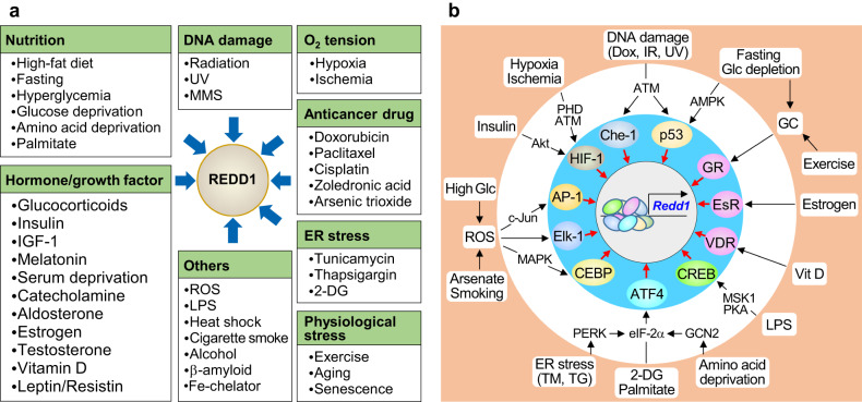
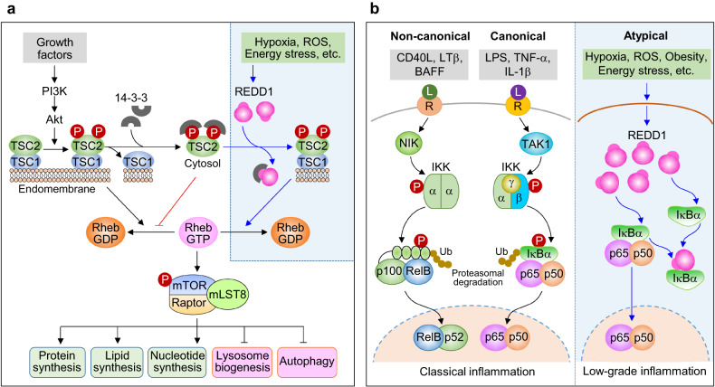
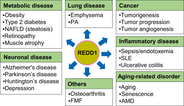
Regulated in development and DNA damage-response 1 (REDD1) is a stress-induced protein that controls various cellular functions, including metabolism, oxidative stress, autophagy, and cell fate, and contributes to the pathogenesis of metabolic and inflammatory disorders, neurodegeneration, and cancer. REDD1 usually exerts deleterious effects, including tumorigenesis, metabolic inflammation, neurodegeneration, and muscle dystrophy; however, it also exhibits protective functions by regulating multiple intrinsic cell activities through either an mTORC1-dependent or -independent mechanism. REDD1 typically regulates mTORC1 signaling, NF-κB activation, and cellular pro-oxidant or antioxidant activity by interacting with 14-3-3 proteins, IκBα, and thioredoxin-interacting protein or 75 kDa glucose-regulated protein, respectively. The diverse functions of REDD1 depend on cell type, cellular context, interaction partners, and cellular localization (e.g., mitochondria, endomembrane, or cytosol). Therefore, comprehensively understanding the molecular mechanisms and biological roles of REDD1 under pathophysiological conditions is of utmost importance. In this review, based on the published literature, we highlight and discuss the molecular mechanisms underlying the REDD1 expression and its actions, biological functions, and pathophysiological roles.


Conflict of interest statement

The authors declare no competing interests.

Figures

Fig. 1
Fig. 1. Multiple stresses and transcription factors induce REDD1 expression.
a Various stresses that stimulate REDD1 expression. 2-DG 2-deoxyglucose, IGF-1 insulin-like growth factor-1, LPS lipopolysaccharide, MMS methyl methane sulfonate, ROS reactive oxygen species, UV ultraviolet. b Transcription factors that induce REDD1 expression. Dox doxorubicin, EsR estrogen receptor, GCN2 general control nonderepressible 2, Glc glucose, GR glucocorticoid receptor, IR irradiation, PHD prolyl hydroxylase domain protein, TG thapsigargin, TM tunicamycin, VDR vitamin D receptor.
Fig. 2
Fig. 2. Models of REDD1-mediated mTORC1 inhibition and atypical NF-κB activation.
a Schematic showing REDD1-mediated mTORC1 inhibition. Growth factor-activated Akt phosphorylates Ser939 and Ser981 in TSC2 (a GTPase-activating protein, GAP) in the endomembrane-bound TSC1/2 complex (active form), facilitates TSC2/14-3-3 association (inactive GAP) in the cytosol, and inhibits GTP hydrolysis of Rheb, resulting in elevated Rheb-GTP levels and mTORC1 activation. Stress-induced REDD1 sequesters 14-3-3, maintains the active TSC1/2 complexes, hydrolyzes Rheb-GTP, and inhibits mTORC1 activity, thereby decreasing catabolism and increasing anabolism. b Schematic showing REDD1-mediated atypical NF-κB activation. NF-κB activation is generally triggered either by the IKKαβ-dependent canonical pathway or the IKKα-mediated noncanonical pathway following the ligation of cytokine or Toll-like receptors (R) via their cognate ligands (L). Furthermore, REDD1 activates the atypical NF-κB pathway by interacting with and sequestering IκBα, liberating NF-κB p65/50 from IκBα, and translocating it to the nucleus.
Fig. 3
Fig. 3. Molecular interaction between the top-scoring hit amino acids of REDD1 and IκBα.
a Trajectories (10 ns) of Ile83:O–Lys219:NZ, Glu85:CD–Lys219:NZ, Asp73:CG–Lys220:NZ, and Asp75:CG–Lys220:NZ. b Details of the interactions between amino acids in IκBα (residues 67−103 in ankyrin repeat domain 1, ANK1) and REDD1 (residues 214–220 in strand β4, indicated with *) or NF-κB p65 (nuclear localization sequence, NLS, 301KRKR304). The solid and dotted lines indicate salt bridges and hydrogen bonds, respectively. c Molecular interaction model showing of REED1 and IκBα. Lys219 and Lys220 in strand β4 of REDD1 are likely to form hydrogen bonds and salt bridges with Asp73, Asp75, Ile83, and Glu85 in ANK1 of IκBα.
Fig. 4
Fig. 4. Models of REDD1-mediated pro-oxidant or antioxidant activity.
a A schematic model of REDD1 as a pro-oxidant. REDD1 and TXNIP are unstable but are stabilized by forming a disulfide bond-mediated dimeric complex between Cys247 of TXINP and Cys32 of thioredoxin (Trx), thereby resulting in the inhibition of the Trx-thioredoxin reductase (TrxR) system coupled with the redox cycle involving peroxiredoxin (Prx), accumulation of cytosolic ROS, and promotion of oxidative stress and autophagy. b A schematic model of REDD1 as an antioxidant. Mitochondria-associated endoplasmic reticulum membranes (MAMs) are stabilized by the protein complexes composed of voltage-dependent anion channel (VDAC), 75 kDa glucose-regulated protein (GRP75), and inositol 1,4,5-trisphosphate receptor (IP3R). MAMs promote mitochondrial function, particularly electron transport chain (ETC) activity coupled with ROS production, leading to mitochondrial APT and ROS production. However, REDD1 disrupts MAM structure by interacting with and sequestering GRP75, resulting in a decrease in mitochondrial ROS production.
Fig. 5
Fig. 5. Various types of diseases associated with REDD1.
AMD age-related macular degeneration, FMF familial Mediterranean fever, NAFLD nonalcoholic fatty liver disease, PH pulmonary hypertension, SLE systemic lupus erythematosus.

References

    1. Alvarez-Garcia O, et al. Regulated in development and DNA damage response 1 deficiency impairs autophagy and mitochondrial biogenesis in articular cartilage and increases the severity of experimental osteoarthritis. Arthritis Rheumatol. 2017;69:1418–1428. doi: 10.1002/art.40104. - DOI - PMC - PubMed
    1. Qiao S, et al. A REDD1/TXNIP pro-oxidant complex regulates ATG4B activity to control stress-induced autophagy and sustain exercise capacity. Nat. Commun. 2015;6:7014. doi: 10.1038/ncomms8014. - DOI - PMC - PubMed
    1. Molitoris JK, et al. Glucocorticoid elevation of dexamethasone-induced gene 2 (Dig2/RTP801/REDD1) protein mediates autophagy in lymphocytes. J. Biol. Chem. 2011;286:30181–30189. doi: 10.1074/jbc.M111.245423. - DOI - PMC - PubMed
    1. Britto FA, et al. REDD1 deletion prevents dexamethasone-induced skeletal muscle atrophy. Am. J. Physiol. Endocrinol. Metab. 2014;307:E983–E993. doi: 10.1152/ajpendo.00234.2014. - DOI - PubMed
    1. Britto FA, et al. Glucocorticoid-dependent REDD1 expression reduces muscle metabolism to enable adaptation under energetic stress. BMC Biol. 2018;16:65. doi: 10.1186/s12915-018-0525-4. - DOI - PMC - PubMed

Publication types

MeSH terms

Substances