. 2023 Oct;30(10):1516-1524.
doi: 10.1038/s41594-023-01081-w. Epub 2023 Aug 31.

Structure of the preholoproteasome reveals late steps in proteasome core particle biogenesis

Affiliations

Structure of the preholoproteasome reveals late steps in proteasome core particle biogenesis

Richard M Walsh Jr et al. Nat Struct Mol Biol. 2023 Oct.

Abstract

Assembly of the proteasome's core particle (CP), a barrel-shaped chamber of four stacked rings, requires five chaperones and five subunit propeptides. Fusion of two half-CP precursors yields a complete structure but remains immature until active site maturation. Here, using Saccharomyces cerevisiae, we report a high-resolution cryogenic electron microscopy structure of preholoproteasome, a post-fusion assembly intermediate. Our data reveal how CP midline-spanning interactions induce local changes in structure, facilitating maturation. Unexpectedly, we find that cleavage may not be sufficient for propeptide release, as residual interactions with chaperones such as Ump1 hold them in place. We evaluated previous models proposing that dynamic conformational changes in chaperones drive CP fusion and autocatalytic activation by comparing preholoproteasome to pre-fusion intermediates. Instead, the data suggest a scaffolding role for the chaperones Ump1 and Pba1/Pba2. Our data clarify key aspects of CP assembly, suggest that undiscovered mechanisms exist to explain CP fusion/activation, and have relevance for diseases of defective CP biogenesis.


Conflict of interest statement

Declaration of Interests

The authors declare that they have no conflict of interest.

Figures

Fig. 1.
Fig. 1.. Proposed consequences of the pre1-1 and pre4-1 mutations.
A) Structure of wild-type CP with the midline-spanning interaction between β4 and β5 highlighted. Pre1-1 results in a β4-S142F mutation. The position of S142 is shown in red. B) Close-up view of β4-S142 which is present at the tightly packed interface between the two subunits. Bulky substitution at this site is predicted to disrupt the β4/β5 interface. C) Structure of wild-type CP showing how β7’s C-terminal region extends across the midline to sit in between β1 and β2. D) β7’s C-terminal region has been proposed to stimulate autocatalytic activation of β1 by stabilizing β1’s catalytic triad (shown in pink), especially Lys52 (numbered as Lys33 in mature CP). Images prepared from PDB: 5CZ4. Dashed lines indicate hydrogen bonds.
Fig. 2.
Fig. 2.. Biochemical and Structural Analysis of pre1-1, 4-1 Proteasomes.
A) Affinity purified material from wild-type and pre1-1, 4-1 was analyzed by SDS-PAGE followed by Coomassie staining. Similar results were seen in 4 experiments. B) The same material was analyzed by native gel electrophoresis followed by immunoblotting with antibodies that recognize CP subunit α5 or immature β5. Note that the preholoproteasome species is of low abundance, and therefore not visualized with the anti-α5 antibody. Asterisk denotes a species in the pre1-1, 4-1 mutant whose identity is unknown. Similar results were seen in 3 experiments. C) Size exclusion chromatography of the purified material from pre1-1, 4-1. Heavier material runs to the left. Arrow indicates the population selected for cryo-EM analysis. Similar results were seen in 2 experiments. mAU indicates the intensity of UV absorption. D). CP (15 nM) enzymatic activity was determined for each of the three active sites using the fluorogenic substrates suc-LLVY-AMC (β5), Z-LLE-AMC (β1), and Boc-LRR-AMC (β2). Data points from biologic duplicates are shown. Similar results were seen in two experiments, each with duplicates. E) Upregulation of Rpn4-mediated new proteasome biogenesis in the pre1-1, 4-1 mutant, as determined by immunoblot analysis of whole cell extracts. Upper panel, anti-Rpn4 antibody. Lower panel, anti-Hog1 antibody (loading control). Similar results were seen in four experiments. F) Cryo-EM structure of the preholoproteasome (3.0 Å) with its associated molecular model.
Fig. 3.
Fig. 3.. Subunit Contacts and Overall Positioning of Ump1 within the Preholoproteasome.
A) Two Ump1 molecules are present within the preholoproteasome, although each appears to be contained within its respective half-CP with a large distance spanning the resolved densities (middle panel). Significant conformational changes in Ump1 before and after CP fusion were not detected (right panel). B) Ump1 spans most of the β-ring, making extensive contacts with β1-β5. C) Detailed contacts between Ump1 and β1, which were not detectable in the prior 13S and pre-15S structures, as these complexes lacked β1.
Fig. 4.
Fig. 4.. The β2 propeptide and C-terminal Extension.
A) The N-terminal 18 residues of the β2 propeptide are well-resolved and remain extensively bound to Ump1. In contrast, the residues closer to the active site (19–29) are poorly resolved, likely reflecting flexibility in this region which in turn suggests that the propeptide has been cleaved. B) The position of β3 is shifted after CP fusion towards the midline through its contacts with the opposing β6 subunit. These contacts are further reflected in the increased resolution of β2’s long C-terminal extension in the preholoproteasome relative to the pre-15S complex. C) A portion of the β1 propeptide (residues 14-19) was resolved in the preholoproteasome, extending directly from the active site Thr20, oriented towards the center of the CP, and passing nearby β2.
Fig. 5.
Fig. 5.. Comparison of the Position and Conformation of Pba1/2 over the Course of CP Assembly.
A) The position of Pba1/2 on the α-ring is shown for 13S, pre-15S, and preholoproteasome assembly intermediates, and appears very similar throughout. B) Overlay of Pba1/2 from the three assembly intermediates shows little evidence of conformational changes during these stages of CP assembly.
Fig. 6.
Fig. 6.. Summary of High-Resolution Structural Analysis of CP Assembly.
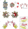
Schematic representation of the known steps and assembly intermediates in CP biogenesis, coupled with their associated high-resolution structures identified here and in ref. . 13S, EMD-23508; pre-15S, EMD-23503; preholoproteasome (this work); and 20S, EMD-23502.

References

    1. Rousseau A & Bertolotti A Regulation of proteasome assembly and activity in health and disease. Nat Rev Mol Cell Biol 19, 697–712 (2018). - PubMed
    1. Chen P & Hochstrasser M Autocatalytic subunit processing couples active site formation in the 20S proteasome to completion of assembly. Cell 86, 961–72 (1996). - PubMed
    1. Jager S, Groll M, Huber R, Wolf DH & Heinemeyer W Proteasome beta-type subunits: unequal roles of propeptides in core particle maturation and a hierarchy of active site function. J Mol Biol 291, 997–1013 (1999). - PubMed
    1. Ramos PC, Hockendorff J, Johnson ES, Varshavsky A & Dohmen RJ Ump1p is required for proper maturation of the 20S proteasome and becomes its substrate upon completion of the assembly. Cell 92, 489–99 (1998). - PubMed
    1. Hirano Y et al. A heterodimeric complex that promotes the assembly of mammalian 20S proteasomes. Nature 437, 1381–5 (2005). - PubMed

Publication types

MeSH terms

Substances

LinkOut - more resources