

Application of ultrasound in the management of TRASH (the radiographic appearance seemed harmless) fractures in preschool children: A review
- PMID: 37653809
- PMCID: PMC10470705
- DOI: 10.1097/MD.0000000000034855
Application of ultrasound in the management of TRASH (the radiographic appearance seemed harmless) fractures in preschool children: A review
Abstract
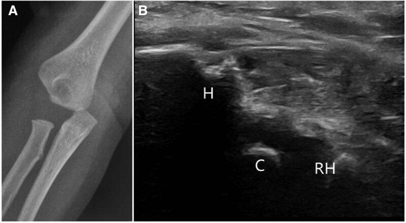
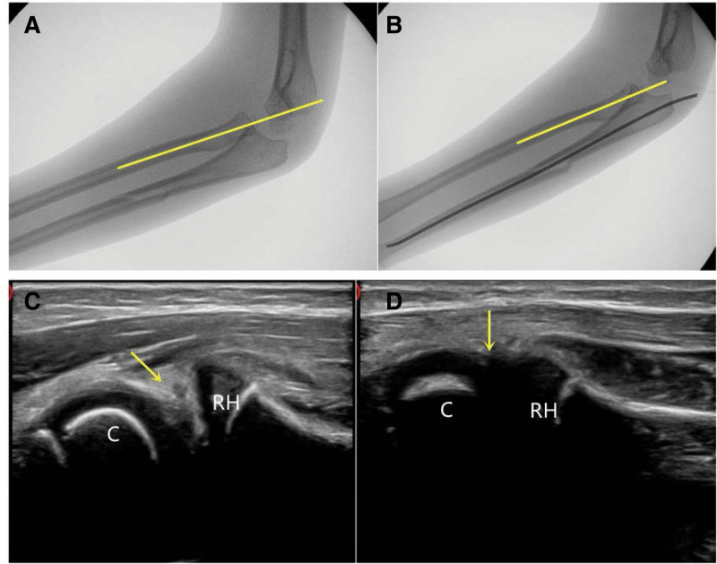
Elbow fracture is one of the most common fractures in preschool children, and the secondary ossification center appears regularly with age increasing. Transphyseal separation of the distal humerus, medial humeral condyle fracture, lateral humeral condyle fracture, radial head osteochondral separation, and Monteggia fracture (minimal ulnar bow type) are difficult to diagnose based on X-ray films alone because of the unossified secondary ossification center or a suspicious non-alignment of the anatomical cartilage of the elbow joint in preschool children. These 5 fractures above are defined as The Radiographic Appearance Seemed Harmless fractures in preschool children (TRASH-PS). The TRASH-PS fractures must be taken into consideration when there is swelling at the injured site without fracture feature on X-ray. Additionally, relevant misdiagnosis or unsuitable management can lead to elbow dysfunction and deformity. Therefore, this work reviews the application of ultrasound in the management of TRASH-PS fractures.
Copyright © 2023 the Author(s). Published by Wolters Kluwer Health, Inc.
Conflict of interest statement
The authors have no conflicts of interest to disclose.
Figures


Similar articles
-
[Ulna osteotomy and joint capsule release and tight for old Monteggia's fracture in children].Zhongguo Gu Shang. 2018 Sep 25;31(9):799-802. doi: 10.3969/j.issn.1003-0034.2018.09.004. Zhongguo Gu Shang. 2018. PMID: 30332870 Chinese.
-
Diagnostic imaging for elbow TRASH lesions in children and usefulness of ultrasonography using standard planes.J Pediatr Orthop B. 2023 Nov 1;32(6):557-564. doi: 10.1097/BPB.0000000000001062. Epub 2023 Jun 8. J Pediatr Orthop B. 2023. PMID: 36847196
-
Significance of the Lateral Humeral Line for Evaluating Radiocapitellar Alignment in Children.J Pediatr Orthop. 2017 Apr/May;37(3):e150-e155. doi: 10.1097/BPO.0000000000000853. J Pediatr Orthop. 2017. PMID: 27603193
-
Complications of pediatric elbow dislocations and monteggia fracture-dislocations.Instr Course Lect. 2015;64:493-8. Instr Course Lect. 2015. PMID: 25745932 Review.
-
Trash Lesions Around the Elbow: A Review of Approach to Diagnosis and Management.Indian J Orthop. 2021 Mar 8;55(3):539-548. doi: 10.1007/s43465-020-00333-x. eCollection 2021 Jun. Indian J Orthop. 2021. PMID: 33995858 Free PMC article. Review.
Cited by
-
The emerging application of ultrasound technology in pediatric bone fractures: Clinical application, related issues and development prospect.Pediatr Discov. 2024 May 28;2(2):e69. doi: 10.1002/pdi3.69. eCollection 2024 Jun. Pediatr Discov. 2024. PMID: 40625891 Free PMC article. Review.
References
-
- Baydar M, Öztürk K, Orman O, et al. . Use of corrective ulnar osteotomy and radial head relocation into preserved annular ligament in the treatment of radiocapitellar instability secondary to pediatric chronic Monteggia fracture-dislocation. J Hand Surg Am. 2022;47:481.e1–9. - PubMed
-
- Waters PM, Beaty J, Kasser J. Elbow “TRASH” (The Radiographic Appearance Seemed Harmless) fractures. J Pediatr Orthop. 2010;30:S77–81.
Publication types
MeSH terms
LinkOut - more resources
Full Text Sources
