

Sonographic reference values for median nerve cross-sectional area: A meta-analysis of data from healthy individuals
- PMID: 37654772
- PMCID: PMC10468154
- DOI: 10.1177/87564793231176009
Sonographic reference values for median nerve cross-sectional area: A meta-analysis of data from healthy individuals
Abstract
Objective: Establish median nerve CSA reference values and identify patient-level factors impacting diagnostic thresholds.
Methods: Studies were identified through a robust search of multiple databases, and quality assessment was conducted using a modified version of the National Institute of Health Study Quality Assessment Tool for Observational Cohort and Cross-Sectional Studies. A meta-analysis was performed to identify normative values stratified by anatomic location. A meta-regression was conducted to examine heterogeneity effects of age, sex, and laterality.
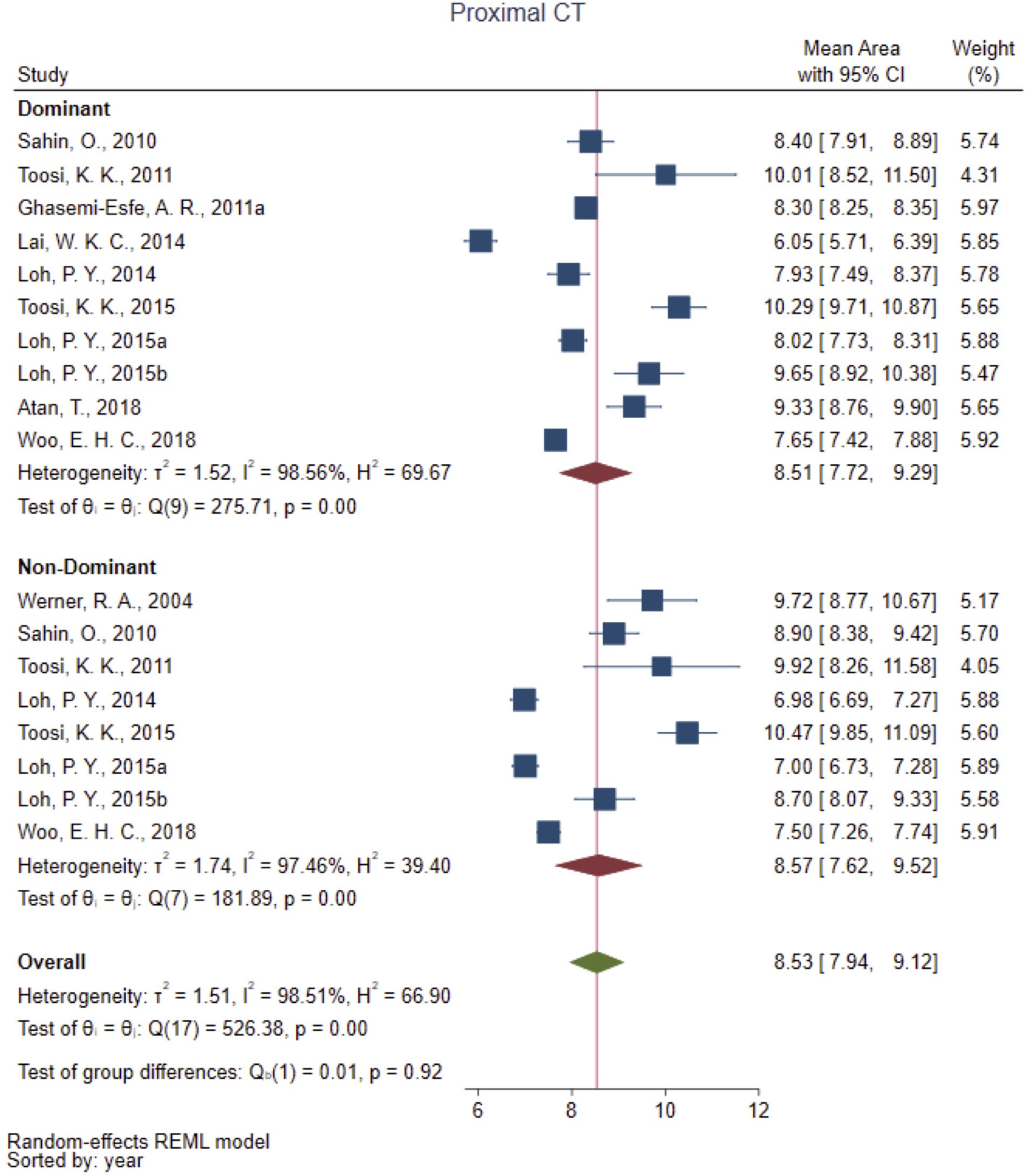
Results: The meta-analysis included 73 studies; 41 (56.2%) were high quality. The median nerve CSA [95% CI] was 6.46mm2 [6.09-6.84], 8.68mm2 [8.22-9.13], and 8.60mm2 [8.23-8.97] at the proximal forearm, the carpal tunnel inlet, and the proximal carpal tunnel, respectively. Age was positively associated with CSA at the level of proximal carpal tunnel (β=0.03mm2, p=0.047). Men (9.42mm2, [8.06-10.78]) had statistically larger proximal tunnel CSA (p = 0.03) as compared to women (7.71mm2, [7.01-8.42]). No difference was noted in laterality.
Conclusion: A reference value for median nerve CSA in the carpal tunnel is 8.60mm2. Adjustments may be required in pediatrics or older adults. The diagnostic threshold of 10.0mm2 for male patients should be cautiously applied as the upper limit of normative averages surpasses this threshold.
Keywords: median nerve; meta-analysis; reference values; ultrasonography.
Figures


Similar articles
-
Sonographic reference values of median nerve cross-sectional area: a protocol for a systematic review and meta-analysis.Syst Rev. 2019 Jan 3;8(1):2. doi: 10.1186/s13643-018-0929-9. Syst Rev. 2019. PMID: 30606255 Free PMC article.
-
Normative Value of the Cross-Sectional Area of the Median Nerve at the Carpal Tunnel Inlet and Distal Forearm in the Singapore Population.J Hand Surg Asian Pac Vol. 2022 Aug;27(4):649-655. doi: 10.1142/S242483552250062X. Epub 2022 Aug 8. J Hand Surg Asian Pac Vol. 2022. PMID: 35965359
-
Median nerve cross-sectional area and MRI diffusion characteristics: normative values at the carpal tunnel.Skeletal Radiol. 2009 Apr;38(4):355-61. doi: 10.1007/s00256-008-0626-1. Epub 2009 Jan 9. Skeletal Radiol. 2009. PMID: 19132371
-
The cross-sectional morphology of median nerve in carpal tunnel of healthy, adult population: A systematic review and meta-analysis.Morphologie. 2023 Mar;107(356):99-115. doi: 10.1016/j.morpho.2022.05.005. Epub 2022 Jun 11. Morphologie. 2023. PMID: 35697557
-
A systematic review: normative reference values of the median nerve cross-sectional area using ultrasonography in healthy individuals.Sci Rep. 2022 Jun 2;12(1):9217. doi: 10.1038/s41598-022-13058-8. Sci Rep. 2022. PMID: 35654926 Free PMC article.
Cited by
-
Cross-Sectional Area and Echogenicity Reference Values for Sonography of Peripheral Nerves in the Lithuanian Population.Diagnostics (Basel). 2024 Jun 28;14(13):1373. doi: 10.3390/diagnostics14131373. Diagnostics (Basel). 2024. PMID: 39001263 Free PMC article.
-
Accuracy of the Standard and Distal-to-Proximal Sequence of the Upper Limb Neurodynamic Test 1 for the Diagnosis of Carpal Tunnel Syndrome: The Role of Side-to-Side Comparisons.J Clin Med. 2024 Nov 25;13(23):7122. doi: 10.3390/jcm13237122. J Clin Med. 2024. PMID: 39685581 Free PMC article.
-
Clinical and Electrodiagnostic Correlations of Ultrasound-Detected Markedly Enlarged Median Nerve at the Wrist.Neurol Int. 2025 Aug 7;17(8):124. doi: 10.3390/neurolint17080124. Neurol Int. 2025. PMID: 40863993 Free PMC article.
References
-
- Fowler JR, Byrne K, Pan T, Goitz RJ (2019) False-positive rates for nerve conduction studies and ultrasound in patients without clinical signs and symptoms of carpal tunnel syndrome. The Journal of Hand Surgery 44:181–185 - PubMed
-
- Wang WL, Buterbaugh K, Kadow TR, Goitz RJ, Fowler JR (2018) A prospective comparison of diagnostic tools for the diagnosis of carpal tunnel syndrome. The Journal of hand surgery 43:833–836. e832 - PubMed
-
- Cartwright MS, Hobson-Webb LD, Boon AJ et al. (2012) Evidence-based guideline: neuromuscular ultrasound for the diagnosis of carpal tunnel syndrome. Muscle Nerve 46:287–293 - PubMed
Grants and funding
LinkOut - more resources
Full Text Sources
Medical