. 2023 Jul 3;12(8):e1271-e1280.
doi: 10.1016/j.eats.2023.03.018. eCollection 2023 Aug.

Ultrasonic Technique for Femoral Tunnel Creation in Anterior Cruciate Ligament Reconstruction

Affiliations

Ultrasonic Technique for Femoral Tunnel Creation in Anterior Cruciate Ligament Reconstruction

Tatsuo Mae et al. Arthrosc Tech. .

Abstract

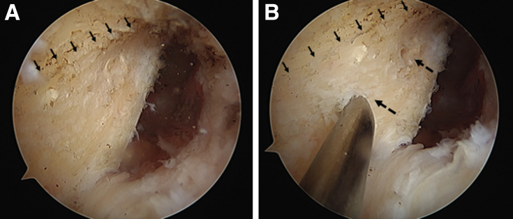
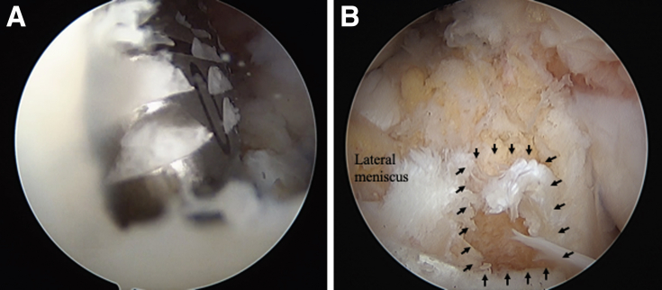
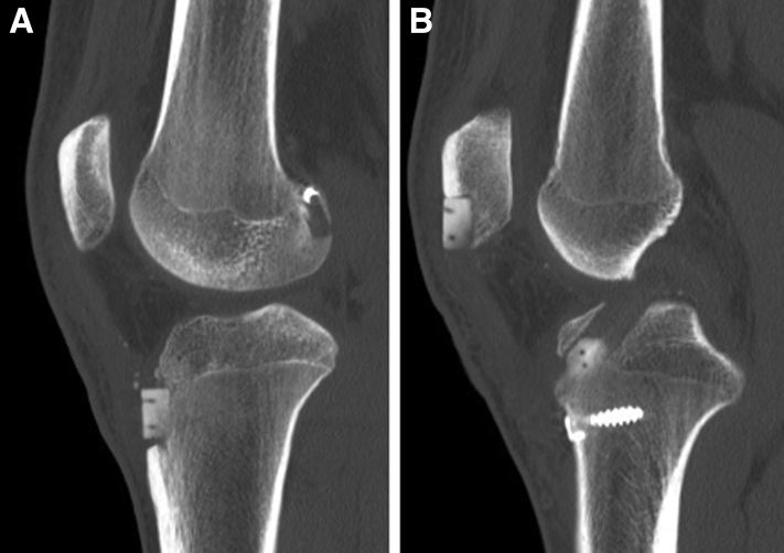
Bone tunnel creation in the anatomical location is essential in anterior cruciate ligament (ACL) reconstruction with an autogenous graft and is commonly performed with a drill bit matched to graft diameter. Anatomic rectangular tunnel ACL reconstruction with a bone-patellar tendon-bone autograft has been developed to anatomically create bone tunnels inside the ACL footprints and has been reported to achieve excellent outcomes. To make the rectangular tunnel, the surgeon needs to dilate 2 adjacent bone tunnels after creation of 2 round tunnels with a drill bit, while the tunnel wall occasionally cracks during dilating. An ultrasonic (US) device was developed with improvement of output power and has been implemented with a rectangular shape blade in the field of arthroscopic surgery. This US device can provide a precise and effective bone cut compared to drills. We introduced this device to clinically create a rectangular tunnel during ACL reconstruction. The US device can be useful for rectangular femoral tunnel creation and can create a precise rectangular femoral tunnel in the ACL footprint.


Figures

Fig 1
Fig 1
Ultrasonic device (ARTHROBEAT; Olympus Medical Systems Corp., Tokyo, Japan). (A) An ultrasonic generator (AUG-100) for production of electric energy. (B) An ultrasonic transducer (ATD-100; blue circle) and a 4 × 5 mm rectangular blade (AB-7718RE45; red circle) attached to the transducer. The transducer converts the electric energy into mechanical vibration transmitted to the rectangular blade. (C) The tip of the 4 × 5-mm rectangle blade.
Fig 2
Fig 2
View of medial wall of lateral femoral condyle from the anteromedial portal (right knee). (A) Anterior cruciate ligament footprint behind the resident’s ridge (arrows). (B) Tunnel locations (dashed arrows) are marked with a microscopic awl behind the ridge (solid arrows).
Fig 3
Fig 3
View of medial wall of lateral femoral condyle from the anteromedial portal (right knee). Two guidewires of 2.4-mm diameter are inserted in parallel into the marked points in the inside-out manner through the far-anteromedial portal.
Fig 4
Fig 4
View of medial wall of lateral femoral condyle from the anteromedial portal (right knee). (A) Two previously inserted guidewires are overdrilled with a 4.5-mm canulated drill. (B) Two round tunnels (arrows) are created.
Fig 5
Fig 5
A 4 × 5-mm rectangle blade is introduced into the joint with a specific cutting guide.
Fig 6
Fig 6
Two round tunnels are excavated into rectangular tunnels with this ultrasonic device.
Fig 7
Fig 7
A rectangular femoral tunnel created with the ultrasonic (US) device, viewing from the anteromedial portal on right knee. (A) The septal wall between tunnels is excavated with the US device. (B) Inside the tunnel with a smooth surface.
Fig 8
Fig 8
Finishing the femoral tunnel creation. (A) A 5 × 10-mm dilator is put into the femoral tunnel to confirm the tunnel size. (B) A femoral rectangular tunnel in the anterior cruciate ligament femoral footprint.
Fig 9
Fig 9
The anterior cruciate ligament tibial footprint is surrounded by anterior horn of lateral meniscus, anterior ridge (dashed arrows), and medial intercondylar ridge (solid arrows). View from the anteromedial portal.
Fig 10
Fig 10
Tibial tunnel creation. (A) Two guidewires are parallelly inserted inside the footprint at a 5-mm distance between the centers of 2 wires. (B) Two round tunnels are created with a 5-mm drill bit overdrilling along the guidewires.
Fig 11
Fig 11
Finishing the tibial tunnel creation. (A) Two tunnels are dilated with a 5 × 10-mm rectangular dilator. (B) A tibial rectangular tunnel (arrows) is viewed from the anteromedial portal.
Fig 12
Fig 12
A guide pin for the interference screw is settled proximally in the femoral tunnel.
Fig 13
Fig 13
A passing pin with a suture is inserted through the femoral tunnel via a far-anteromedial portal to pass the leading suture.
Fig 14
Fig 14
Suture passage. (A) The distal suture is caught through the tibial tunnel with a suture manipulator. (B) The suture for graft passage runs from tibial tunnel to femoral tunnel.
Fig 15
Fig 15
Graft is fixed with an interference screw, keeping the tendon-bone interface of graft adjusted to the femoral tunnel aperture, to replicate the direct insertion of the native anterior cruciate ligament attachment.
Fig 16
Fig 16
Anterior cruciate ligament graft after graft fixation. View from the anteromedial portal.
Fig 17
Fig 17
Two-week postoperative computer tomography. (A) Rectangular femoral tunnel is located posterior on the lateral femoral condyle. (B) Tibial tunnel runs from anterior-distal to posterior-proximal. Bone plug from the graft is located in the tunnel.
Fig 18
Fig 18
Two-week postoperative magnetic resonance images (T2-weight). (A) Sagittal view of anterior cruciate ligament (ACL) graft. (B) Oblique coronal view of ACL graft.

Similar articles

References

    1. Shino K., Nakata K., Nakamura N., Toritsuka Y., Nakagawa S., Horibe S. Anatomically oriented anterior cruciate ligament reconstruction with a bone–patellar tendon–bone graft via rectangular socket and tunnel: A snug-fit and impingement-free grafting technique. Arthroscopy. 2005;21:1402–e1-e5. - PubMed
    1. Shino K., Nakata K., Nakamura N., et al. Rectangular tunnel double-bundle anterior cruciate ligament reconstruction with bone-patellar tendon-bone graft to mimic natural fiber arrangement. Arthroscopy. 2008;24:1178–1183. - PubMed
    1. Tachibana Y., Shino K., Mae T., Iuchi R., Take Y., Nakagawa S. Anatomic rectangular tunnels identified with the arthroscopic landmarks result in excellent outcomes in ACL reconstruction with a BTB graft. Knee Surg Sports Traumatol Arthrosc. 2019;27:2680–2690. - PubMed
    1. Ai X.M., Ho L.C., Yang N.Y., Han L.L., Lu J.J., Yue X. A comparative study of ultrasonic scalpel (US) versus conventional metal clips for closure of the cystic duct in laparoscopic cholecystectomy (LC): A meta-analysis. Medicine (Baltimore) 2018;97 - PMC - PubMed
    1. Amaral J.F. The experimental development of an ultrasonically activated scalpel for laparoscopic use. Surg Laparosc Endosc. 1994;4:92–99. - PubMed

LinkOut - more resources