Review
. 2023 Jun 28;1(5):403-413.
doi: 10.1021/cbmi.3c00045. eCollection 2023 Aug 28.

Switchable and Functional Fluorophores for Multidimensional Single-Molecule Localization Microscopy

Affiliations
Review

Switchable and Functional Fluorophores for Multidimensional Single-Molecule Localization Microscopy

Yunshu Liu et al. Chem Biomed Imaging. .

Abstract

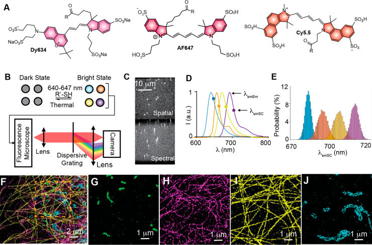
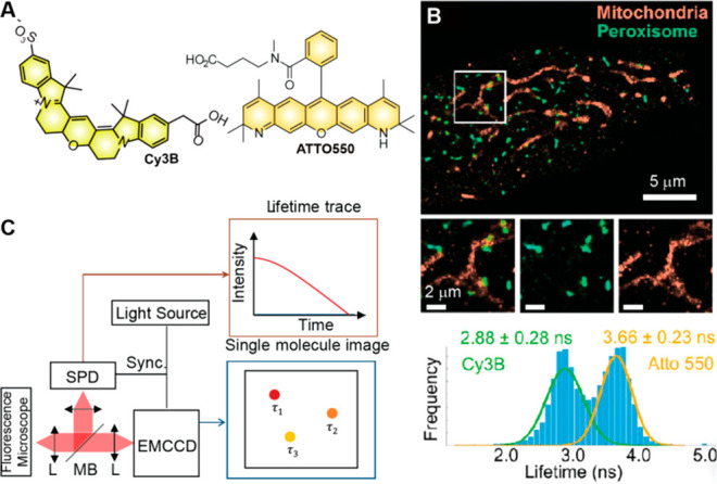
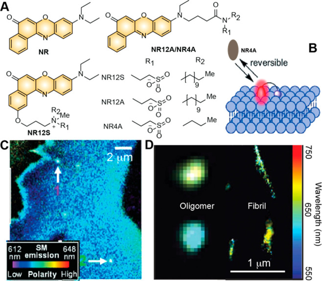
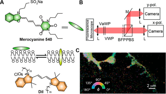
Multidimensional single-molecule localization microscopy (mSMLM) represents a paradigm shift in the realm of super-resolution microscopy techniques. It affords the simultaneous detection of single-molecule spatial locations at the nanoscale and functional information by interrogating the emission properties of switchable fluorophores. The latter is finely tuned to report its local environment through carefully manipulated laser illumination and single-molecule detection strategies. This Perspective highlights recent strides in mSMLM with a focus on fluorophore designs and their integration into mSMLM imaging systems. Particular interests are the accomplishments in simultaneous multiplexed super-resolution imaging, nanoscale polarity and hydrophobicity mapping, and single-molecule orientational imaging. Challenges and prospects in mSMLM are also discussed, which include the development of more vibrant and functional fluorescent probes, the optimization of optical implementation to judiciously utilize the photon budget, and the advancement of imaging analysis and machine learning techniques.


Conflict of interest statement

The authors declare no competing financial interest.

Figures

Figure 1
Figure 1
(A) Representative molecular structures of far-red photoswitchable cyanine dyes for multiplexed sSMLM; R in dye structures indicates the labeling site to attach biomolecules. (B–E) Concept of parallel multiplexed sSMLM: (B) a set of photoswitchable fluorophores (shown as blue, orange, yellow, and purple circles) can simultaneously photoswitch under similar imaging conditions; their single-molecule emission signals are captured by a typical fluorescence microscope coupled with a dispersive element (grating shown as an example) to provide spatial and spectral images (C) of every single molecule; simulated emission spectra (D) and histogram of λsmSC probability distributions (E) of four hypothetical far-red photoswitchable fluorophores demonstrate the concept of single-molecule spectral discrimination based on statistically resolved λsmSC distribution regardless of spectral overlaps with λsmEm separations about 10 nm. (F–J) Representative four-color simultaneous multiplexed sSMLM overlaid image (F) in a PtK2 cell with its peroxisome (G), vimentin (H), tubulin (I), mitochondria (J) structures separately displayed and labeled with Dy634, DL650, CF660C, and CF680, respectively. Images in (F)–(J) are adapted with permission from ref (18). Copyright 2015 Springer Nature.
Figure 2
Figure 2
(A) Representative pair of photoswitchable fluorophores Cy3B and ATTO550 used for simultaneous FL-SMLM imaging. (B) Top: FL-SMLM images of a fixed HeLa cell labeled with Cy3B (green) and Atto550 (orange). Bottom: Histogram (bars) and double Gaussian fitting (green and orange curves) of the fluorescence lifetime distributions of Cy3B and ATTO550 measured in the HeLa cell. (C) Schematic illustration of an FL-SMLM system; L: lens; MB: magnetic base; Images in (B) were adaptedfrom ref (27). Copyright 2022 American Chemical Society.
Figure 3
Figure 3
(A) Structures of NR and its derivatives designed for super-resolution polarity and hydrophobicity mapping using sPAINT. (B) Principle of sPAINT with NR4A: the single-molecule fluorescence blinkings signals are generated by the transient interactions between NR4A and the lipid bilayers. (C) sPAINT image of a live COS-7 cell with the λsmEm changes on the cell membranes; the arrow-pointed area in (C) highlights the observed pits with significant bathochromic shifts in λsmEm compared with those detected in the membrane. (D) sPAINT image of individual aβ oligomers and fibrils. Images in (C) and (D) were adapted with permission from ref (10), Copyright 2019 Wiley-VCH; and ref (8), Copyright 2016 Springer Nature, respectively.
Figure 4
Figure 4
(A) Structures of orientation-sensitive fluorophores MC540 and DiI and an illustration of the fluorophore orientation changes from parallel to perpendicular when interacting with lipid bilayers. (B) Simplified optical detection scheme for SMOLM; VaWP: variable wave plate; VWP: voltex wave plate; BFP: back focal plane; PBS: polarizing beam splitter. (C) Representative SMOLM image of a fixed HEK-293T cell imaged that was color-coded by the detected azimuthal angles (ϕ) of each single MC540 molecule. Image in (C) was adapted with permission from ref (15). Copyright 2023 Springer Nature.
Figure 5
Figure 5
(A) Concept of stroboscopic illumination for SMdM which excites the fluorophores using paired pulses of stroboscopic illumination, synchronized with the camera acquisition. (B) Structures of SMdM fluorophores BTF1-BTF3 and BODIPY-TMR alkyne. (C) Representative SMdM image of a live COS-7 cell using BODIPY-TMR alkyne; arrows indicate the nanodomains with lower diffusivity; inset in (C) shows the distributions of single-molecule displacements for the nanodomains (left) and other parts of the plasma membrane (right) together with the maximum-likelihood fittings for diffusivity distributions. Images in (C) were adapted from ref (14). Copyright 2020 American Chemical Society.
Figure 6
Figure 6
(A) Structures of TTT and TTC isomers of a merocyanine chromophore. (B) Distinct λsmSC populations were observed for the merocyanine TTC and TTT isomers. (C) Plot showing the solvent-dependent ratios of the TTT versus the TTC isomers. (D) Structures and four conformational isomers of three BODIPY chromophores observed using sSMLM; (E) a representative scatterplot shows distinct λsmSC populations of BODIPY chromophores from the ttt and ttc versus ctc and ctt isomers. (F−H) Histogram of λsmSC probability distributions of Rh101 (F), pentacene (G), and AF647 (H) respectively. Images in (B) and (C) were adapted from ref (51). Copyright 2017 American Chemical Society. Image in (E) was adapted from ref (50). Copyright 2019 American Chemical Society. Images in (F)–(H) were adapted ref (39). Copyright 2021 American Chemical Society, respectively.

Similar articles

Cited by

References

    1. Rust M. J.; Bates M.; Zhuang X. Sub-Diffraction-Limit Imaging by Stochastic Optical Reconstruction Microscopy (STORM). Nat. Methods 2006, 3, 793–796. 10.1038/nmeth929. - DOI - PMC - PubMed
    1. Betzig E.; Patterson G. H.; Sougrat R.; Lindwasser O. W.; Olenych S.; Bonifacino J. S.; Davidson M. W.; Lippincott-Schwartz J.; Hess H. F. Imaging Intracellular Fluorescent Proteins at Nanometer Resolution. Science 2006, 313, 1642.10.1126/science.1127344. - DOI - PubMed
    1. Jungmann R.; Avendano M. S.; Woehrstein J. B.; Dai M.; Shih W. M.; Yin P. Multiplexed 3D Cellular Super-Resolution Imaging with DNA-PAINT and Exchange-PAINT. Nat. Methods 2014, 11, 313–318. 10.1038/nmeth.2835. - DOI - PMC - PubMed
    1. Moerner W. E. Single-Molecule Spectroscopy, Imaging, and Photocontrol: Foundations for Super-Resolution Microscopy (Nobel Lecture). Angew. Chem., Int. Ed. 2015, 54, 8067–8093. 10.1002/anie.201501949. - DOI - PubMed
    1. Gustafsson M. G. L. Nonlinear Structured-Illumination Microscopy: Wide-Field Fluorescence Imaging with Theoretically Unlimited Resolution. Proc. Natl. Acad. Sci. U. S. A. 2005, 102, 13081–13086. 10.1073/pnas.0406877102. - DOI - PMC - PubMed