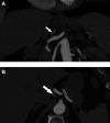
Case report: Isolated dissection of the left gastric artery: an unusual cause of acute abdominal pain
- PMID: 37655216
- PMCID: PMC10466798
- DOI: 10.3389/fcvm.2023.1240853
Case report: Isolated dissection of the left gastric artery: an unusual cause of acute abdominal pain
Abstract
Spontaneous and isolated dissection of the left gastric artery is a rare occurrence, with only a handful of cases reported in the medical literature. Clinical presentation may mimic more common intra-abdominal pathologies; however, it is imperative to identify this condition promptly due to its potential serious consequences. This underscores the importance of maintaining a high level of clinical suspicion and including this pathology in the differential diagnosis of patients presenting with acute abdominal symptoms. Hence, this case report aims to increase awareness among clinicians about the importance of identifying and treating this rare condition promptly. A 69-year-old female experienced severe epigastric pain while attending a yoga class, prompting her admission to the emergency department 24 h later due to the persistence of her symptoms. Following imaging work-up utilizing computed tomography angiography (CTA), she was diagnosed with a dissection of the left gastric artery. Notably, there was no associated aneurysm or any evidence of ischemia in the esophageal or gastric wall. Conservative management, including low-dose aspirin and blood pressure control, was implemented. After 6 months of follow-up, CTA demonstrated expansion of the true lumen and the absence of secondary aneurysm formation, leading to discontinuation of aspirin. The management of spontaneous dissection of visceral arteries is primarily determined by the presence of complications and organ ischemia. In the case of uncomplicated visceral artery dissections, first-line treatment comprises surveillance and antiaggregation. Nevertheless, the optimal duration of antiplatelet therapy and the necessity for long-term follow-up remain unclear. Endovascular or surgical interventions should be reserved for patients exhibiting deteriorating symptoms or complications, and the decision to pursue these interventions should be made on a case-by-case basis.
Keywords: conservative treatment; contrast-enhanced computed tomography; left gastric artery dissection; sudden epigastric pain; visceral artery dissection.
© 2023 Frey, Engelberger and Psathas.
Conflict of interest statement
The authors declare that the research was conducted in the absence of any commercial or financial relationships that could be construed as a potential conflict of interest.
Figures


Similar articles
-
A Case of Spontaneous Isolated Celiac Artery Dissection with Pseudoaneurysm Formation.Cureus. 2017 Aug 27;9(8):e1616. doi: 10.7759/cureus.1616. Cureus. 2017. PMID: 29104834 Free PMC article.
-
Unusual presentation and treatment of isolated spontaneous gastric artery dissection.Clin Exp Emerg Med. 2016 Jun 30;3(2):112-115. doi: 10.15441/ceem.15.029. eCollection 2016 Jun. Clin Exp Emerg Med. 2016. PMID: 27752628 Free PMC article.
-
Spontaneous isolated left gastric artery dissection: unusual visceral artery dissection.Acute Med Surg. 2016 Feb 26;3(4):369-371. doi: 10.1002/ams2.191. eCollection 2016 Oct. Acute Med Surg. 2016. PMID: 29123814 Free PMC article.
-
Nonoperative management of isolated celiac and superior mesenteric artery dissection: case report and review of the literature.Vascular. 2009 Nov-Dec;17(6):359-64. doi: 10.2310/6670.2009.00053. Vascular. 2009. PMID: 19909685 Review.
-
Spontaneous dissection of the celiac trunk: a rare cause of abdominal pain--case report and review of the literature.Acta Gastroenterol Belg. 2013 Sep;76(3):335-9. Acta Gastroenterol Belg. 2013. PMID: 24261029 Review.
References
-
- Yoshimi S, Tatsuya K, Kotani T, Sakiko H, Kuniyasu H, Shigeyuki M, et al. Isolated spontaneous dissection of the splanchnic arteries in the era of multidetector computed tomography. Matsushita MJ. (2015) 54:79–85.
Publication types
LinkOut - more resources
Full Text Sources

