

Development of a rapid lateral flow assay for detection of anti-coccidioidal antibodies
- PMID: 37655868
- PMCID: PMC10512788
- DOI: 10.1128/jcm.00631-23
Development of a rapid lateral flow assay for detection of anti-coccidioidal antibodies
Abstract
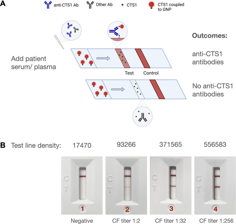
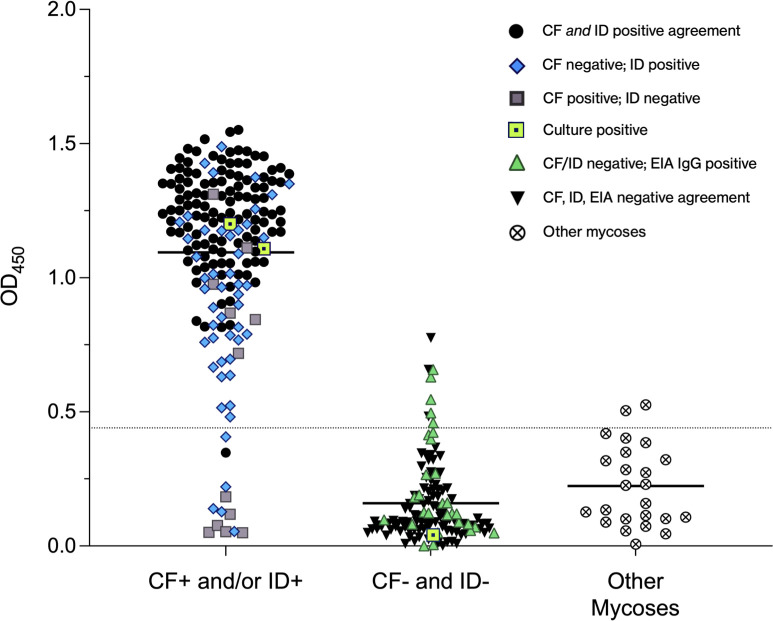
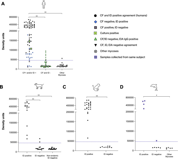
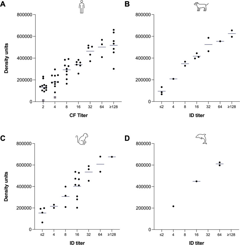
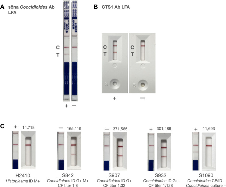
Coccidioides spp. are dimorphic fungi that are capable of infecting human and non-human mammals and can cause diverse manifestations of coccidioidomycosis or Valley fever (VF). In combination with clinical symptoms and radiographic findings, antibody-based diagnostic tests are often used to diagnose and monitor patients with VF. Chitinase 1 (CTS1) has previously been identified as the seroreactive antigen used in these diagnostic assays to detect anticoccidial IgG. Here, an indirect enzyme-linked immunosorbent assay to detect IgG to CTS1 demonstrated 165 of 178 (92.7%) patients with a positive result by immunodiffusion (ID) and/or complement fixation (CF) had antibodies to the single antigen CTS1. We then developed a rapid antibody lateral flow assay (LFA) to detect anti-CTS1 antibodies. Out of 143 samples tested, the LFA showed 92.9% positive percent agreement [95% confidence interval (CI), 84.3%-96.9%] and 97.7% negative percent agreement (95% CI, 87.9%-99.6%) with ID and CF assays. Serum or plasma from canines, macaques, and dolphins was also tested by the CTS1 LFA. Test line densities of the CTS1 LFA correlated in a linear manner with the reported CF and ID titers for human and non-human samples, respectively. This 10-min point-of-care test for the rapid detection of anti-coccidioidal antibodies could help to inform healthcare providers in real-time, potentially improving the efficiency of healthcare delivery.
Keywords: LFA; Valley fever; coccidioidomycosis; diagnostic.
Conflict of interest statement
F.J.G., T.E.G., and D.F.L. are inventors on an unlicensed patent application involving the LFA evaluated in this report. All other authors declare no conflict of interest.
Figures
Similar articles
-
Two lateral flow assays for detection of anti-coccidioidal antibodies show similar performance to immunodiffusion in dogs with coccidioidomycosis.Am J Vet Res. 2024 Mar 25;85(6):ajvr.23.12.0272. doi: 10.2460/ajvr.23.12.0272. Print 2024 Jun 1. Am J Vet Res. 2024. PMID: 38531155 Free PMC article.
-
Evaluation of a commercially available, point-of-care Coccidioides antibody lateral flow assay to aid in rapid diagnosis of coccidioidomycosis in dogs.Med Mycol. 2020 Apr 1;58(3):328-332. doi: 10.1093/mmy/myz067. Med Mycol. 2020. PMID: 31212311
-
Disruption of the gene which encodes a serodiagnostic antigen and chitinase of the human fungal pathogen Coccidioides immitis.Infect Immun. 2000 Oct;68(10):5830-8. doi: 10.1128/IAI.68.10.5830-5838.2000. Infect Immun. 2000. PMID: 10992492 Free PMC article.
-
Serologic studies in coccidioidomycosis.Semin Respir Infect. 2001 Dec;16(4):242-50. doi: 10.1053/srin.2001.29315. Semin Respir Infect. 2001. PMID: 11740825 Review.
-
Review of Clinical and Laboratory Diagnostics for Coccidioidomycosis.J Clin Microbiol. 2023 May 23;61(5):e0158122. doi: 10.1128/jcm.01581-22. Epub 2023 Mar 8. J Clin Microbiol. 2023. PMID: 36883820 Free PMC article. Review.
Cited by
-
Antigenic Relatedness between Mannans from Coccidioides immitis and Coccidioides posadasii Spherules and Mycelia.J Fungi (Basel). 2024 Jan 23;10(2):89. doi: 10.3390/jof10020089. J Fungi (Basel). 2024. PMID: 38392761 Free PMC article.
-
Two lateral flow assays for detection of anti-coccidioidal antibodies show similar performance to immunodiffusion in dogs with coccidioidomycosis.Am J Vet Res. 2024 Mar 25;85(6):ajvr.23.12.0272. doi: 10.2460/ajvr.23.12.0272. Print 2024 Jun 1. Am J Vet Res. 2024. PMID: 38531155 Free PMC article.
-
Improving Coccidioidomycosis Testing for Emergency Department Patients With Suspect Community-Acquired Pneumonia: Analysis of Provider Attitudes and the Effect of a Targeted Intervention.Open Forum Infect Dis. 2024 Aug 12;11(8):ofae461. doi: 10.1093/ofid/ofae461. eCollection 2024 Aug. Open Forum Infect Dis. 2024. PMID: 39192992 Free PMC article.
-
Practical Guidance for Clinical Microbiology Laboratories: Antibody and antigen detection methods for dimorphic fungal infections.Clin Microbiol Rev. 2025 Jun 12;38(2):e0000520. doi: 10.1128/cmr.00005-20. Epub 2025 May 21. Clin Microbiol Rev. 2025. PMID: 40396718 Review.
References
-
- Kolivras KN, Johnson PS, Comrie AC, Yool SR. 2001. Environmental variability and coccidioidomycosis (valley fever). Aerobiologia (Bologna) 17:31–42. doi:10.1023/A:1007619813435 - DOI
-
- Fisher MC, Koenig GL, White TJ, Taylor JW. 2002. Molecular and phenotypic description of coccidioides posadasii sp. nov., previously recognized as the non-California population of coccidioides immitis. Mycologia 94:73–84. - PubMed
-
- Del Rocío Reyes-Montes M, Pérez-Huitrón MA, Ocaña-Monroy JL, Frías-De-León MG, Martínez-Herrera E, Arenas R, Duarte-Escalante E. 2016. The habitat of Coccidioides spp. and the role of animals as reservoirs and disseminators in nature. BMC Infect Dis 16:550. doi:10.1186/s12879-016-1902-7 - DOI - PMC - PubMed
Publication types
MeSH terms
Substances
Grants and funding
LinkOut - more resources
Full Text Sources
Medical
Research Materials
Miscellaneous
