. 2023 Sep 1;11(5):e0148723.
doi: 10.1128/spectrum.01487-23. Online ahead of print.

High selectivity of the hyperthermophilic subtilase propeptide domain toward inhibition of its cognate protease

Affiliations

High selectivity of the hyperthermophilic subtilase propeptide domain toward inhibition of its cognate protease

Miha Bahun et al. Microbiol Spectr. .

Abstract

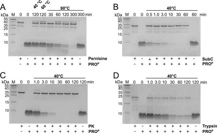
Microbial extracellular subtilases are highly active proteolytic enzymes commonly used in commercial applications. These subtilases are synthesized in their inactive proform, which matures into the active protease under the control of the propeptide domain. In mesophilic bacterial prosubtilases, the propeptide functions as both an obligatory chaperone and an inhibitor of the subtilase catalytic domain. In contrast, the propeptides of hyperthermophilic archaeal prosubtilases act mainly as tight inhibitors and are not essential for subtilase folding. It is unclear whether this stronger inhibitory activity of hyperthermophilic propeptides results in their higher selectivity toward their cognate subtilases, in contrast to promiscuous mesophilic propeptides. Here, we showed that the propeptide of pernisine, a hyperthermostable archaeal subtilase, strongly interacts with and inhibits pernisine, but not the homologous subtilisin Carlsberg and proteinase K. Instead, the pernisine propeptide was readily degraded by subtilisin Carlsberg and proteinase K. In addition, the catalytic domain of unprocessed propernisine was also susceptible to degradation but became proteolytically stable after autoprocessing of propernisine into the inactive, noncovalent complex propeptide:pernisine. This allowed efficient transactivation of the autoprocessed complex propeptide:pernisine through degradation of pernisine propeptide by subtilisin Carlsberg and proteinase K at mesophilic temperature. Moreover, we demonstrated that active pernisine molecules are inhibited by the propeptide that is released after pernisine-catalyzed degradation of the unprocessed propernisine catalytic domain. This highlights the high inhibitory potency of the hyperthermophilic propeptide toward its cognate subtilase and its importance in regulating subtilase maturation, to prevent the degradation of the unprocessed subtilase precursors by the prematurely activated molecules. IMPORTANCE Many microorganisms secrete proteases into their environment to degrade protein substrates for their growth. The important group of these extracellular enzymes are subtilases, which are also widely used in practical applications. These subtilases are inhibited by their propeptide domain, which is degraded during the prosubtilase maturation process. Here, we showed that the propeptide of pernisine, a prion-degrading subtilase from the hyperthermophilic archaeon, strongly inhibits pernisine with extraordinarily high binding affinity. This interaction proved to be highly selective, as pernisine propeptide was rapidly degraded by mesophilic pernisine homologs. This in turn allowed rapid transactivation of propernisine by mesophilic subtilases at lower temperatures, which might simplify the procedures for preparation of active pernisine for commercial use. The results reported in this study suggest that the hyperthermophilic subtilase propeptide evolved to function as tight and selective regulator of maturation of the associated prosubtilase to prevent its premature activation under high temperatures.

Keywords: enzyme activation; enzyme inhibition; hyperthermophile; propeptide; protein stability; subtilase.


Conflict of interest statement

The authors declare no conflict of interest.

Figures

Fig 1
Fig 1
Inhibitory activity of pernisine propeptide against different subtilases. Hydrolysis of Suc-AAPF-pNA (0.3 mM) by pernisine (2 nM), SubC (1 nM), and PK (2 nM) was recorded in the absence (black lines) or presence of different concentrations of PROP (turquoise lines) at 25°C or 50°C, as indicated. PROP was mixed with Suc-AAPF-pNA before addition of the proteases, as described in Materials and Methods. The absorbance signal at 410 nm (A 410) was normalized to the highest (final) A 410 values of the reactions without PROP. SubC, subtilisin Carlsberg; PK, proteinase K; PROP, pernisine propeptide.
Fig 2
Fig 2
Analysis of complex formation between PROP and different subtilases. (A) Proteolytic activity of unmodified (black circles) or PMSF-modified (turquoise circles) subtilases (2 nM) pernisine, subtilisin Carlsberg (SubC), and proteinase K (PK), measured by Suc-AAPF-pNA (0.3 mM) hydrolysis at 25°C in 20 mM Tris (pH 8.0), 1 mM CaCl2. (B) SEC chromatograms of pernisine, SubC, and PK, either alone (black lines) or in the presence of PROP (orange lines) at room temperature. Apparent molecular weights are indicated above the corresponding peaks. Proteins from the SEC fractions corresponding to each chromatographic peak (A–E) were precipitated with trichloroacetic acid and resolved with Tricine-SDS-PAGE on 15% polyacrylamide gel. The chromatographic peaks and their corresponding SDS-PAGE lanes are labeled with the same letters. Black letters, subtilases without PROP; orange letters, subtilases preincubated with PROP before SEC. Lane M, protein marker with molecular masses (kDa) indicated next to the gel. Pernisine-PMSF, pernisine inhibited by PMSF; SubC-PMSF, subtilisin Carlsberg inhibited by PMSF; PK-PMSF, proteinase K inhibited by PMSF; PROP, pernisine propeptide.
Fig 3
Fig 3
Analysis of interactions between PROP and different subtilases. (A) ITC thermograms of successive PROP injections into the solutions of pernisine, SubC, PK, and the PMSF-inhibited SubC and PK (SubC-PMSF and PK-PMSF, respectively). As indicated, ITC experiments were conducted at 25°C or 50°C. Integrated data from the pernisine titration with PROP are shown in the insets. SubC, subtilisin Carlsberg; PK, proteinase K. (B) Progress curves of Suc-AAPL-pNA hydrolysis by pernisine at different PROP concentrations at 25°C or 50°C, as indicated. Values of absorbance at 410 nm (A 410) are shown as black dots, and fits to experimental data are shown as red lines. Data were fitted as described in the Materials and Methods.
Fig 4
Fig 4
Structural analysis of PROP interaction with subtilases. (A) Comparison of amino acid sequences of pernisine, SubC, and PK propeptide domains (PROP, PROC, and PROK, respectively). The sequences were aligned using Clustal Omega (25, 26) and visualized with Jalview (27). The amino acid residues that are conserved between all three sequences are shaded in darker blue. The residues conserved between only two sequences are shaded in lighter blue. Amino acid numbering is indicated at the end of each row. (B) 3D models of the propeptide domains of pernisine (magenta), SubC (orange), and PK (cyan) in complex with their corresponding catalytic domains (gray). The N′- and C′-termini of the propeptides are indicated. The arrows indicate the β-strands that form the β-sheet (β1–β4). The additional β-strand that is present only in PK is marked with β*. The side chains of the amino acid residues forming the Ser-His-Asp catalytic triad in the active site of the subtilase are shown as red sticks, with the corresponding residues named. (C) Interaction interface between PROP and different subtilases (pernisine, SubC, and PK, as indicated). PROP is shown in magenta, the two α-helices (α3 and α4) and the two β-strands of the “upper” part of the subtilase catalytic domain (gray) that form the interface with PROP are shown in blue. The unique loop between the α3 and downstream β-strand in pernisine is shown in orange. The amino acid residues on the subtilase domain that form the hydrophobic pocket for the protruding leucine of the propeptide (L34′; red sticks) are labeled and shown with yellow sticks. The two β-strands of PROP upstream and downstream of L34′ are labeled as β2′ and β3′, respectively, and correspond to the β2′- and β3′-strands of panel (B). Amino acid numbering begins with the N-terminus of PROP and N-termini of the catalytic domains of pernisine/SubC/PK. Models were generated using the ColabFold program (28) and visualized using the VMD software (29). PROP, pernisine propeptide; PROC, subtilisin Carlsberg propeptide; PROK, proteinase K propeptide; SubC, subtilisin Carlsberg; PK, proteinase K.
Fig 5
Fig 5
Degradation of free PROP by different proteases. PROP (25 µM) was incubated with (A) pernisine (2.5 µM), (B) SubC (2.5 µM), (C) PK (2.5 µM), and (D) trypsin (2.5 µM) at the indicated temperatures. Reactions were stopped at the indicated times with TCA and resolved by Tricine-SDS-PAGE on 15% polyacrylamide gels. The presence (+) or absence (−) of PROP and protease in each reaction is indicated below the gels. Lane M, protein markers with molecular masses (kDa) next to the gels. SubC, subtilisin Carlsberg; PK, proteinase K; PROP, pernisine propeptide.
Fig 6
Fig 6
Degradation of PROP in complex with pernisine catalytic domain by different proteases. (A−C) The unprocessed and autoprocessed propernisine forms (proPerUP and proPerAP, respectively) were incubated with (A) SubC, (B) PK, (C) trypsin, and (E) pernisine. The concentrations of each protein in the reactions were 0.8 µM. Reactions were incubated at 40°C (A−C) or 90°C (E), stopped at the indicated times with TCA and resolved by Tricine-SDS-PAGE on 15% polyacrylamide gels. The presence (+) or absence (−) of active protease and proPerUP or proPerAP in each reaction is indicated below the gels. (D) proPerUP and proPerAP incubated at 40°C or 90°C for 120 min without addition of the active proteases. Lane M, protein markers with molecular masses (kDa) next to the gels. The individual pernisine propeptide (P; gray) and catalytic (CD; yellow) domains, and their complex, are assigned to their corresponding SDS-PAGE bands with their respective molecular weights, as shown schematically next to the gels. The catalytic domain of pernisine is shown in both the untruncated (38 kDa) and truncated (37 kDa) forms. The N-termini indicated were determined by Edman degradation. (F) Activity of 2 nM pernisine was measured at 90°C, using the Suc-AAPF-pNA as substrate, in the absence (black line) or presence of 25 nM proPerUP (light gray line) and 25 nM proPerAP (dark gray line). Orange dots, activity of 2 nM pernisine in the presence of 25 nM BSA. The absorbance signal at 410 nm (A 410) was normalized to the highest (final) A 410 value of the control reaction that contained only pernisine and substrate. (G) N-terminal and C-terminal amino acid sequences of the propernisine domains. The sequences corresponding to PROP and pernisine catalytic domain are underlined with gray and yellow lines, respectively. The terminal amino acid residues of each domain and of Ser12 are numbered. (H) The chromatogram of proPerAP (turquoise) is compared with chromatograms of pernisine (gray line) and pernisine mixed with pernisine propeptide (PROP) (black line) that were analyzed with the same column. Proteins from the fractions corresponding to the different chromatographic peaks (A, proPerAP; B, pernisine with PROP; C, pernisine) were precipitated with trichloroacetic acid and resolved by Tricine-SDS-PAGE on 15% polyacrylamide gel. The SDS-PAGE lanes are labeled with the same letters as the corresponding chromatographic peaks. The protein markers with the indicated molecular masses (kDa) are in the second lane from the left. The N-terminal amino acid sequences of the proteins in individual SDS-PAGE bands were determined by Edman degradation and are shown next to the gel. SEC and SDS-PAGE data from pernisine and pernisine-PROP samples used to visualize this panel are the same as in Fig. 2B. SubC, subtilisin Carlsberg; PK, proteinase K; proPerUP, unprocessed propernisine; proPerAP, autoprocessed propernisine; BSA, bovine serum albumin.
Fig 7
Fig 7
Conformational stabilities of unprocessed and autoprocessed propernisine. (A) The far-UV (left panel) and near-UV (right panel) CD spectra. Black lines, proPerUP; turquoise lines, proPerAP. The CD spectra were recorded in 10 mM Tris (pH 8.0), 10 mM CaCl2 at 20°C. (B) Intrinsic fluorescence spectra of proPerUP and proPerAP at different pH values, as indicated on the diagrams. Top diagrams: black solid line, pH 8; black dotted line, pH 4; orange solid line, pH 3; orange dotted line, pH 2. Bottom diagrams: black solid line, pH 8; black dotted line, pH 10; orange solid line, pH 12. (C) Differential scanning fluorimetry of the proPerUP (black dots) and proPerAP (turquoise dots) in 50 mM citrate buffer (pH 5.0), 2.5 mM CaCl2. SYPRO Orange dye was used as the fluorophore, as described in Materials and Methods. Data are means ± standard deviation of three replicates. proPerUP, unprocessed propernisine; proPerAP, autoprocessed propernisine.
Fig 8
Fig 8
Transactivation of propernisine by different proteases. (A) Proteolytic activities of pernisine after its preincubation without (black bars) or with (turquoise bars) the active proteases (SubC, PK, pernisine, or trypsin) at 40°C or 90°C, as indicated. The concentrations of individual proteins in the preincubation reactions were 1 µM. At the indicated time points, the preincubation reactions were frozen at −20°C until their proteolytic activities were determined. Of note, the reactions that were preincubated at 40°C and contained SubC, PK, or trypsin were exposed to 90°C for 60 s before freezing to denature these mesophilic proteases. Proteolytic activities in each preincubated sample were determined at 90°C using azocasein as substrate, as described in the Materials and Methods. Error bars are standard deviations of the three independently preincubated samples. (B) Tricine-SDS-PAGE analysis of propernisine after 300 min incubation without or with the active proteases (SubC, PK, pernisine, or trypsin) at 40°C or 90°C, as indicated above the gel. The proteases SubC, PK, the individual pernisine propeptide (P; gray), and catalytic (CD; yellow) domains, and their complex, are assigned to the corresponding SDS-PAGE bands with their respective molecular weights shown schematically next to the gels. Lane M, protein markers with molecular masses (kDa) shown next to the gels. SubC, subtilisin Carlsberg; PK, proteinase K.
Fig 9
Fig 9
Schematic representation of propernisine activation pathways. The catalytic domain of unprocessed propernisine (CD; shown in yellow, outlined in dashed line) is proteolytically unstable and can be degraded at low temperatures (40°C) by mesophilic subtilases (SubC and PK; shown in blue). The propeptide domain of unprocessed propernisine (P; shown in gray) is also hydrolyzed by SubC and PK. In the absence of SubC or PK, propernisine undergoes autoprocessing (i.e., cleavage of the peptide linker between the catalytic domain and the propeptide shown in magenta) at either low (40°C) or high (90°C) temperature. After autoprocessing, the catalytic domain is stabilized (shown in orange, outlined by a solid line). The autoprocessed propernisine can then be converted to the active, mature pernisine (M; shown in dark orange, outlined by a solid line) either by autocatalytic degradation of its own propeptide at high temperature (90°C) or by transactivation by SubC or PK at low temperature (40°C). The mature pernisine can be reversibly inhibited by the propeptide that is released from the unprocessed propernisine after the mature pernisine-mediated degradation of the unstable propernisine catalytic domain.

Similar articles

References

    1. Siezen RJ, Leunissen JAM. 1997. Subtilases: the superfamily of subtilisin-like serine proteases. Protein Sci 6:501–523. doi:10.1002/pro.5560060301 - DOI - PMC - PubMed
    1. Yin S, Li M, Rao X, Yao X, Zhong Q, Wang M, Wang J, Peng Y, Tang J, Hu F, Zhao Y. 2016. Subtilisin-like protease-1 secreted through type IV secretion system contributes to high virulence of Streptococcus suis 2. Sci Rep 6. doi:10.1038/srep27369 - DOI - PMC - PubMed
    1. Razzaq A, Shamsi S, Ali A, Ali Q, Sajjad M, Malik A, Ashraf M. 2019. Microbial proteases applications. Front Bioeng Biotechnol 7. doi:10.3389/fbioe.2019.00110 - DOI - PMC - PubMed
    1. Shinde U, Thomas G. 2011. Insights from bacterial subtilases into the mechanisms of Intramolecular chaperone-mediated activation of furin. Methods Mol Biol 768:59–106. doi:10.1007/978-1-61779-204-5_4 - DOI - PMC - PubMed
    1. Azrin NAM, Ali MSM, Rahman R, Oslan SN, Noor NDM. 2022. Versatility of subtilisin: a review on structure, characteristics, and applications. Biotechnol Appl Biochem 69:2599–2616. doi:10.1002/bab.2309 - DOI - PubMed

LinkOut - more resources