. 2023 Sep 26;205(9):e0017123.
doi: 10.1128/jb.00171-23. Epub 2023 Sep 1.

Maintenance of heme homeostasis in Staphylococcus aureus through post-translational regulation of glutamyl-tRNA reductase

Affiliations

Maintenance of heme homeostasis in Staphylococcus aureus through post-translational regulation of glutamyl-tRNA reductase

Catherine S Leasure et al. J Bacteriol. .

Abstract

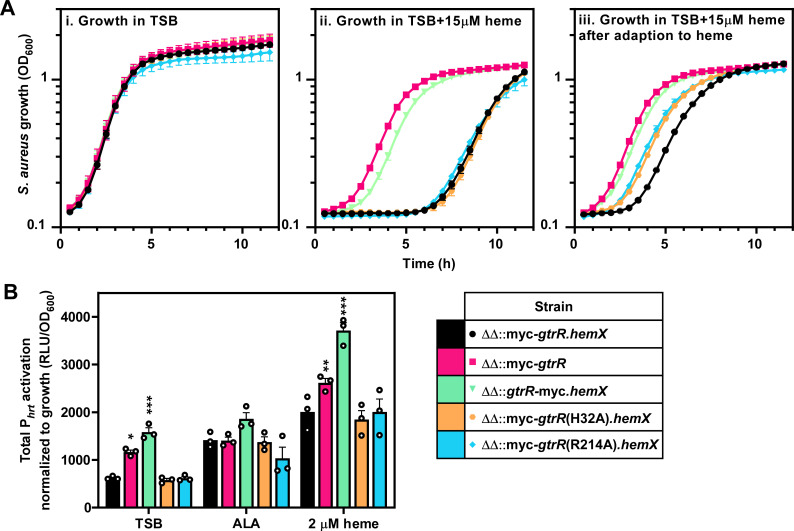
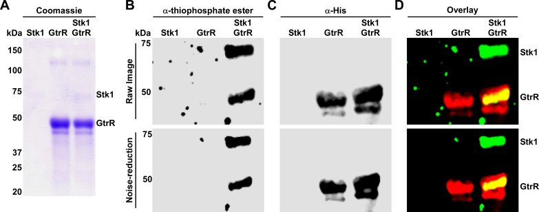
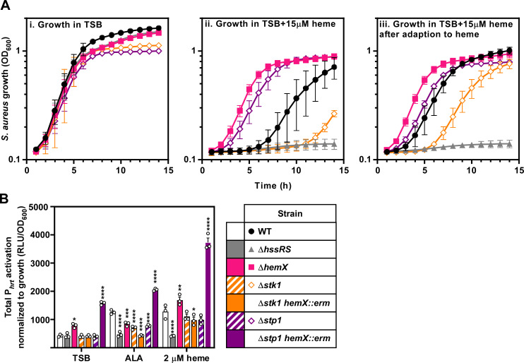
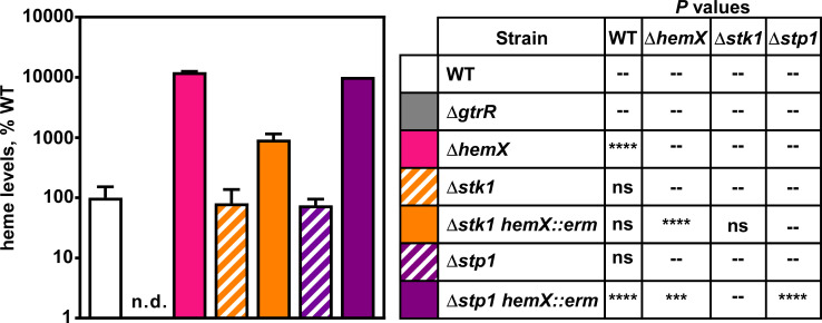
Staphylococcus aureus is an important human pathogen responsible for a variety of infections including skin and soft tissue infections, endocarditis, and sepsis. The combination of increasing antibiotic resistance in this pathogen and the lack of an efficacious vaccine underscores the importance of understanding how S. aureus maintains metabolic homeostasis in a variety of environments, particularly during infection. Within the host, S. aureus must regulate cellular levels of the cofactor heme to support enzymatic activities without encountering heme toxicity. Glutamyl tRNA reductase (GtrR), the enzyme catalyzing the first committed step in heme synthesis, is an important regulatory node of heme synthesis in Bacteria, Archaea, and Plantae. In many organisms, heme status negatively regulates the abundance of GtrR, controlling flux through the heme synthesis pathway. We identified two residues within GtrR, H32 and R214, that are important for GtrR-heme binding. However, in strains expressing either GtrRH32A or GtrRR214A, heme homeostasis was not perturbed, suggesting an alternative mechanism of heme synthesis regulation occurs in S. aureus. In this regard, we report that heme synthesis is regulated through phosphorylation and dephosphorylation of GtrR by the serine/threonine kinase Stk1 and the phosphatase Stp1, respectively. Taken together, these results suggest that the mechanisms governing staphylococcal heme synthesis integrate both the availability of heme and the growth status of the cell. IMPORTANCE Staphylococcus aureus represents a significant threat to human health. Heme is an iron-containing enzymatic cofactor that can be toxic at elevated levels. During infection, S. aureus must control heme levels to replicate and survive within the hostile host environment. We identified residues within a heme biosynthetic enzyme that are critical for heme binding in vitro; however, abrogation of heme binding is not sufficient to perturb heme homeostasis within S. aureus. This marks a divergence from previously reported mechanisms of heme-dependent regulation of the highly conserved enzyme glutamyl tRNA reductase (GtrR). Additionally, we link cell growth arrest to the modulation of heme levels through the post-translational regulation of GtrR by the kinase Stk1 and the phosphatase Stp1.

Keywords: Staphylococcus aureus; heme synthesis.


Conflict of interest statement

The authors declare no conflict of interest.

Figures

Fig 1
Fig 1
S. aureus GtrR binds heme in vitro. (A) Absorption spectra of heme in solution and in the presence of purified WT GtrR in dialysis buffer. The spectrum corresponding to 50 µM free heme is shown as a solid pink line. Increasing concentrations of heme (5–50 µM, in grayscale from light to dark) were incubated in the presence of 5 µM purified protein. (B and C) Absorption spectra of heme in dialysis buffer (B) and after reduction with the addition of excess dithionite (C). Absorbance values at wavelengths below 500 nm are plotted on the left Y axis and absorbance values above 500 nm are plotted on the right Y axis for visualization of α/β bands.
Fig 2
Fig 2
Conservation, domain organization, and homology model of S. aureus GtrR. (A) Homology model of a S. aureus GtrR homodimer generated using SWISS-MODEL with GtrR from Arabidopsis thaliana as a template (61, 62). Residues selected for mutagenesis are highlighted. On the left monomer, domains are colored to match the domain organization modeled in B. On the right, amino acids are colored according to the degree of conservation. A scale of variable (teal) to conserved (purple) residues is indicated in the figure. (B) Domain organization (bottom bar: Catalytic, pink; NADPH binding, green; Spinal Helix, light orange; Dimerization, dark orange) and conservation of S. aureus GtrR primary sequence compared to increasing taxonomic levels (top bars, colored as in A).
Fig 3
Fig 3
Histidine 32 and arginine 214 are individually important for GtrR binding to heme in vitro. Absorption spectra of increasing concentrations of heme (5–50 µM) in the presence of 5 µM purified GtrRH32A (A) or GtrRR214A (B). The spectrum corresponding to 50 µM free heme is shown as a solid pink line. Heme concentrations ranging from 5 to 50 µM of heme are graphed in grayscale from light to dark.
Fig 4
Fig 4
GtrR variants expressed in a S. aureus ΔgtrR ΔhemX mutant are catalytically active and heme produced is utilized in cellular hemoproteins. (A) S. aureus strains were serially diluted and spot plated on TSA. After growth, hydrogen peroxide was pipetted onto the resulting colonies. Colony morphology and qualitative catalase activity are summarized on the left (A). Strains with wild-type colony size are annotated as “WT” and small colony variants “SCV.” (B) Catalase activity of strains used in (A) was quantified using a Catalase Assay Kit. Bars are representative of the averages from three independent experiments performed in biological triplicate with error bars representing the SEM. Significance was calculated in Graphpad Prism 8 using the Brown-Forsythe and Welch ANOVA tests and Dunnett’s T3 multiple comparisons tests with individual variances computed for each comparison with WT; **P < 0.01; non-significant comparisons are not shown.
Fig 5
Fig 5
Disruption of heme binding to GtrR does not result in activation of the HssRS two-component system. (A) Growth as measured by OD600 was monitored over time for S. aureus strains grown in TSB containing either vehicle (i) or 15 µM heme (ii, iii). Prior to the measured growth, the strains were pre-grown to stationary phase in TSB containing vehicle (i, ii) or 2 µM heme (iii). (B) Total activation of the hrtAB promoter (Phrt) over 20 h of strains grown in TSB containing 2 mM δ-aminolevulinic acid, 2 µM heme, or vehicle. Time course data used to determine total promoter activation are graphed in Fig. S3. (A and B) Graphs represent the averages of means from three independent experiments, performed in biological triplicate ± SEM. Significance was calculated using two-way ANOVA with Dunnett’s test for multiple comparisons between ΔΔ::myc-gtrR.hemX and each strain, with *P < 0.05; **P < 0.005; ***P = 0.001; non-significant comparisons are not shown.
Fig 6
Fig 6
GtrR is a substrate of Stk1 kinase. The cytoplasmic kinase domain of S. aureus Stk1 was expressed and purified as a GST-tagged fusion and incubated with purified His-tagged GtrR in the presence of the ATP analog ATPγS. Following PNBM alkylation, the labeled substrates were separated by SDS-PAGE, (A) coomassie stained, and immunoblotting used to detect (B) phosphorylated protein and (C) His-tagged GtrR. Phosphorylated protein and His-tagged GtrR were detected using an anti-thiophosphate ester and anti-His antibodies, respectively. An overlay of the His and thiophosphate ester signals is shown in (D). Bands corresponding to phosphorylated Stk1 and GtrR are annotated. (B–D) Raw images with noise were processed using noise reduction software to improve background clarity. Both the raw (top) and processed images (bottom) are shown for each immunoblot.
Fig 7
Fig 7
Stk1 and Stp1 impact heme stress signaling within S. aureus. (A) Growth as measured by OD600 was monitored over time for S. aureus strains grown in TSB containing either vehicle (i) or 15 µM heme (ii, iii). Prior to the measured growth, the strains were pre-grown to stationary phase in TSB containing vehicle (i, ii) or 2 µM heme (iii). (B) Total activation of luciferase expression from the hrtAB promoter (Phrt) from strains grown in TSB containing 2 mM δ-aminolevulinic acid (ALA), 2 µM heme, or vehicle. Time course data used to determine total promoter activation are graphed in Fig. S4. (A and B) Graphed are the averages of means from three independent experiments performed in biological triplicate ± SEM. Significance was calculated using two-way ANOVA with Tukey’s test for multiple comparisons using the comparisons listed in Table 3.
Fig 8
Fig 8
Stk1 is important for full heme synthesis in a hemX mutant. Heme was quantified by HPLC in S. aureus WT and mutant strains. Data are representative of the averages of means normalized to the amount of heme measured in WT from two independent experiments performed in biological triplicate ± SEM. “n.d.” indicates that no signal was detected. Significance was calculated in Graphpad Prism 8 using a two-way ANOVA with Welch’s test for multiple comparisons between the strain pairs listed in the table on the right with ***P = 0.001; ****P < 0.0001; ns, non-significant; --, comparison not made. No comparison was made between WT and ΔgtrR since Welch’s test cannot be performed on samples with identical values.

References

    1. Choby JE, Grunenwald CM, Celis AI, Gerdes SY, DuBois JL, Skaar EP, Kline KA, Richardson A, Heinrichs D. 2018. Staphylococcus aureus hemX modulates glutamyl-tRNA reductase abundance to regulate heme biosynthesis. mBio 9:e02287-17. doi: 10.1128/mBio.02287-17 - DOI - PMC - PubMed
    1. Ponka P. 1999. Cell biology of Heme. Am J Med Sci 318:241–256. doi: 10.1097/00000441-199910000-00004 - DOI - PubMed
    1. Celis AI, DuBois JL. 2019. Making and breaking heme. Curr Opin Struct Biol 59:19–28. doi: 10.1016/j.sbi.2019.01.006 - DOI - PMC - PubMed
    1. Dailey HA, Dailey TA, Gerdes S, Jahn D, Jahn M, O’Brian MR, Warren MJ. 2017. Prokaryotic heme biosynthesis: multiple pathways to a common essential product. Microbiol Mol Biol Rev 81:e00048-16. doi: 10.1128/MMBR.00048-16 - DOI - PMC - PubMed
    1. Stauff DL, Skaar EP. 2009. The heme sensor system of staphylococcus aureus. Contrib Microbiol 16:120–135. doi: 10.1159/000219376 - DOI - PMC - PubMed

Publication types

MeSH terms