Review
. 2023 Nov;146(5):663-683.
doi: 10.1007/s00401-023-02629-2. Epub 2023 Sep 1.

Microglial contribution to the pathology of neurodevelopmental disorders in humans

Affiliations
Review

Microglial contribution to the pathology of neurodevelopmental disorders in humans

Rugile Matuleviciute et al. Acta Neuropathol. 2023 Nov.

Abstract

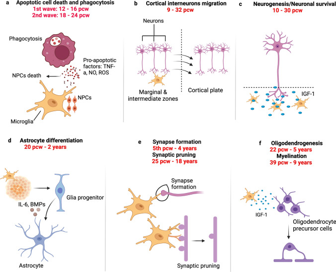
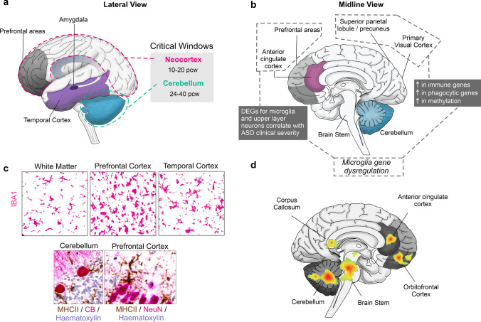
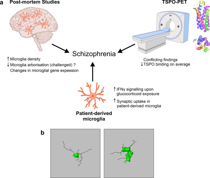
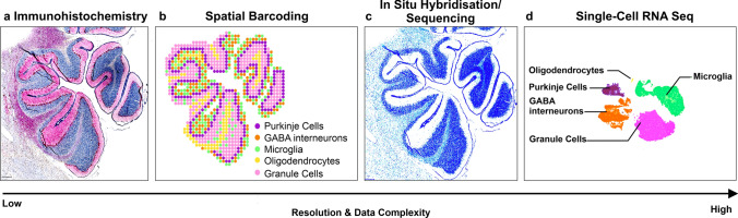
Microglia are the brain's resident macrophages, which guide various developmental processes crucial for brain maturation, activity, and plasticity. Microglial progenitors enter the telencephalic wall by the 4th postconceptional week and colonise the fetal brain in a manner that spatiotemporally tracks key neurodevelopmental processes in humans. However, much of what we know about how microglia shape neurodevelopment comes from rodent studies. Multiple differences exist between human and rodent microglia warranting further focus on the human condition, particularly as microglia are emerging as critically involved in the pathological signature of various cognitive and neurodevelopmental disorders. In this article, we review the evidence supporting microglial involvement in basic neurodevelopmental processes by focusing on the human species. We next concur on the neuropathological evidence demonstrating whether and how microglia contribute to the aetiology of two neurodevelopmental disorders: autism spectrum conditions and schizophrenia. Next, we highlight how recent technologies have revolutionised our understanding of microglial biology with a focus on how these tools can help us elucidate at unprecedented resolution the links between microglia and neurodevelopmental disorders. We conclude by reviewing which current treatment approaches have shown most promise towards targeting microglia in neurodevelopmental disorders and suggest novel avenues for future consideration.

Keywords: Autism spectrum conditions; Human microglia; Human-induced pluripotent stem cells; Neurodevelopmental disorders; Neurodevelopmental models; Schizophrenia; Spatial transcriptomics.


Figures

Fig. 1
Fig. 1
Main functions of microglia in the developing human CNS. a Microglia regulate the number of neuronal cells, notably by engaging in active phagocytosis of progenitor cells and promoting the apoptosis of differentiated cells [17, 158]. b Microglia regulate neuronal migratory processes within the neocortex [17]. c Microglia also release factors (such as insulin growth factor 1 (IGF-1), that support the growth and survival of neurons [17]. d Microglia impact onto astrocyte differentiation via direct cell-to-cell communication, secretion of microglia-derived and trophic factors [5, 135]. e Microglia mediate synapse elimination for instance via the engulfment of presynaptic inputs [119, 121]. f Microglia promote the proliferation and differentiation of oligodendrocyte precursor cells, and they interact with oligodendrocytes to mediate their maturation and survival, as well as contribute to maintaining the myelination status of the CNS [77]
Fig. 2
Fig. 2
Microglial findings in ASCs. a Lateral view of the human brain with the critical/sensitive windows for ASC development, which include the cerebellum and the neocortex [209]. b Microglial gene dysregulation observed in ASC samples from the prefrontal and cingulate [203] as well as the primary visual areas and the superior parietal lobule [49]. c Microglial densities (IBA1+, magenta) are unchanged, higher, or lower in these brain areas compared to typically developing controls (top panel). In the cerebellum and neocortex, neuroinflammatory processes are heightened and microglia express MHC-II (brown) and cup Purkinje Calbindin+ cells in the cerebellum (CB+, magenta) and neurons in the neocortex (NeuN+, magenta) (bottom panel). Scale bars: 25 μm. d Areas of the brain demonstrated to show hyperactivation linked to microglial TPSO signal. Most areas show hyperactivation except the cerebellum and the cingulate which show both hyper and hypoactivation depending on the study [175, 187, 223]
Fig. 3
Fig. 3
Microglial impairments in SZ. a Post-mortem studies report changes in microglial density [52, 89, 197], arborization and gene expression [80, 139, 156, 176, 222] in patients with SZ compared to controls. TSPO-PET shows conflicting findings, however, there is a suggestion of decreased TSPO binding in grey matter in patients with SZ [108, 109, 153, 154]. TSPO structure representation is shown on the top right (structure adapted from Guo et al. [57]). HiPSC and patient monocyte microglia show increased IFN-γ signalling upon glucocorticoid exposure [211] and increased synaptic uptake compared to control lines, respectively [172]. b Drawing of 3D microglial morphology in a control brain (left) and a schizophrenia brain (right). Images adapted and redrawn from De Picker et al. [31]'s original confocal stack images, scale bar: 10 μm
Fig. 4
Fig. 4
Type of data acquired using spatial methods. a Immunohistochemistry (IHC) of the cerebellum tagged with Calbindin for Purkinje cell identification, IBA1 for microglia and haematoxylin for all nuclei. b Spatial transcriptomics schematisation with cell populations approximated topographically in the same section. c In situ hybridisation (ISH)/ in situ sequencing (ISS) example in the cerebellum showing schematisation of transcripts against myelin basic protein. d Single-cell RNA seq schematisation of cell populations that can be identified in the cerebellum. Overall, spatial barcoding offers a lower resolution, whereas ISS/ISH offer a higher resolution. The complexity of the data increases with the resolution. Relative to IHC, the number of identified cells would need to be larger going from IHC to spatial barcoding and ISS/ISH spatial methods, to scRNA-seq. Conceptually, this would place the two spatial technologies at the heart of modern-day developmental science, at the intersection between histology and single-cell sequencing

Similar articles

Cited by

References

    1. Abud EM, Ramirez RN, Martinez ES, Healy LM, Nguyen CHH, Newman SA, Yeromin AV, Scarfone VM, Marsh SE, Fimbres C, Caraway CA, Fote GM, Madany AM, Agrawal A, Kayed R, Gylys KH, Cahalan MD, Cummings BJ, Antel JP, Mortazavi A, Carson MJ, Poon WW, Blurton-Jones M. iPSC-derived human microglia-like cells to study neurological diseases. Neuron. 2017;94:278–293.e9. doi: 10.1016/j.neuron.2017.03.042. - DOI - PMC - PubMed
    1. Agans R, Rigsbee L, Kenche H, Michail S, Khamis HJ, Paliy O. Distal gut microbiota of adolescent children is different from that of adults. FEMS Microbiol Ecol. 2011;77:404–412. doi: 10.1111/j.1574-6941.2011.01120.x. - DOI - PMC - PubMed
    1. Almehmadi KA, Tsilioni I, Theoharides TC. Increased expression of miR-155p5 in amygdala of children with autism spectrum disorder. Autism Res. 2020;13:18–23. doi: 10.1002/aur.2205. - DOI - PubMed
    1. American Psychiatric Association (2013) Diagnostic and Statistical Manual of Mental Disorders, Fifth Edition. American Psychiatric Association
    1. Antony JM, Paquin A, Nutt SL, Kaplan DR, Miller FD. Endogenous microglia regulate development of embryonic cortical precursor cells. J Neurosci Res. 2011;89:286–298. doi: 10.1002/jnr.22533. - DOI - PubMed

Publication types