Review
. 2024 Jan;109(1):81-99.
doi: 10.1113/EP090761. Epub 2023 Sep 1.

The atypical 'hippocampal' glutamate receptor coupled to phospholipase D that controls stretch-sensitivity in primary mechanosensory nerve endings is homomeric purely metabotropic GluK2

Affiliations
Review

The atypical 'hippocampal' glutamate receptor coupled to phospholipase D that controls stretch-sensitivity in primary mechanosensory nerve endings is homomeric purely metabotropic GluK2

Karen J Thompson et al. Exp Physiol. 2024 Jan.

Abstract

A metabotropic glutamate receptor coupled to phospholipase D (PLD-mGluR) was discovered in the hippocampus over three decades ago. Its pharmacology and direct linkage to PLD activation are well established and indicate it is a highly atypical glutamate receptor. A receptor with the same pharmacology is present in spindle primary sensory terminals where its blockade can totally abolish, and its activation can double, the normal stretch-evoked firing. We report here the first identification of this PLD-mGluR protein, by capitalizing on its expression in primary mechanosensory terminals, developing an enriched source, pharmacological profiling to identify an optimal ligand, and then functionalizing it as a molecular tool. Evidence from immunofluorescence, western and far-western blotting indicates PLD-mGluR is homomeric GluK2, since GluK2 is the only glutamate receptor protein/receptor subunit present in spindle mechanosensory terminals. Its expression was also found in the lanceolate palisade ending of hair follicle, also known to contain the PLD-mGluR. Finally, in a mouse model with ionotropic function ablated in the GluK2 subunit, spindle glutamatergic responses were still present, confirming it acts purely metabotropically. We conclude the PLD-mGluR is a homomeric GluK2 kainate receptor signalling purely metabotropically and it is common to other, perhaps all, primary mechanosensory endings.

Keywords: GluK2; PLD-mGluR; glutamate receptor; kainate receptor; mechanosensation; muscle spindle.


Conflict of interest statement

H.E.S., B.A.H., L.M.B. and D.B. were employees of Eli Lilly at the time the work was done, and Eli Lilly joint‐funded the scholarship for S.W.’s contribution to this study.

Figures

FIGURE 1
FIGURE 1
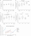
Whole nerve electroneurograms of stretch‐evoked responses from rat lumbrical muscles: firing enhanced by glutamate and inhibited by PCCG‐13. (a) Top: trapezoidal stretch‐hold‐release profile (1 mm stretch, ∼10% increase in length) used for all electrophysiology experiments. Middle: normal stretch response (cyan), and enhanced firing (black) in glutamate (1 mM, 1 h). Bottom: mean firing frequency profile for these responses, showing the glutamate‐mediated enhancement of firing (rolling average, sampled every 10 ms). (b) Initial 500 ms of hold phase firing, showing (top) glutamate (1 mM, 1 h) reversibly enhances and (bottom) PCCG‐13 (10 μM, 3 h) reversibly inhibits firing. These are illustrative repetitions of full experiments published earlier (Bewick et al., 2005). NB: action potential (AP) amplitude variation reflects the long duration of experiments (up to 14 h), reconnecting the nerve for each recording and AP summation in whole nerve recordings, so cannot be used to infer drug action.
FIGURE 2
FIGURE 2
Further pharmacological characterization of PLD‐mGluR in muscle spindles. (a) Ligands without modulatory action included ibotenate (gp I mGluR agonist), l‐CCG‐I (gp II mGluR agonist), DCG‐IV (gp II mGluR and NMDA‐R agonist), ACPD (gp I, II and III mGluR agonist), and LY341495 (gp I, II and III mGluR antagonist at 100 μM). (b–d) In contrast, (b) quisqualate (AMPA, KAR and gp I mGluR agonist), (c) t‐ADA and (d) l‐CSA (weak gp I agonists) increase stretch‐evoked firing. (e) Partial dose–response relationships for effective agonists. Rank order of potency: l‐CSA > t‐ADA > quisqualate > kainate > glutamate. Kynurenic acid (KA, 1 mM) substantially inhibits the excitation by l‐CSA. (a–d) Individual data expressed relative to control grand mean; statistical analysis of raw data: one‐way ANOVA with replicates, post‐hoc pairwise paired Student's t‐test, significant differences shown after Benjamini–Hochberg correction method for multiple tests.
FIGURE 3
FIGURE 3
The ‘inert’ enantiomer (R)‐3,5‐DHPG inhibits the PLD‐mGluR. The racemic (RS)‐3,5‐DHPG mixture blocks enhancement of stretch‐evoked firing in spindles by exogenous glutamate (Bewick et al., 2005). (a) Applied alone, it tended to decrease firing, but did not reach significance (m = 5 muscles; r = 5 rats). (b) (S)‐DHPG had no effect on firing up to 200 μM (m = 5, r = 5). (c) (R)‐DHPG (inactive enantiomer against gp I‐III mGluRs) significantly decreased it even at 1 μM (m = 7, r = 5), reversing slowly if at all. (d) 200 μM (S)‐DHPG blocked 1 mM glutamate‐mediated enhancement (m = 7, r = 4), suggesting it may be a neutral antagonist. Box‐and‐whisker plots in (a–d): individual data expressed relative to control grand mean; statistical analysis of raw data: (a–c) one‐way ANOVA with replicates, post‐hoc pairwise paired t‐tests, significant differences shown after Benjamini–Hochberg correction method for multiple tests; (d) paired t‐tests versus relevant controls (controls not shown).
FIGURE 4
FIGURE 4
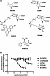
Kainate functionalization and screening ZCZ90 for receptor activity in rat primary cortical neurones. (a) Top: adding a triazole group to kainate produced ZCZ90. Middle: the triazole group enabled a linker side chain to attach either fluorescein (ZCZ172) or biotin (ZCZ180) for use in fluorescence imaging or far‐western blotting, respectively. Bottom: configuration of the structurally highly constrained PLD‐mGluR specific antagonist PCCG‐13, for comparison. (b) FLIPR assay screening of PLD‐mGluR ligands and the functionalized kainate analogue ZCZ90 against glutamate receptors in primary cultures of rat cortical neurones (all n = 3). S‐DHPG (gp I agonist) and LY379268 (potent gp II agonist) both reduced Ca2+ oscillation frequency (EC50 336 ± 213 nM and 1.5 ± 0.27 nM respectively; both P < 0.0001; two‐way ANOVA). However, the PLD‐mGluR agonists ZCZ‐90, l‐CSA and the enantiomer (R)‐DHPG had no effect (all P > 0.5, two‐way ANOVA) indicating they do not activate classical mGluRs. Both kainate and ZCZ90 are PLD‐mGluR agonists, blocked by PCCG‐13 (10 μM; Zanato et al., 2014).
FIGURE 5
FIGURE 5
Kainate derivatives ZCZ180 and ZCZ172 label muscle spindle blots and sensory endings, respectively. (a) ZCZ180, the biotinylated kainate derivative, labelled an approximately 120 kDa protein (dashed box) in tissue homogenates of both spindle‐rich material (S), and the hippocampus (H; positive control). (b) In fixed, squashed rat deep lumbrical muscle whole mounts, fluorescently tagged kainate derivative ZCZ172 (green) labelled the sensory nerve endings of muscle spindles, colocalizing (merged) with immunolabelling for synaptophysin (red) in synaptic‐like vesicles in the endings. (c) In contrast, in the same preparations, there was no ZCZ172 signal (green) above background at motor nerve terminals (arrows) on nearby extrafusal fibres. There are three motor terminals in this field identified by anti‐synaptophysin immunofluorescence (red), two (red, upper and lower right) out of the focal plane, on the fibre edges, and one en face (centre) in the plane of focus. Note, the high background ZCZ172 labelling reflects the minimal wash, as its binding is readily reversible, unlike antibodies. Images are representative of three separate experiments. Scale bar = 10 μm.
FIGURE 6
FIGURE 6
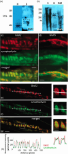
Muscle spindle primary sensory terminals express only GluK2; GluK5 is in intrafusal muscle fibre nuclei. (a, b) Western blots of spindle‐rich homogenate contained bands (boxed areas) for (a) GluK2 (113 kDa; n = 5) and (b) GluK5 (123 kDa; n = 4). The spindle (S) GluK2 band was slightly heavier than that in the hippocampus (H) positive control. DM, spindle‐free lateral deep masseter (negative control); kDa, kilodalton, marker lane. (c) In teased rat lumbrical whole mount preparations, spindle sensory terminals (green synaptophysin labelling) are brightly labelled by GluK2 antibodies (red). (d) However, GluK5 antibodies (red) labelled only the nuclei of muscle spindle intrafusal muscle fibres. Merged images (lower panels) show these spatial relationships more clearly. (e–g) Confocal optical sections showed a halo of enrichment for GluK2 at the edges of the terminal. Intensity quantification of a line along the central long axis of the terminal helix (white line connecting two circles) for GluK2 (e, red) and synaptophysin (f, green) showed GluK2 labelling present throughout the terminal, but it also extended beyond that of synaptophysin (h), more clearly seen in the enlargements (right). Thus, GluK2 is expressed throughout the terminal, possibly within the SLVs, but particularly highly on the terminal surface. Scale bar = 10 μm main images, 4 μm insets.
FIGURE 7
FIGURE 7
mGluR5 is expressed on non‐spindle sensory axons. (a) A band for mGluR5 was present in spindle homogenates (S) but at a substantially lower molecular mass than in hippocampus (H, positive control). No bands were detected in lateral deep masseter (DM; negative control). (b) In whole‐mounts, the mGluR5 antibody (red) labelled thin, unmyelinated axons (arrow), presumptive nociceptive sensory axons, near spindle capsules but not primary mechanosensory terminals (green, arrow). The merged image shows the separate locations more clearly. Scale bar = 10 μm.
FIGURE 8
FIGURE 8
Glutamate‐induced modulation of muscle spindle firing is still present in mice with ablated ionotropic GluK2 function. (a) Schematic diagram showing homologous replacement of the MD2 exon of Grik2 with a Neo‐cassette to disrupt GluK2 ionotropic function (only relevant portion of Grik2 shown). (b) Mean whole‐nerve action potential (spike) counts from mouse soleus muscle at hold‐phase stretch of 1 mm (∼10% initial length) in GluK2‐Neo mice (n = 9, m = 5) and wild‐type (n = 8, m = 5) littermates. Both genotypes displayed repeatable responses to the first four stretches (group 1 on x‐axis). Also, unloading muscles (60 min), minimizing stretch‐evoked release of endogenous glutamate, markedly inhibited their firing (group 2; both P < 0.01). Both recovered strongly (group 3) in the next series, as stretch re‐established endogenous release levels. Adding exogenous glutamate (100 μM) during unloading prevented the unloading‐induced reduction (WT P = 0.7; Neo P = 0.5). There was no further enhancement to futher increase in glutamate (1 mM) in either genotype. Overall, in both genotypes, glutamate sensitivity in both directions (reduced endogenous release reduced firing, 100 μM exogenous glutamate prevented this) remained. Since GluK2‐Neo ablates ionotropic function, the remaining responses seem most likely to reflect continued metabotropic signalling.
FIGURE 9
FIGURE 9
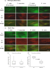
Normal GluK2 expression in soleus muscle spindles of GluK2‐Neo mice but C‐terminus immunolabelling is disrupted. (a, b) Wild‐type (a) and GluK2‐Neo (b) mouse soleus muscle‐spindle primary endings displayed GluK2 immunolabelling with antibodies directed at either N‐terminus (rGRIK2, column 1A, green) or C‐terminus (rGluR6/7, column 3A, green) epitopes, and both colocalized (yellow, 1C, 3C) with anti‐synaptophysin labelling (red, 1B, 3B), a marker for the primary sensory terminals. In both genotypes, labelling was low/absent if primary antibody was pre‐incubated with blocking peptide (rGRIK2, A and B column 2) or the primary antibody was omitted (rGluR6/7, A and B column 4). Scale bar = 100 μm. Thus, full‐length GluK2 is expressed in both genotypes. (c) However, while N‐terminus‐directed antibody (upstream of the insertion) labelling was not different from that in wild‐type mice (n = 9), the intensity with C‐terminus‐directed antibodies (downstream of the insertion) was approximately 50% lower in GluK2‐Neo mice (n = 7, P < 0.009, two‐tailed t test for 7 df and α = 0.025 with Bonferroni correction, carried out on arctan transformed data), indicating the intracellular epitope recognized by the antibody was disrupted.
FIGURE 10
FIGURE 10
Primary afferent terminals in other mechanosensory organs also exhibit GluK2 immunoreactivity. Adult male Wistar rat ear hair follicle afferents (synaptophysin, green) labelled with antibodies to GluK2 (red). As for spindle primary afferents, the great majority of individual terminals exhibited a halo of red GluK2 labelling surrounding the green core (merged, and enlargement of boxed area), again suggesting a surface membrane accumulation in the terminal. Scale bar = 10 μm (main image) and 2 μm (enlarged).

Similar articles

Cited by

References

    1. Albani‐Torregrossa, S. , Attucci, S. , Marinozzi, M. , Pellicciari, R. , Moroni, F. , & Pellegrini‐Giampietro, D. E. (1999). Antagonist pharmacology of metabotropic glutamate receptors coupled to phospholipase D activation in adult rat hippocampus: Focus on (2R,1'S,2'R,3'S)‐2‐(2'‐carboxy‐3'‐phenylcyclopropyl)glycine versus 3, 5‐dihydroxyphenylglycine. Molecular Pharmacology, 55, 699–707. - PubMed
    1. Banks, R. W. (2006). An allometric analysis of the number of muscle spindles in mammalian skeletal muscles. Journal of Anatomy, 208(6), 753–768. - PMC - PubMed
    1. Banks, R. W. , Bewick, G. S. , Reid, B. , & Richardson, C. (2002). Evidence for activity‐dependent modulation of sensory‐terminal excitability in spindles by glutamate release from synaptic‐like vesicles. Advances in Experimental Medicine and Biology, 508, 13–18. - PubMed
    1. Banks, R. W. , Cahusac, P. M. B. , Graca, A. , Kain, N. , Shenton, F. , Singh, P. , Njå, A. , Simon, A. , Watson, S. , Slater, C. R. , & Bewick, G. S. (2013). Glutamatergic modulation of synaptic‐like vesicle recycling in mechanosensory lanceolate nerve terminals of mammalian hair follicles. The Journal of Physiology, 591(10), 2523–2540. - PMC - PubMed
    1. Béhé, P. , Stern, P. , Wyllie, D. J. , Nassar, M. , Schoepfer, R. , & Colquhoun, D. (1995). Determination of NMDA NR1 subunit copy number in recombinant NMDA receptors. Proceedings of the Royal Society B: Biological Sciences, 262, 205–213. - PubMed

Publication types