. 2023 Sep 26;42(9):113058.
doi: 10.1016/j.celrep.2023.113058. Epub 2023 Aug 31.

System-wide mapping of peptide-GPCR interactions in C. elegans

Affiliations

System-wide mapping of peptide-GPCR interactions in C. elegans

Isabel Beets et al. Cell Rep. .

Abstract

Neuropeptides and peptide hormones are ancient, widespread signaling molecules that underpin almost all brain functions. They constitute a broad ligand-receptor network, mainly by binding to G protein-coupled receptors (GPCRs). However, the organization of the peptidergic network and roles of many peptides remain elusive, as our insight into peptide-receptor interactions is limited and many peptide GPCRs are still orphan receptors. Here we report a genome-wide peptide-GPCR interaction map in Caenorhabditis elegans. By reverse pharmacology screening of over 55,384 possible interactions, we identify 461 cognate peptide-GPCR couples that uncover a broad signaling network with specific and complex combinatorial interactions encoded across and within single peptidergic genes. These interactions provide insights into peptide functions and evolution. Combining our dataset with phylogenetic analysis supports peptide-receptor co-evolution and conservation of at least 14 bilaterian peptidergic systems in C. elegans. This resource lays a foundation for system-wide analysis of the peptidergic network.

Keywords: C. elegans; CP: Cell biology; CP: Neuroscience; GPCR; interactome; neuropeptide; reverse pharmacology.


Conflict of interest statement

Declaration of interests The authors declare no competing interests.

Figures
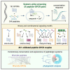
Graphical abstract
Graphical abstract
Figure 1
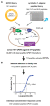
Figure 1. Systematic ligand screening of peptide-GPCR candidates in C. elegans
(A) Platform for identifying peptide ligands of C. elegans GPCRs. Candidate receptors are cloned under control of the cytomegalovirus immediate-early promoter (pCMV) for heterologous expression in CHO cells. Each GPCR is expressed together with the calcium-activated photoprotein aequorin and the promiscuous human Gα16 subunit and is screened with a synthetic library of C. elegans FLP and NLP peptides. Upon GPCR activation, phospholipase Cβ (PLCβ) is activated and hydrolyzes phosphatidylinositol bisphosphate (PIP2) into diacylglycerol (DAG) and inositol trisphosphate (IP3), which activates IP3-dependent calcium channels, resulting in calcium release from intracellular storage sites and increased luminescence. Screening of 55,384 individual peptide-GPCR interactions identified putative ligands for 114 out of 161 GPCRs. See also Figure S1, Tables S1–S4, and Data S1. (B) For each GPCR, putative hits are iteratively selected and each interaction is validated by measuring concentration-response curves, from which half-maximal effective concentrations (EC50) values are calculated. Out of 776 putative hits, 459 pairs show concentration-dependent GPCR activation. See Figures S2–S7 and Tables S4–S6 for tested and validated couples.
Figure 2
Figure 2. System-wide characterization of peptide GPCRs identifies a complex network of ligand-receptor interactions
(A) Cumulative frequency plot for log(EC50) values of 459 validated peptide-GPCR couples. (B) Venn diagram of peptides in the synthetic library that have been biochemically isolated and/ or for which a receptor has been identified. (C) Heatmap of log10(EC50) values for validated peptide-GPCR pairs. When a GPCR interacts with multiple peptides from a single peptide-encoding gene, only the log(EC50) value of the most potent ligand is included. Histograms indicate the number of interactions for each GPCR (top) or peptide precursor (right). See also Figures S2–S8 and Tables S1, S6, and S7.
Figure 3
Figure 3. The promiscuous receptor DMSR-7 inhibits cAMP signaling in response to diverse RFamide peptides
(A) Continuous TEVC recordings from untreated (left) and PTX-injected (right) Xenopus laevis oocytes expressing DMSR-7, mGIRK1, and mGIRK2 and treated with 100 nM FLP-1-6 peptide. FLP-1-6 activates DMSR-7, resulting in a robust stimulation of K+ current, which is blocked in PTX-injected oocytes. (B) The percentage GIRK activation for at least seven oocytes expressing DMSR-7 or the Gi/o-coupled muscarinic M2 receptor, mGIRK1, and mGIRK2 with or without PTX treatment. M2 was exposed to acetylcholine and DMSR-7 to FLP-1-6 (100 nM). Boxplots indicate 25th (lower boundary), 50th (central line), and 75th (upper boundary) percentiles. Whiskers show minimum and maximum values. (C and D) Mean percentage GIRK activation for oocytes injected with DMSR-7, mGIRK1, and mGIRK2 (C), or with mGIRK1/2 alone (D), in response to different peptides at 1 μM(n ≥ 5). NLP-3-1 does not activate DMSR-7 (Table S6) and was included as a negative control. (E and F) Activation of DMSR-7 by diverse FLP ligands (10 μM) significantly decreases cAMP levels in HEK cells (E). None of the peptides affect cAMP signaling in cells transfected with a control vector (F). Data are shown as relative luminescence after normalization to the ligand-free control (n ≥ 6). (G and H) Activation of DMSR-7 does not affect calcium levels in CHO cells in the absence of the Gα16 protein. Values are reported as the ratio of the total calcium response in cells transfected with DMSR-7 (G) or empty vector (H), and challenged with peptides (10 μM), BSA (negative control), or ATP (positive control) (n ≥ 6). Error bars indicate SEM. Significance was assessed by Mann-Whitney test (A), Kruskal-Wallis with Dunn’s test (C, D, G, and H), and one-way ANOVA with Dunnett’s test (E and F). ****p < 0.0001, ***p < 0.001, **p < 0.01, *p < 0.05; ns, not significant. See also Figure S8.
Figure 4
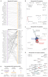
Figure 4. Specific and combinatorial interactions in the peptidergic network
(A) Bipartite graphs of NLP and FLP peptide-GPCR networks. Nodes represent peptides (purple) and receptors (orange). Edges between two nodes depict peptide-GPCR couples with sub-micromolar EC50 values (EC50 < 1 μM) identified in this resource. Asterisks indicate receptors in both NLP and FLP networks. (B and C) Monopartite peptide projections of the NLP (B) and FLP (C) bipartite peptide-GPCR networks. Nodes are sized by the total number of connections (degree) and colored by module. (D) Monopartite receptor projection of the FLP bipartite network. Nodes are sized by degree and colored by module. (E) Different types of peptide-receptor interaction motifs in the peptidergic network. See also Figures S9–S12.
Figure 5
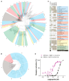
Figure 5. Peptide-GPCR pairs support conservation of bilaterian peptidergic systems and expansions of peptide-GPCR families in nematodes
(A) Maximum-likelihood tree of bilaterian rhodopsin peptide GPCRs. Subtrees are numbered according to the receptor families in (C). Subtrees comprising bilaterian families with nematode representatives are indicated in blue; those without nematode representatives are in red. Subtrees containing only protostomian sequences are colored yellow and nematode-specific subtrees are green. Node support values at the root of all delineated subtrees are above 0.95 unless depicted otherwise. Atypical peptide receptors (angiotensin, bradykinin, and chemokine receptors) were used as outgroup (gray). (B) Maximum-likelihood tree of bilaterian secretin receptors. Color-coding, numbering, and node support values as in (A). Adhesion and cadherin receptors were used as outgroup. (C) Inferred evolutionary relationships for nematode peptide-receptor systems. Names of peptidergic systems are assigned according to the classification by Mirabeau and Joly and Elphick et al. and color-coded as in (A). Gray squares mark the presence of a receptor ortholog. A receptor is considered present in a nematode clade when it is positioned inside a well-supported subtree (branch support value >0.95). The number of C. elegans receptors in each family is depicted in the left column and squares are colored green if at least one receptor is paired with a peptide ligand. (D) Elevenin-like SNET-1 (LDCRKFSFAPACRGIML; C3–C12) and calcitonin/DH31-like NLP-73 (NRQCLLNAGLSQGCDFSDLLHAQTQARKFMSFAGPamide; C4–C14) peptides activate the elevenin and calcitonin/DH31 receptor orthologs NPR-34 and SEB-2. Calcium responses of CHO cells expressing NPR-34 or SEB-2 are shown relative (%) to the highest value (100% activation) after normalization to the total calcium response. Error bars represent SEM (n ≥ 6). See also Data S1 and Tables S6, S8, and S9.

References

    1. Yañez-Guerra LA, Thiel D, Jékely G. Premetazoan origin of neuropeptide signaling. Mol Biol Evol. 2022;39:msac051. doi: 10.1093/molbev/msac051. - DOI - PMC - PubMed
    1. Schoofs L, De Loof A, Van Hiel MB. Neuropeptides as regulators of behavior in insects. Annu Rev Entomol. 2017;62:35–52. doi: 10.1146/annurev-ento-031616-035500. - DOI - PubMed
    1. Van Damme S, De Fruyt N, Watteyne J, Kenis S, Peymen K, Schoofs L, Beets I. Neuromodulatory pathways in learning and memory: Lessons from invertebrates. J Neuroendocrinol. 2021;33:e12911. doi: 10.1111/jne.12911. - DOI - PubMed
    1. Nässel DR, Zandawala M. Endocrine cybernetics: neuropeptides as molecular switches in behavioural decisions. Open Biol. 2022;12:220174. doi: 10.1098/rsob.220174. - DOI - PMC - PubMed
    1. Van Sinay E, Mirabeau O, Depuydt G, Van Hiel MB, Peymen K, Watteyne J, Zels S, Schoofs L, Beets I. Evolutionarily conserved TRH neuropeptide pathway regulates growth in Caenorhabditis elegans. Proc Natl Acad Sci USA. 2017;114:E4065–E4074. doi: 10.1073/pnas.1617392114. - DOI - PMC - PubMed

Publication types

LinkOut - more resources