. 2023 Sep;9(35):eadh8619.
doi: 10.1126/sciadv.adh8619. Epub 2023 Sep 1.

OLED catheters for inner-body phototherapy: A case of type 2 diabetes mellitus improved via duodenal photobiomodulation

Affiliations

OLED catheters for inner-body phototherapy: A case of type 2 diabetes mellitus improved via duodenal photobiomodulation

Jee Hoon Sim et al. Sci Adv. 2023 Sep.

Abstract

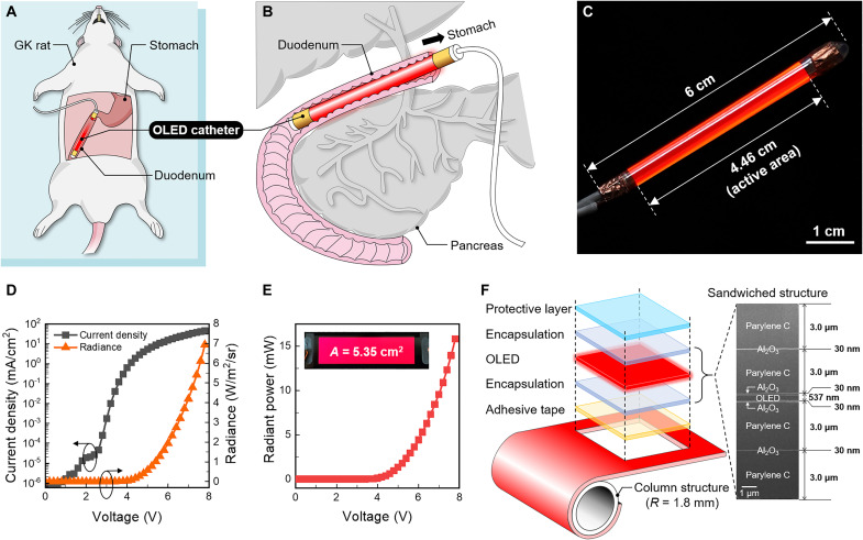
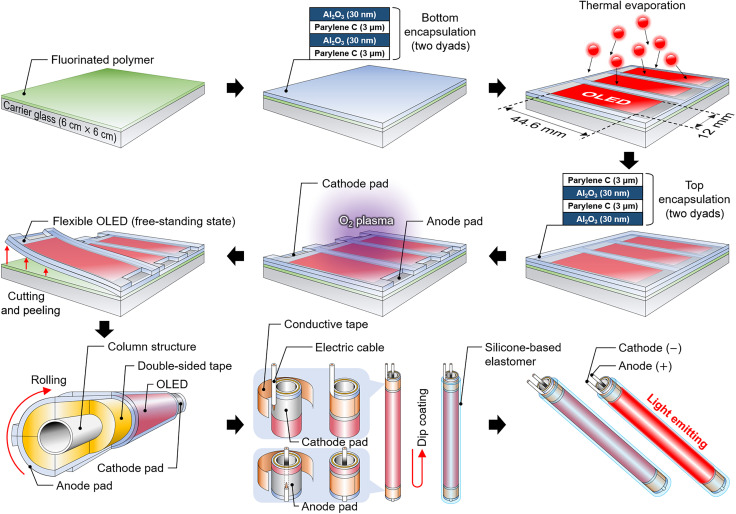
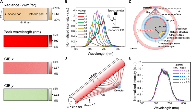
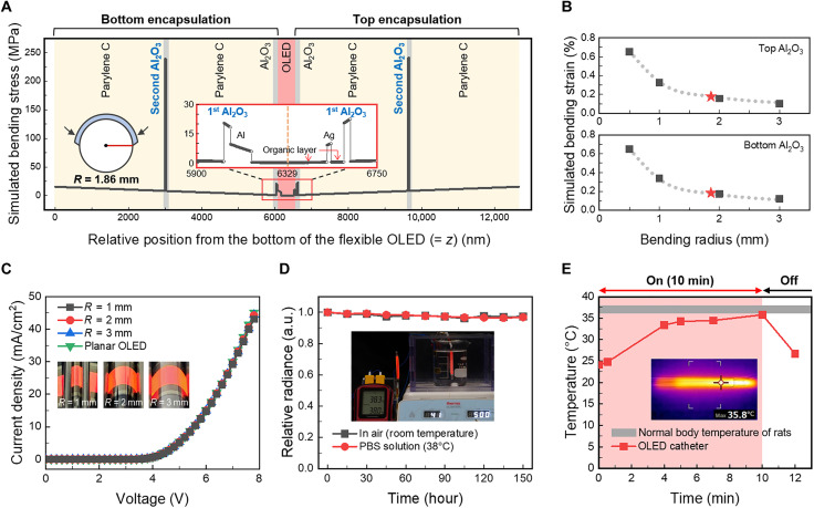
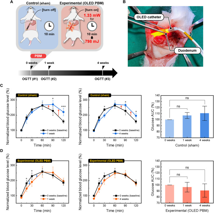
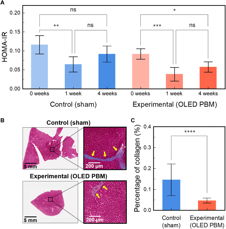
Phototherapeutics has shown promise in treating various diseases without surgical or drug interventions. However, it is challenging to use it in inner-body applications due to the limited light penetration depth through the skin. Therefore, we propose an organic light-emitting diode (OLED) catheter as an effective photobiomodulation (PBM) platform useful for tubular organs such as duodenums. A fully encapsulated highly flexible OLED is mounted over a round columnar structure, producing axially uniform illumination without local hotspots. The biocompatible and airtight OLED catheter can operate in aqueous environments for extended periods, meeting the essential requirements for inner-body medical applications. In a diabetic Goto-Kakizaki (GK) rat model, the red OLED catheter delivering 798 mJ of energy is shown to reduce hyperglycemia and insulin resistance compared to the sham group. Results are further supported by the subdued liver fibrosis, illustrating the immense potential of the OLED-catheter-based internal PBM for the treatment of type 2 diabetes and other diseases yet to be identified.


Figures

Fig. 1.
Fig. 1.. Overview of the proposed OLED catheter.
Schematic diagrams of (A) the phototherapy process proposed in this study and (B) an OLED catheter inserted into the duodenum of a GK rat. (C) A photograph of the actual light emission of the OLED catheter. (D) The current density (J)–voltage (V)–radiance (R) and (E) radiant power characteristics of the OLED catheter. (F) Overall configuration of the OLED catheter and focused ion beam scanning electron microscope cross-sectional image of the flexible OLED.
Fig. 2.
Fig. 2.. Fabrication process of the OLED catheter.
Flexible OLED fabrication, rolling, and dip-coating process.
Fig. 3.
Fig. 3.. Optical properties and design of the OLED catheter.
(A) Contour plots showing the uniformity of the flexible OLED in planar state: radiance, peak wavelength, and color coordinates. (B) Distribution of angular spectra of the flexible OLED in planar state. The optical simulation design of OLED catheter in (C) two-dimensional (2D) and (D) 3D view. (E) Measured (meas.) and simulated (sim.) spectral results of the OLED catheter according to distance (d). a.u., arbitrary units; CIE, commission on illumination.
Fig. 4.
Fig. 4.. Mechanical and thermal stability of the OLED catheter.
(A) Simulated stress distribution plot of the flexible OLED with bending radius of 1.86 mm, and the z axis represents the relative position with respect to the bottom of the device. (B) Simulated bending strain plot of the flexible OLED according to the bending radius [R = 0.5, 1, 1.86 (shown as a red-colored star; the strain value at the bending radius of the OLED attached onto the catheter), 2, 3 mm]. (C) J-V characteristics of the flexible OLEDs according to the bending radius (R = 1, 2, and 3 mm). (D) Lifetime test of the OLED catheter in PBS aqueous solution for 150 hours (photograph of inset corresponds to the actual measurement setup). (E) Temperature behavior of the OLED catheter during continuous operation in ambient air for 10 min. (The temperature changes of the duodenum induced by the OLED catheter in the actual experimental environment is shown in fig. S8.)
Fig. 5.
Fig. 5.. Duodenal PBM with an OLED catheter improves blood glucose levels in diabetic rats.
(A) Schematic illustration of in vivo experimental process showing duodenal PBM with an OLED catheter in a rat model and experimental design to evaluate efficacy through OGTT. (B) Photograph of duodenal PBM with OLED catheter as duodenal insertion. Glycemic curve during OGTT and normalized AUC in (C) sham control group (n = 4) and in (D) OLED duodenal PBM group (n = 5) showing the values at baseline, after 1 week, and 4 weeks from baseline (the arrows indicate the trends on the serum glucose level after 1 and 4 weeks from baseline). The data are presented as the median ± IQR. Ordinary one-way analysis of variance (ANOVA) with the Bonferroni’s post hoc test was used for statistical analysis. *P < 0.05 and ***P < 0.005. ns, not significant.
Fig. 6.
Fig. 6.. OLED PBM reduces IR and liver fibrosis in diabetic rats.
(A) HOMA-IR was measured at baseline (0 weeks), 1 week after the PBM, and 4 weeks after the PBM. (B) Histopathological image (MT staining) of a liver section showing the collagen deposition, which is the indicator of liver fibrosis. The blue area corresponds to collagen as indicated by the yellow arrows, and the pink area corresponds to cytoplasm. Zoomed area (right, black lined box) was acquired at ×4 magnification from the whole slice image (left, ×0.24). (C) Percentage of collagen deposition [=“blue” area/(blue + “pink” area)] in the liver section of the OLED PBM group (n = 5) versus the sham group (n = 4). *P < 0.05 and ***P < 0.005. ns, not significant. The data are presented as the median ± IQR. Ordinary one-way ANOVA with the Bonferroni’s post hoc test and unpaired t test were used for statistical analysis.

Similar articles

Cited by

References

    1. G. Hong, X. Gan, C. Leonhardt, Z. Zhang, J. Seibert, J. M. Busch, S. Bräse, A brief history of OLEDs—Emitter development and industry milestones. Adv. Mater. 33, 2005630 (2021). - PubMed
    1. J. Song, H. Lee, E. G. Jeong, K. C. Choi, S. Yoo, Organic light-emitting diodes: Pushing toward the limits and beyond. Adv. Mater. 32, 1907539 (2020). - PubMed
    1. C. Murawski, M. C. Gather, Emerging biomedical applications of organic light-emitting diodes. Adv. Opt. Mater. 9, 2100269 (2021).
    1. Y. Jeon, H. Choi, M. Lim, S. Choi, H. Kim, J. H. Kwon, K. Park, K. C. Choi, A wearable photobiomodulation patch using a flexible red-wavelength OLED and its in vitro differential cell proliferation effects. Adv. Mater. Technol. 3, 1700391 (2018).
    1. S. K. Attili, A. Lesar, A. McNeill, M. Camacho-Lopez, H. Moseley, S. Ibbotson, I. D. W. Samuel, J. Ferguson, An open pilot study of ambulatory photodynamic therapy using a wearable low-irradiance organic light-emitting diode light source in the treatment of nonmelanoma skin cancer. Br. J. Dermatol. 161, 170–173 (2009). - PubMed