. 2023 Sep;9(35):eadg5234.
doi: 10.1126/sciadv.adg5234. Epub 2023 Sep 1.

Mettl3-catalyzed m6A regulates histone modifier and modification expression in self-renewing somatic tissue

Affiliations

Mettl3-catalyzed m6A regulates histone modifier and modification expression in self-renewing somatic tissue

Alexandra M Maldonado López et al. Sci Adv. 2023 Sep.

Abstract

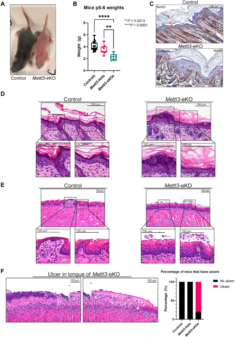
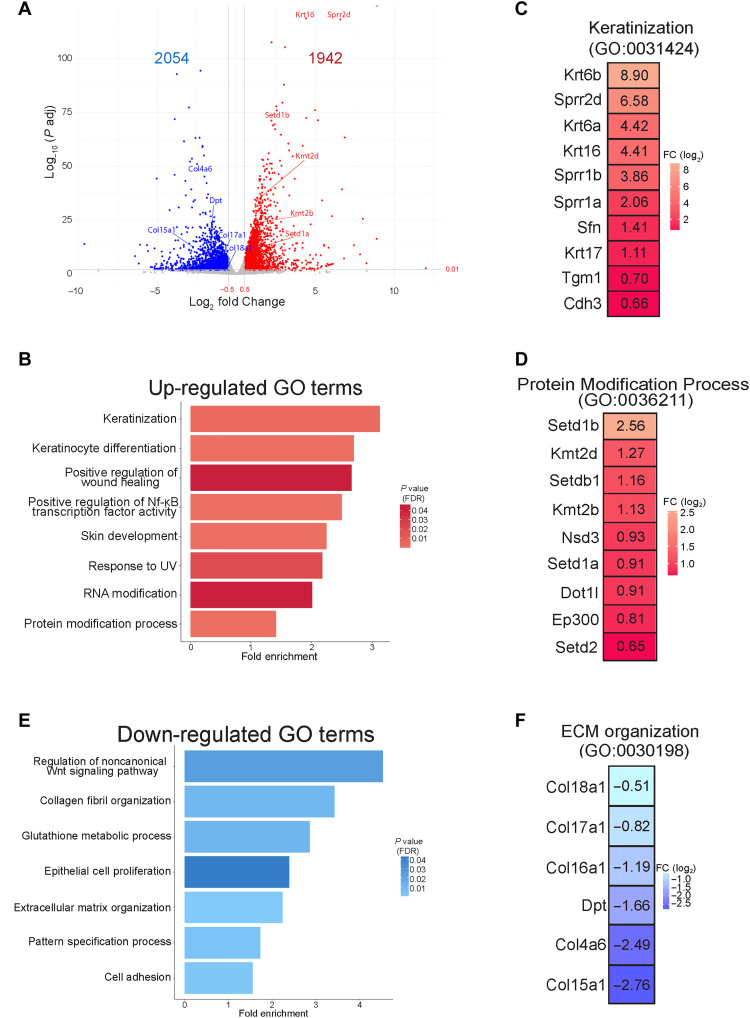
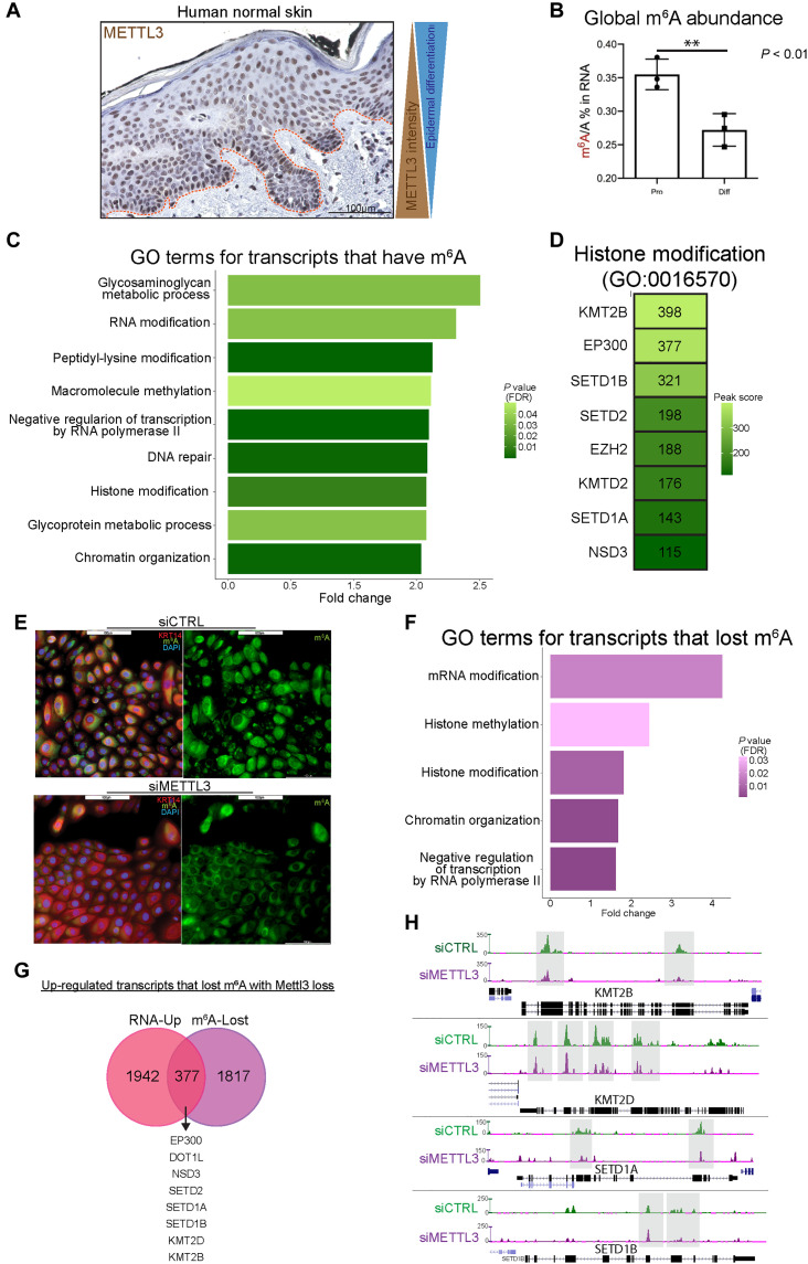
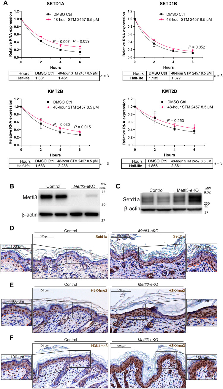
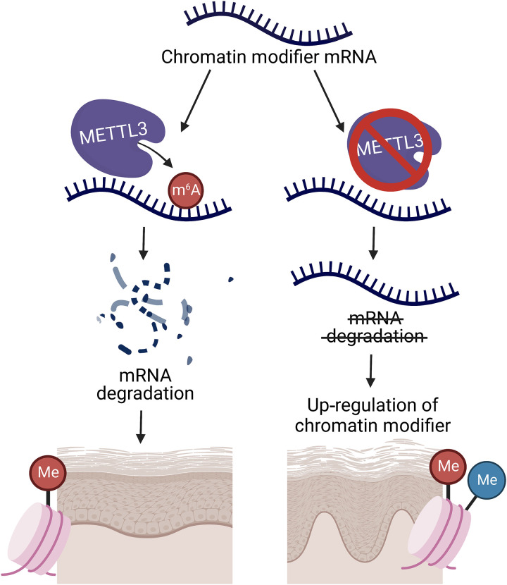
N6-methyladenosine (m6A) is the most abundant modification on messenger RNAs (mRNAs) and is catalyzed by methyltransferase-like protein 3 (Mettl3). To understand the role of m6A in a self-renewing somatic tissue, we deleted Mettl3 in epidermal progenitors in vivo. Mice lacking Mettl3 demonstrate marked features of dysfunctional development and self-renewal, including a loss of hair follicle morphogenesis and impaired cell adhesion and polarity associated with oral ulcerations. We show that Mettl3 promotes the m6A-mediated degradation of mRNAs encoding critical histone modifying enzymes. Depletion of Mettl3 results in the loss of m6A on these mRNAs and increases their expression and associated modifications, resulting in widespread gene expression abnormalities that mirror the gross phenotypic abnormalities. Collectively, these results have identified an additional layer of gene regulation within epithelial tissues, revealing an essential role for m6A in the regulation of chromatin modifiers, and underscoring a critical role for Mettl3-catalyzed m6A in proper epithelial development and self-renewal.


Figures

Fig. 1.
Fig. 1.. Loss of Mettl3 leads to grossly abnormal epidermal and tongue development.
(A) Postnatal day 6 (P6) Mettl3-eKO mice display a complete absence of hair and are half the size of WT control littermates. (B) Mettl3-eKO mice weigh less than controls and Mettl3-heterozygotes (“Hets”). (C) IHC of murine dorsal skin. The dotted orange line demarcates the epidermis-dermis junction. The orange arrow signals the nucleus of the keratinocyte in which Mettl3 is found. Controls have Mettl3 present throughout the epidermis, but the staining is gone in the Mettl3-eKO epidermis. (D) Mettl3-eKO skin displays malformed hair follicles (silver arrows), as well as loss of cell polarity in the basal epidermal cells along with thicker granular and cornified layers. (E) Mettl3-eKO oral epithelium of the tongue has malformed filiform papillae and tastebuds. (F) Mettl3-eKO tongues also display large ulcers in the posterior dorsal aspect.
Fig. 2.
Fig. 2.. Mettl3 loss promotes marked transcriptional dysregulation.
(A) RNA-seq was performed on epidermis from Mettl3-eKO P6 mice (n = 3) versus control littermates (n = 3). A log2 fold change of ±0.5 and an adjusted P value of <0.01 was done to visualize the significant changes. (B) Biological Process Terms Gene Ontology (GO:BP) analysis of up-regulated transcripts in Mettl3-eKO mice. UV, ultraviolet. (C) Highly significant up-regulated transcripts in Mettl3-eKO mice from the “keratinization” category. (D) Highly significant transcripts from the up-regulated “protein modification process” category. (E) GO:BP analysis of down-regulated transcripts in Mettl3-eKO mice. (F) Highly significant down-regulated transcripts of the down-regulated extracellular matrix (ECM) organization category.
Fig. 3.
Fig. 3.. Mettl3-mediated m6A dynamically regulates chromatin modifier mRNAs in epithelia.
(A) IHC of Mettl3 in normal human skin, where above the dotted line is the epidermis. (B) Quantitative analysis of the m6A level by liquid chromatography–tandem mass spectrometry done in proliferating versus differentiated neonatal human epidermal keratinocytes (NHEKs) demonstrates reduced global m6A with differentiation. (C) GO analysis of transcripts which contain m6A peaks enriched in epidermal progenitors. (D) Representative transcripts of the “histone modification” GO:BP category. DAPI, 4′,6-diamidino-2-phenylindole. (E) Mettl3 siRNA reduces visible m6A in NHEKs. (F) GO analysis of transcripts which contain m6A peaks that are lost with siRNA depletion of Mettl3. (G) Overlap of the up-regulated transcripts by RNA-seq (pink) and transcripts that lost m6A peaks with Mettl3 depletion (purple). (H) UCSC Genome Browser on Human (GRCh37/hg19) tracks from the m6A-seq analysis to visualize the decrease of m6A peaks in representative “histone modification” transcripts in control (green) and Mettl3-depleted NHEKs (purple).
Fig. 4.
Fig. 4.. Mettl3 loss increases mRNA half-life and expression of chromatin modifiers.
(A) mRNA turnover assay to measure the mRNA half-life transcripts in epidermal progenitors demonstrates prolonged half-life of SETD1A, SETD1B, and KMT2B transcripts with METTL3 depletion. KMT2D transcripts shows a trend of prolonged half-life with METTL3 depletion. Transcripts were normalized by glyceraldehyde-3-phosphate dehydrogenase. DMSO, dimethyl sulfoxide. (B) Immunoblotting of Mettl3 in control versus Mettl3-eKO P6 mouse epidermis. (C) High-molecular weight Western blot for Setd1a in P6 mouse epidermis. (D) IHC for Setd1a in P6 mouse epidermis. (E) IHC for H3K4me2 in P6 mouse epidermis. (F) IHC for H3K4me3 in P6 mouse epidermis.
Fig. 5.
Fig. 5.. Schematic of Mettl3-mediated m6A gene regulation in self-renewing stratifying epithelial tissues.
Loss of the Mettl3-m6A epitranscriptome promotes the up-regulation of chromatin modifiers by reducing mRNA degradation to promote increased mRNA half-life. This up-regulation of chromatin modifiers and, in turn, their associated histone modifications leads to dysfunctional epidermal development and differentiation.

References

    1. A. Baroni, E. Buommino, V. de Gregorio, E. Ruocco, V. Ruocco, R. Wolf, Structure and function of the epidermis related to barrier properties. Clin. Dermatol. 30, 257–262 (2012). - PubMed
    1. Y. A. Miroshnikova, I. Cohen, E. Ezhkova, S. A. Wickstrom, Epigenetic gene regulation, chromatin structure, and force-induced chromatin remodelling in epidermal development and homeostasis. Curr. Opin. Genet. Dev. 55, 46–51 (2019). - PMC - PubMed
    1. P. Rousselle, E. Gentilhomme, Y. Neveux, Markers of epidermal proliferation and differentiation, in Agache’s Measuring the Skin, P. Humbert, F. Fanian, H. Maibach, P. Agache, Eds. (Springer International Publishing, 2017), pp. 397–405.
    1. E. M. Novoa, C. E. Mason, J. S. Mattick, Charting the unknown epitranscriptome. Nat. Rev. Mol. Cell Biol. 18, 339–340 (2017). - PubMed
    1. C. J. Lewis, T. Pan, A. Kalsotra, RNA modifications and structures cooperate to guide RNA-protein interactions. Nat. Rev. Mol. Cell Biol. 18, 202–210 (2017). - PMC - PubMed