. 2023 Sep 1;13(1):14384.
doi: 10.1038/s41598-023-41247-6.

Analysis of flow-induced transcriptional response and cell alignment of different sources of endothelial cells used in vascular tissue engineering

Affiliations

Analysis of flow-induced transcriptional response and cell alignment of different sources of endothelial cells used in vascular tissue engineering

Diana M Rojas-González et al. Sci Rep. .

Abstract

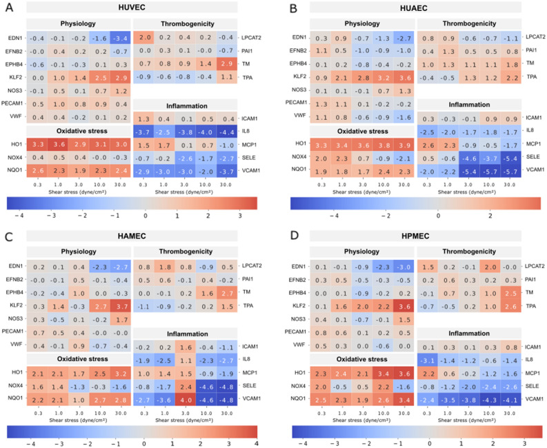
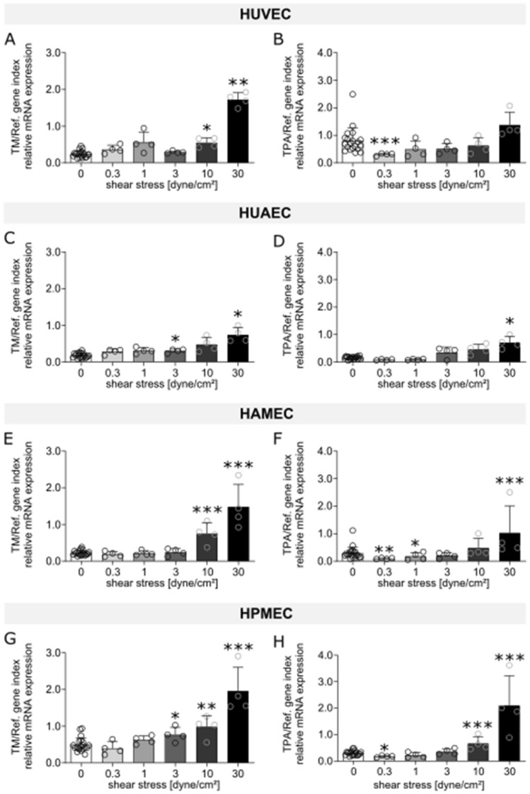
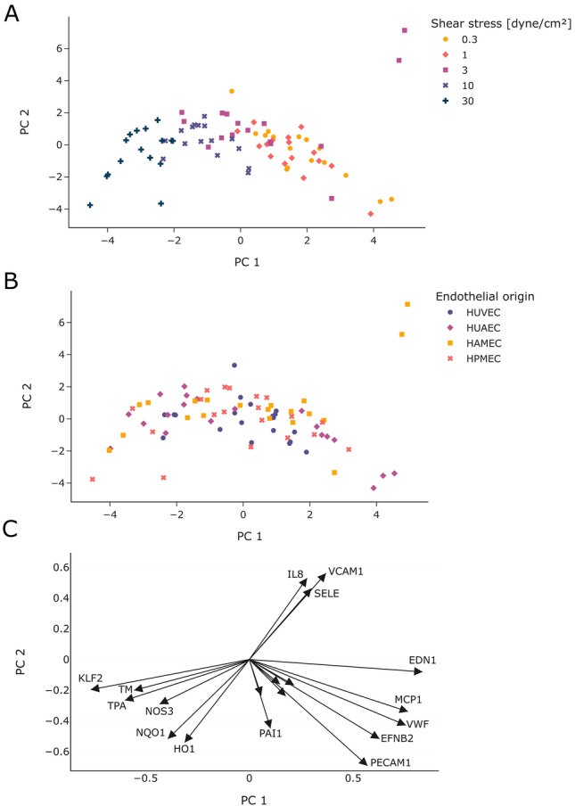
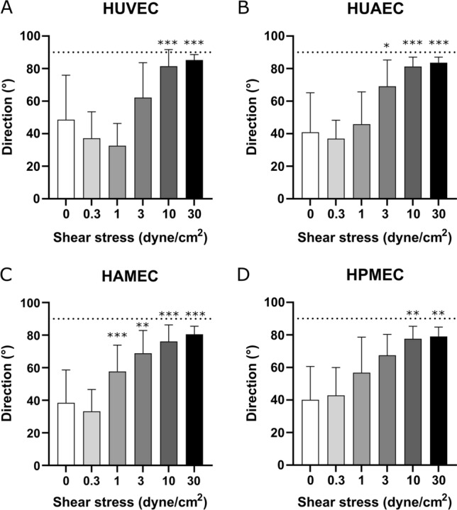
Endothelialization of tissue-engineered vascular grafts has proven crucial for implant functionality and thus clinical outcome, however, the choice of endothelial cells (ECs) is often driven by availability rather than by the type of vessel to be replaced. In this work we studied the response to flow of different human ECs with the aim of examining whether their response in vitro is dictated by their original in vivo conditions. Arterial, venous, and microvascular ECs were cultured under shear stress (SS) of 0, 0.3, 3, 1, 10, and 30 dyne/cm2 for 24 h. Regulation of flow-induced marker KLF2 was similar across the different ECs. Upregulation of anti-thrombotic markers, TM and TPA, was mainly seen at higher SS. Cell elongation and alignment was observed for the different ECs at 10 and 30 dyne/cm2 while at lower SS cells maintained a random orientation. Downregulation of pro-inflammatory factors SELE, IL8, and VCAM1 and up-regulation of anti-oxidant markers NQO1 and HO1 was present even at SS for which cell alignment was not observed. Our results evidenced similarities in the response to flow among the different ECs, suggesting that the maintenance of the resting state in vitro is not dictated by the SS typical of the tissue of origin and that absence of flow-induced cell orientation does not necessarily correlate with a pro-inflammatory state of the ECs. These results support the use of ECs from easily accessible sources for in vitro vascular tissue engineering independently from the target vessel.


Conflict of interest statement

The authors declare no competing interests.

Figures

Figure 1
Figure 1
Heat maps depicting fold changes for all measured markers in HUVEC (A), HUAEC (B), HAMEC (C), and HPMEC (D). The numbers represent the fold change in log2 scale of the relative mRNA expression levels of the samples normalized to their corresponding static controls. Heatmaps were created using the seaborn package in python (v3.11, https://seaborn.pydata.org/index.html).
Figure 2
Figure 2
mRNA expression of markers related to endothelial cell physiology (KLF2, EDN1, NOS3) for HUVEC (AC), HUAEC (DF), HAMEC (GI), and HPMEC (JL) at the different magnitudes of shear stress. Statistically significant differences to the static control are indicated by asterisks (*p < 0.05, **p < 0.01 and ***p < 0.001).
Figure 3
Figure 3
mRNA expression of markers related to thrombogenicity (TM, TPA) for HUVEC (A,B), HUAEC (C,D), HAMEC (E,F), and HPMEC (G,H) at the different magnitudes of shear stress. Statistically significant differences to the static control are indicated by asterisks (*p < 0.05, **p < 0.01 and ***p < 0.001).
Figure 4
Figure 4
mRNA expression of markers related to inflammation (MCP1, SELE, VCAM1) for HUVEC (AC), HUAEC (DF), HAMEC (GI), and HPMEC (JL) at the different magnitudes of shear stress. Statistically significant differences to the static control are indicated by asterisks (*p < 0.05, **p < 0.01 and ***p < 0.001).
Figure 5
Figure 5
mRNA expression of markers related to oxidative stress (HO1, NQO1) for HUVEC (A,B), HUAEC (C,D), HAMEC (E,F), and HPMEC (G,H) at the different magnitudes of shear stress. Statistically significant differences to the static control are indicated by asterisks (*p < 0.05, **p < 0.01 and ***p < 0.001).
Figure 6
Figure 6
Comparison of mRNA expression levels across endothelial cells from different vascular beds exposed to 0 and 30 dyne/cm2. Statistically significant differences across different ECs types are indicated by asterisks (*p < 0.05, **p < 0.01 and ***p < 0.001).
Figure 7
Figure 7
Principal component analysis of mRNA expression data. The results are presented as scattered plots where each symbol represents one sample. Samples were colored for the different shear stress levels (A) or the different endothelial cell type (B). The loadings for the different genes were plotted as arrows (C).
Figure 8
Figure 8
Phase contrast images of the different types of endothelial cells after 24 h of static culture (AD) and flow exposure at shear stress magnitudes of 0.3 (EH), 1 (IL), 3 (MP), 10 (QT), and 30 dyne/cm2 (UX). Scale bar: 100 µm.
Figure 9
Figure 9
Analysis on preferred cell direction at the different shear stress magnitudes for HUVEC (A), HUAEC (B), HAMEC (C), and HPMEC (D). Statistically significant differences to the static control are indicated by asterisks (*p < 0.05, **p < 0.01 and ***p < 0.001). Dotted line indicates 90°.

References

    1. Pashneh-Tala S, MacNeil S, Claeyssens F. The tissue-engineered vascular graft-past, present, and future. Tissue Eng. Part B. 2016;22:68–100. doi: 10.1089/ten.teb.2015.0100. - DOI - PMC - PubMed
    1. Chang WG, Niklason LE. A short discourse on vascular tissue engineering. npj Regener. Med. 2017;2:7. doi: 10.1038/s41536-017-0011-6. - DOI - PMC - PubMed
    1. Jouda H, Larrea Murillo L, Wang T. Current progress in vascular engineering and its clinical applications. Cells. 2022;11:493. doi: 10.3390/cells11030493. - DOI - PMC - PubMed
    1. Gupta P, Mandal BB. Tissue-engineered vascular grafts: Emerging trends and technologies. Adv. Func. Mater. 2021;31:2100027. doi: 10.1002/adfm.202100027. - DOI
    1. Zilla P, Deutsch M, Bezuidenhout D, Davies NH, Pennel T. Progressive reinvention or destination lost? Half a century of cardiovascular tissue engineering. Front. Cardiovasc. Med. 2020;7:00159. doi: 10.3389/fcvm.2020.00159. - DOI - PMC - PubMed

Publication types

LinkOut - more resources