Observational Study
. 2023 Dec;152(6):1619-1633.e11.
doi: 10.1016/j.jaci.2023.07.022. Epub 2023 Sep 1.

Intestinal microbiome and metabolome signatures in patients with chronic granulomatous disease

Affiliations
Observational Study

Intestinal microbiome and metabolome signatures in patients with chronic granulomatous disease

Prabha Chandrasekaran et al. J Allergy Clin Immunol. 2023 Dec.

Abstract

Background: Chronic granulomatous disease (CGD) is caused by defects in any 1 of the 6 subunits forming the nicotinamide adenine dinucleotide phosphate oxidase complex 2 (NOX2), leading to severely reduced or absent phagocyte-derived reactive oxygen species production. Almost 50% of patients with CGD have inflammatory bowel disease (CGD-IBD). While conventional IBD therapies can treat CGD-IBD, their benefits must be weighed against the risk of infection. Understanding the impact of NOX2 defects on the intestinal microbiota may lead to the identification of novel CGD-IBD treatments.

Objective: We sought to identify microbiome and metabolome signatures that can distinguish individuals with CGD and CGD-IBD.

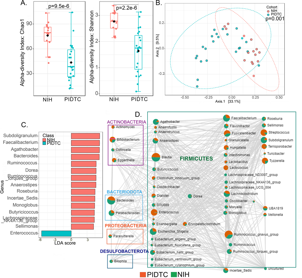
Methods: We conducted a cross-sectional observational study of 79 patients with CGD, 8 pathogenic variant carriers, and 19 healthy controls followed at the National Institutes of Health Clinical Center. We profiled the intestinal microbiome (amplicon sequencing) and stool metabolome, and validated our findings in a second cohort of 36 patients with CGD recruited through the Primary Immune Deficiency Treatment Consortium.

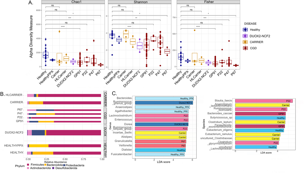
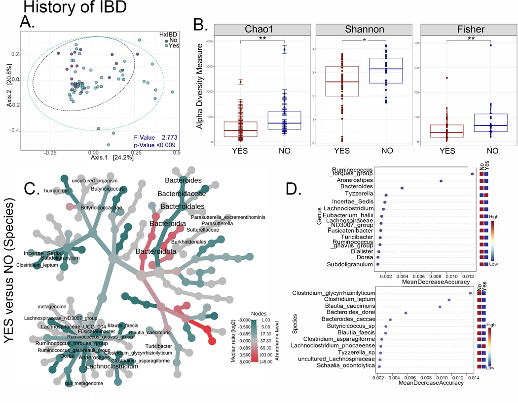
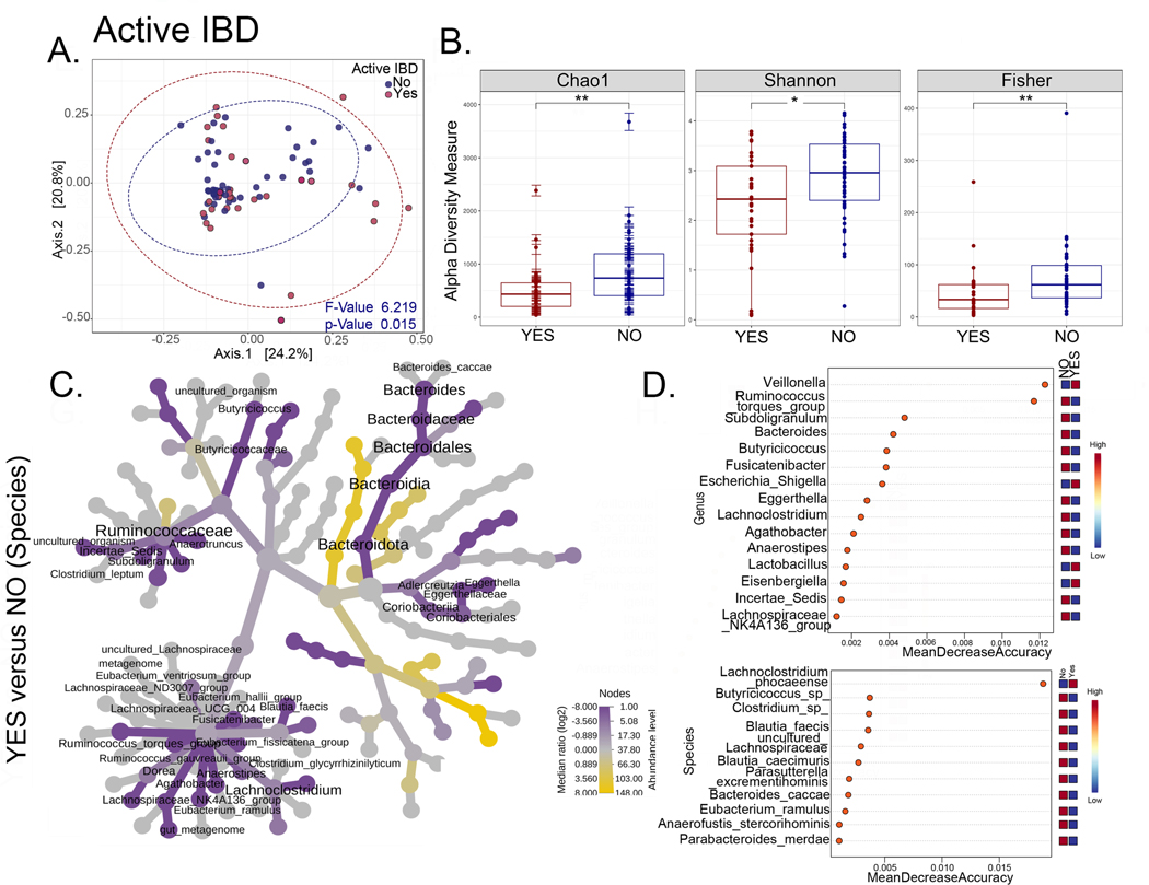
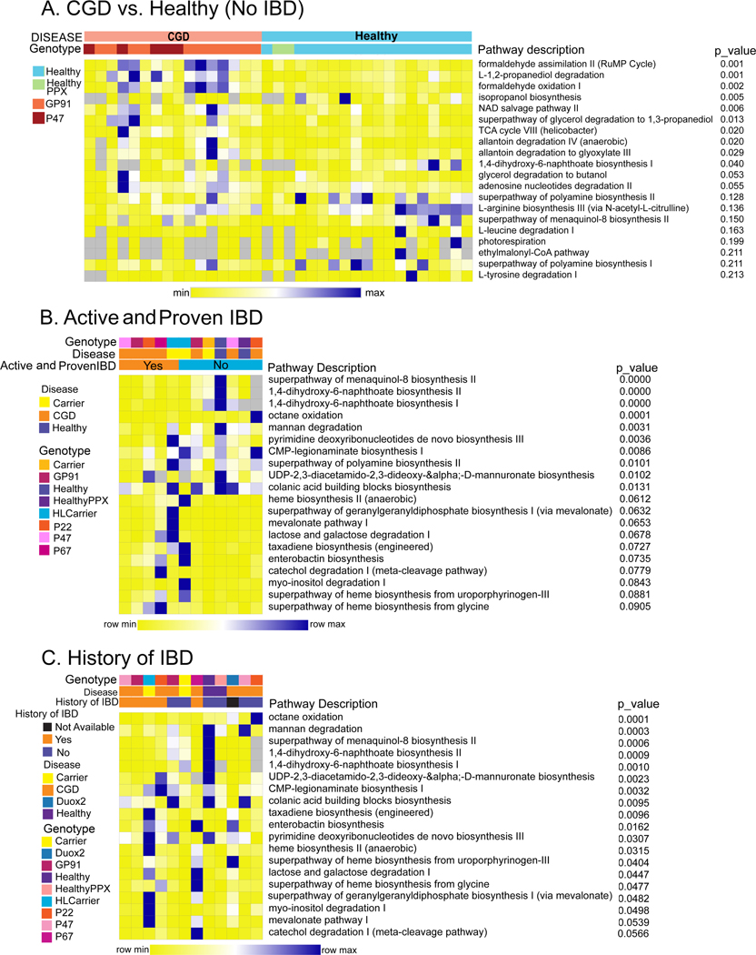
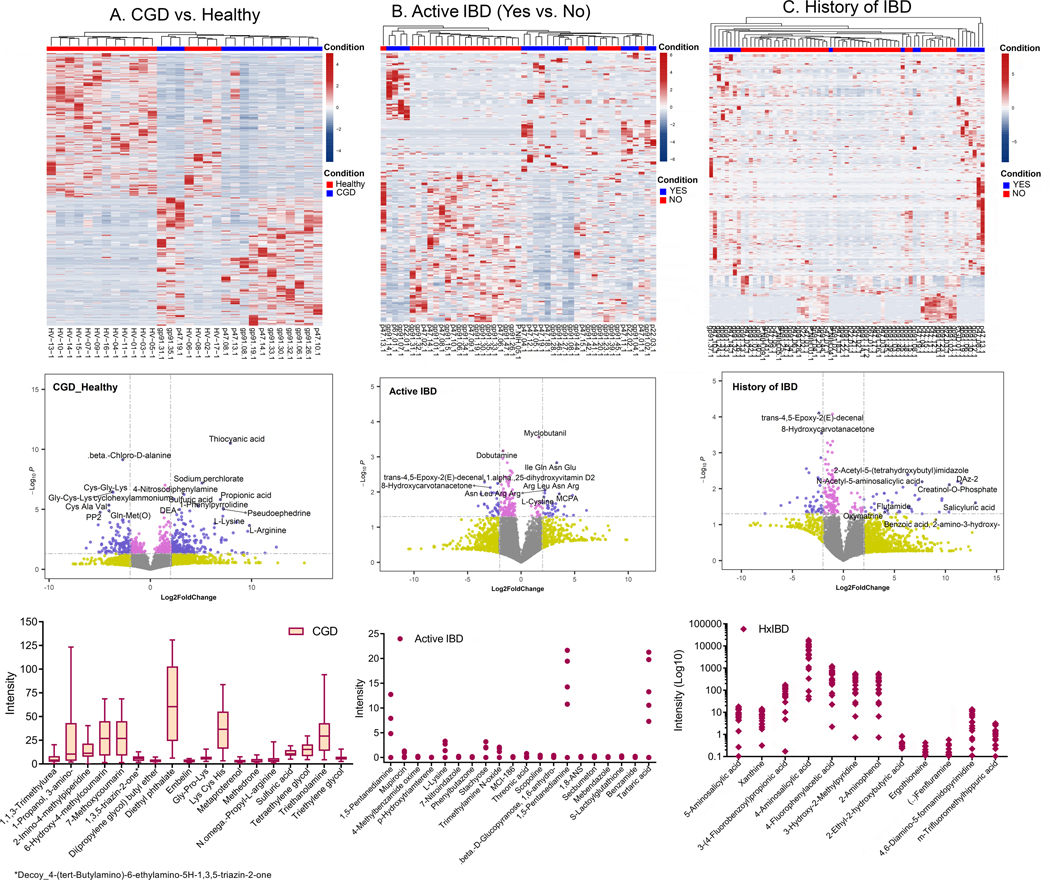
Results: We identified distinct intestinal microbiome and metabolome profiles in patients with CGD compared to healthy individuals. We observed enrichment for Erysipelatoclostridium spp, Sellimonas spp, and Lachnoclostridium spp in CGD stool samples. Despite differences in bacterial alpha and beta diversity between the 2 cohorts, several taxa correlated significantly between both cohorts. We further demonstrated that patients with CGD-IBD have a distinct microbiome and metabolome profile compared to patients without CGD-IBD.

Conclusion: Intestinal microbiome and metabolome signatures distinguished patients with CGD and CGD-IBD, and identified potential biomarkers and therapeutic targets.

Trial registration: ClinicalTrials.gov NCT02082353.

Keywords: CGD; Chronic granulomatous disease; IBD; NADPH oxidase; dysbiosis; inborn errors of immunity; inflammatory bowel disease; intestinal inflammation; metabolome; microbiome; primary immune deficiency.


Figures

Figure 1.
Figure 1.. Association of CGD genotype with microbiome signature.
(A) Alpha diversity (within-group variations) analyses (Chao1, Shannon, Fisher) comparing different genotypes (Healthy [n= 17]; Healthy PPX [i.e. healthy patients on infection prophylaxis with TMP-SMX for recurrent cystitis, n=2]; Carriers [CYBB−/+, n=4]; HL [highly lyonized, n=2] carriers; DUOX2-NCF2 [DUOX2−/+ and NCF2−/+ digenic heterozygous carriers, n=2]; GP91 [CYBB0/−, n=48], P22 [CYBA−/−, n=5], P47 [NCF1−/−, n=23], and P67 [NCF2−/−, n=2] CGD patients. * = p<0.05, **=p<0.01, ***=p<0.001, ****=p<0.0001, ns = non-significant. (B) Bar graphs showing relative abundance of bacterial phyla. (C) Linear discriminant analysis (LDA) score determined by the LEfSe analysis for identification of biomarkers, showing significantly enriched taxa in specific genotypes (p<0.05).
Figure 2.
Figure 2.. Intestinal microbiome signatures distinguish patients with CGD from healthy individuals.
(A-G) Comparisons for the CGD group (without a history of IBD and only on prophylactic antimicrobials, n=16) with the Healthy group (n=17). (A) Alpha diversity (within group variations) analyses (Chao1, Shannon, Fisher; * = p<0.05, **=p<0.01, ***=p<0.001, ****=p<0.0001, ns = non-significant) for CGD compared to Healthy. (B) Relative abundance of amplicon sequence variants in both groups presented at phylum level. (C) PCoA plot of beta diversity (between-group variations) based on weighted Unifrac distances for the two comparison groups with p values determined by Analysis of similarities (ANOSIM; p<0.003) and PERMDISP test for significant differences in dispersion (p =0.034). (D) Heat tree depicting the phylogenetic relationship and significant differential abundance (p<0.05) of bacterial genera between CGD and Healthy groups (red = higher abundance; blue = lower abundance). (E) Top differentially abundant family and species as identified by edgeR analysis (p values for all comparisons are <0.0001). (F) Linear discriminant analysis (LDA) score determined by the LEfSe analysis showing biomarkers at genus and species levels that are present or absent in the experimental groups shown. (G) Random forest analysis where genera and species with highest discriminatory power between CGD and Healthy groups are listed (red = high abundance in the experimental group, blue = low abundance in the experimental group).
Figure 3.
Figure 3.. Comparison of microbiome signatures between patients with CGD from NIH CC and PIDTC cohorts.
(A-D) Comparisons for the CGD group (without a history of IBD and not on any medications other than prophylactic antimicrobials) between the NIH CC (n=16) and PIDTC cohorts (n=23). (A) Alpha diversity (within group variations) analyses (Chao1 and Shannon) at genus level. (B) PCoA plot of beta diversity (between group variations) based on weighted Unifrac distances for the two comparison groups with p values determined by permutational MANOVA (PERMANOVA, p=0.001). (C) Linear discriminant analysis (LDA) score determined by the LEfSe analysis for biomarker identification, showing the defining genera of the cohorts. (D) Correlation at genus level of the two cohorts is depicted as the network. Each node represents a taxon and the size of the node corresponds to the number of connections. Two taxa connected by an edge when p<0.05 and the correlation threshold >0.3. Within each taxon circle, green represents distribution in NIH CC cohort and red in PIDTC cohort.
Figure 4.
Figure 4.. The intestinal microbiome distinguishes patients with CGD and a history of IBD.
(A-D) Comparisons for the NIH CC CGD group with or without the history of IBD (Yes n=54, No n=25). (A) PCoA plots of beta diversity based on weighted Unifrac distances for the two comparison groups with p values determined by permutational MANOVA (PERMANOVA, p<0.009). (B) Alpha diversity plots (Chao1, Shannon, Fisher; * = p<0.05, **=p<0.01) comparing patients with CGD, with and without a history of IBD. (C) Heat tree depicting phylogenetic relationship and significant differential abundance (p<0.05) of bacterial genera for the comparison groups. (D) Random forest plots where genera and species with highest discriminatory power between patients with CGD with vs. without a history of IBD are shown (red = high abundance, blue = low abundance).
Figure 5.
Figure 5.. The intestinal microbiome distinguishes patients with CGD and active IBD.
(A-D) Comparisons for the NIH CC CGD group with or without active IBD (Yes n=33 and No n=46). (A) PCoA plots of beta diversity based on weighted Unifrac distances for the two comparison groups with p values determined by permutational MANOVA (PERMANOVA, p=0.015). (B) Alpha diversity plots (Chao1, Shannon, Fisher; * = p<0.05, **=p<0.01). (C) Heat tree depicting phylogenetic relationship and significant differential abundance (p<0.05) of bacterial genera for the comparison groups. (D) Random forest plots where genera and species with highest discriminatory power between patients with CGD with vs. without a active IBD are shown (red = high abundance, blue = low abundance).
Figure 6.
Figure 6.. Distinct functional profiles of the intestinal microbiome in patients with CGD from the NIH CC cohort.
Heatmap of significantly different functional profiles inferred by PICRUSt2 analysis performed to identify the pathways associated with changes in amplicon sequence variants (blue represents higher abundance and yellow represents lower abundance). The relative abundance was normalized to a Z-score and utilized to generate the heatmaps. (A) Pathway comparisons of the NIH CC CGD group (no history of IBD and only on prophylactic antimicrobials, n=16) with Healthy (n=17). (B) Pathway comparisons of patients with vs. without active and endoscopically proven CGD-IBD (Yes n=33 and No n=46). (C) Pathway comparisons of patients with vs. without a history of CGD-IBD (Yes n=54 and No n=25). The predicted p-values values are shown alongside the heatmap.
Figure 7.
Figure 7.. Distinct metabolome profiles in patients with CGD from the NIH CC cohort.
Top to Bottom: Heatmap of the metabolomic profile, volcano plot displaying high metabolic diversity between the comparison groups, and the plots for the intensity measurements of the metabolites that are expressed only in the indicated groups, which are potential biomarkers. (A) Comparison of CGD group (without a history of IBD and only on prophylactic antimicrobials, n=14) to Healthy (n=16), (B) comparison within the CGD group of those with (Yes n=30) vs. without active IBD (No n=36), and (C) comparison within the CGD group of those with (Yes n=50) vs. without a history of IBD (No n=19). In the heatmaps, the rows display the metabolites and the columns represent the samples (blue = decreased, red = increased). The brightness of each color corresponds to the magnitude of the difference when compared with the average value. The lines in the volcano plots indicate the significance cut-off for the p-value (-log10 P value of 1.3013 corresponding to p<0.05) and fold change (log2 Fold Change >2, log2 Fold Change <−2). All metabolites that are significant and over an absolute log2 Fold Change of 2 are shown in violet and those that are significant but have an expression change less than an absolute log2 Fold Change of 2 are shown in pink. The bar plot indicates metabolites identified only in the CGD group, the dot plot indicates metabolites identified only in CGD patients with active IBD, and the diamond plot indicates metabolites identified only in patients with CGD and a history of IBD.

Similar articles

Cited by

References

    1. Mittal M, Siddiqui MR, Tran K, Reddy SP, Malik AB. Reactive oxygen species in inflammation and tissue injury. Antioxid Redox Signal 2014; 20:1126–67. - PMC - PubMed
    1. Dhillon SS, Fattouh R, Elkadri A, Xu W, Murchie R, Walters T, et al. Variants in nicotinamide adenine dinucleotide phosphate oxidase complex components determine susceptibility to very early onset inflammatory bowel disease. Gastroenterology 2014; 147:680–9 e2. - PubMed
    1. Falcone EL, Holland SM. Gastrointestinal Complications in Chronic Granulomatous Disease. Methods Mol Biol 2019; 1982:573–86. - PubMed
    1. Magnani A, Brosselin P, Beaute J, de Vergnes N, Mouy R, Debre M, et al. Inflammatory manifestations in a single-center cohort of patients with chronic granulomatous disease. J Allergy Clin Immunol 2014; 134:655–62 e8. - PubMed
    1. Segal BH, Leto TL, Gallin JI, Malech HL, Holland SM. Genetic, biochemical, and clinical features of chronic granulomatous disease. Medicine (Baltimore) 2000; 79:170–200. - PubMed

Publication types

Associated data