

Development of a burst wave lithotripsy system for noninvasive fragmentation of ureteroliths in pet cats
- PMID: 37660015
- PMCID: PMC10474658
- DOI: 10.1186/s12917-023-03705-1
Development of a burst wave lithotripsy system for noninvasive fragmentation of ureteroliths in pet cats
Abstract
Background: Upper urinary tract stones are increasingly prevalent in pet cats and are difficult to manage. Surgical procedures to address obstructing ureteroliths have short- and long-term complications, and medical therapies (e.g., fluid diuresis and smooth muscle relaxants) are infrequently effective. Burst wave lithotripsy is a non-invasive, ultrasound-guided, handheld focused ultrasound technology to disintegrate urinary stones, which is now undergoing human clinical trials in awake unanesthetized subjects.
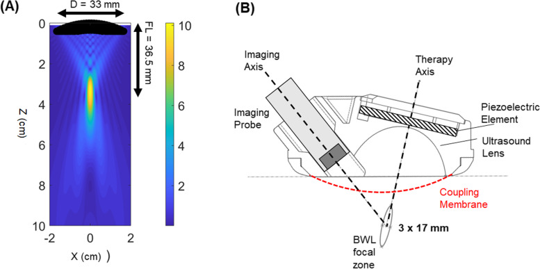
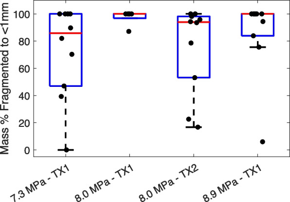
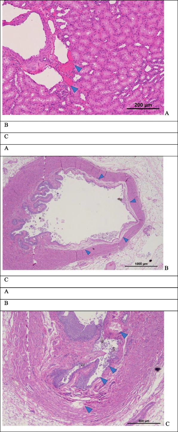
Results: In this study, we designed and performed in vitro testing of a modified burst wave lithotripsy system to noninvasively fragment stones in cats. The design accounted for differences in anatomic scale, acoustic window, skin-to-stone depth, and stone size. Prototypes were fabricated and tested in a benchtop model using 35 natural calcium oxalate monohydrate stones from cats. In an initial experiment, burst wave lithotripsy was performed using peak ultrasound pressures of 7.3 (n = 10), 8.0 (n = 5), or 8.9 MPa (n = 10) for up to 30 min. Fourteen of 25 stones fragmented to < 1 mm within the 30 min. In a second experiment, burst wave lithotripsy was performed using a second transducer and peak ultrasound pressure of 8.0 MPa (n = 10) for up to 50 min. In the second experiment, 9 of 10 stones fragmented to < 1 mm within the 50 min. Across both experiments, an average of 73-97% of stone mass could be reduced to fragments < 1 mm. A third experiment found negligible injury with in vivo exposure of kidneys and ureters in a porcine animal model.
Conclusions: These data support further evaluation of burst wave lithotripsy as a noninvasive intervention for obstructing ureteroliths in cats.
© 2023. BioMed Central Ltd., part of Springer Nature.
Conflict of interest statement
The authors declare no competing interests.
Figures


Similar articles
-
Evaluation of Renal Stone Comminution and Injury by Burst Wave Lithotripsy in a Pig Model.J Endourol. 2019 Oct;33(10):787-792. doi: 10.1089/end.2018.0886. Epub 2019 May 27. J Endourol. 2019. PMID: 31016998 Free PMC article.
-
Fragmentation of urinary calculi in vitro by burst wave lithotripsy.J Urol. 2015 Jan;193(1):338-44. doi: 10.1016/j.juro.2014.08.009. Epub 2014 Aug 9. J Urol. 2015. PMID: 25111910 Free PMC article.
-
Combined Burst Wave Lithotripsy and Ultrasonic Propulsion for Improved Urinary Stone Fragmentation.J Endourol. 2018 Apr;32(4):344-349. doi: 10.1089/end.2017.0675. Epub 2018 Mar 20. J Endourol. 2018. PMID: 29433329 Free PMC article.
-
Burst wave lithotripsy and acoustic manipulation of stones.Curr Opin Urol. 2020 Mar;30(2):149-156. doi: 10.1097/MOU.0000000000000727. Curr Opin Urol. 2020. PMID: 31905177 Free PMC article. Review.
-
Electrohydraulic and extracorporeal shock-wave lithotripsy.Vet Clin North Am Small Anim Pract. 1999 Jan;29(1):293-302, xv. doi: 10.1016/s0195-5616(99)50017-x. Vet Clin North Am Small Anim Pract. 1999. PMID: 10028164 Review.
Cited by
-
Burst wave lithotripsy - a paradigm shift: inferences from a scoping review.World J Urol. 2025 Apr 25;43(1):250. doi: 10.1007/s00345-025-05645-x. World J Urol. 2025. PMID: 40278907 Free PMC article.
-
Application of novel burst wave lithotripsy and ultrasonic propulsion technology for the treatment of ureteral calculi in a bottlenose dolphin (Tursiops truncatus) and renal calculi in a harbor seal (Phoca vitulina).Urolithiasis. 2024 Jan 8;52(1):21. doi: 10.1007/s00240-023-01515-6. Urolithiasis. 2024. PMID: 38189835 Free PMC article.
References
MeSH terms
Substances
Grants and funding
LinkOut - more resources
Full Text Sources
Miscellaneous
