

The prefrontal cortex of the bottlenose dolphin (Tursiops truncatus Montagu, 1821): a tractography study and comparison with the human
- PMID: 37660322
- PMCID: PMC10517040
- DOI: 10.1007/s00429-023-02699-8
The prefrontal cortex of the bottlenose dolphin (Tursiops truncatus Montagu, 1821): a tractography study and comparison with the human
Abstract
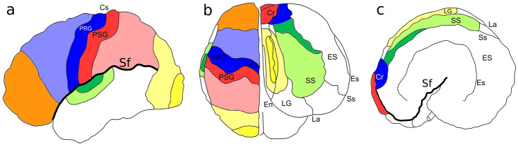
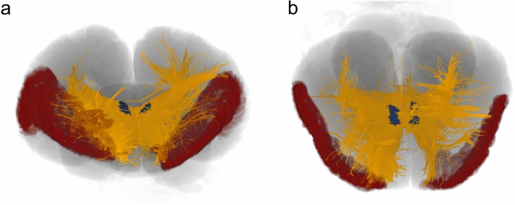
Cetaceans are well known for their remarkable cognitive abilities including self-recognition, sound imitation and decision making. In other mammals, the prefrontal cortex (PFC) takes a key role in such cognitive feats. In cetaceans, however, a PFC could up to now not be discerned based on its usual topography. Classical in vivo methods like tract tracing are legally not possible to perform in Cetacea, leaving diffusion-weighted imaging (DWI) as the most viable alternative. This is the first investigation focussed on the identification of the cetacean PFC homologue. In our study, we applied the constrained spherical deconvolution (CSD) algorithm on 3 T DWI scans of three formalin-fixed brains of bottlenose dolphins (Tursiops truncatus) and compared the obtained results to human brains, using the same methodology. We first identified fibres related to the medio-dorsal thalamic nuclei (MD) and then seeded the obtained putative PFC in the dolphin as well as the known PFC in humans. Our results outlined the dolphin PFC in areas not previously studied, in the cranio-lateral, ectolateral and opercular gyri, and furthermore demonstrated a similar connectivity pattern between the human and dolphin PFC. The antero-lateral rotation of the PFC, like in other areas, might be the result of the telescoping process which occurred in these animals during evolution.
Keywords: Brain evolution; CSD; Cetacean; DWI; Dolphin; PFC.
© 2023. The Author(s).
Conflict of interest statement
The authors declare no competing interests.
Figures


Similar articles
-
Diffusion tractography reveals pervasive asymmetry of cerebral white matter tracts in the bottlenose dolphin (Tursiops truncatus).Brain Struct Funct. 2018 May;223(4):1697-1711. doi: 10.1007/s00429-017-1525-9. Epub 2017 Nov 30. Brain Struct Funct. 2018. PMID: 29189908 Free PMC article.
-
Cetacean morbillivirus in coastal Indo-Pacific bottlenose dolphins, Western Australia.Emerg Infect Dis. 2014 Apr;20(4):666-70. doi: 10.3201/eid2004.131714. Emerg Infect Dis. 2014. PMID: 24656203 Free PMC article.
-
Constrained spherical deconvolution on diffusion-weighted images of dolphin brains.Magn Reson Imaging. 2024 May;108:104-110. doi: 10.1016/j.mri.2024.02.002. Epub 2024 Feb 8. Magn Reson Imaging. 2024. PMID: 38336113
-
The Gulf of Ambracia's Common Bottlenose Dolphins, Tursiops truncatus: A Highly Dense and yet Threatened Population.Adv Mar Biol. 2016;75:259-296. doi: 10.1016/bs.amb.2016.07.002. Epub 2016 Aug 25. Adv Mar Biol. 2016. PMID: 27770987 Review.
-
An update on the development of a bottlenose dolphin, Tursiops truncatus, immune reagent toolkit.Vet Immunol Immunopathol. 2024 Jun;272:110769. doi: 10.1016/j.vetimm.2024.110769. Epub 2024 Apr 29. Vet Immunol Immunopathol. 2024. PMID: 38703558 Review.
Cited by
-
Beyond the surface: how ex-vivo diffusion-weighted imaging reveals large animal brain microstructure and connectivity.Front Neurosci. 2024 Jun 26;18:1411982. doi: 10.3389/fnins.2024.1411982. eCollection 2024. Front Neurosci. 2024. PMID: 38988768 Free PMC article. Review.
-
Neuroanatomy of the Cetacean Sensory Systems.Animals (Basel). 2023 Dec 23;14(1):66. doi: 10.3390/ani14010066. Animals (Basel). 2023. PMID: 38200796 Free PMC article. Review.
-
"Of Marine Mammal Neuroscience and Men": Needs and Perspectives in Marine Mammal Neuroscience.J Comp Neurol. 2025 Jul;533(7):e70067. doi: 10.1002/cne.70067. J Comp Neurol. 2025. PMID: 40629534 Free PMC article. Review.
-
Distribution of calcium-binding proteins immunoreactivity in the bottlenose dolphin entorhinal cortex.Front Neuroanat. 2024 Feb 5;18:1321025. doi: 10.3389/fnana.2024.1321025. eCollection 2024. Front Neuroanat. 2024. PMID: 38379680 Free PMC article.
-
Meningeal Lymphatic and Glymphatic Structures in a Pelagic Delphinid (Delphinus delphis).Animals (Basel). 2025 Mar 4;15(5):729. doi: 10.3390/ani15050729. Animals (Basel). 2025. PMID: 40076012 Free PMC article.
References
-
- Arrigo A, Calamuneri A, Mormina E. Biomedical imaging. SmGroup: Dover; 2016. Diffusion MRI: from principles to modelling and clinical applications; pp. 1–18.
MeSH terms
Grants and funding
LinkOut - more resources
Full Text Sources
Miscellaneous
